Hostname: page-component-848d4c4894-x24gv Total loading time: 0 Render date: 2024-05-10T07:57:46.027Z Has data issue: false hasContentIssue false

Lorentz Transmission Electron Microscopy Imaging of Magnetic Textures in MnBi

Published online by Cambridge University Press:  30 July 2021

Núria Bagués
Affiliation:
Center for Electron Microscopy and Analysis, The Ohio State University, Columbus, OH, USA, COLUMBUS, Ohio, United States
Brandi L. Wooten
Affiliation:
Department of Materials Science and Engineering, The Ohio State University, Columbus, OH, USA, Columbus, Ohio, United States
Bin He
Affiliation:
Max Planck Insitute for Chemical Phisics of Solids, Dresden, Gemany, United States
Brian C. Sales
Affiliation:
Materials Science and Technology Division, Oak Ridge National Lab, Oak Ridge, TN, USA, United States
Joseph Heremans
Affiliation:
Department of Mechanical and Aerospace Engineering, The Ohio State University, Columbus, OH, USA, United States
David McComb
Affiliation:
Center for Electron Microscopy and Analysis, The Ohio State University, Columbus, OH, USA, COLUMBUS, Ohio, United States

Abstract

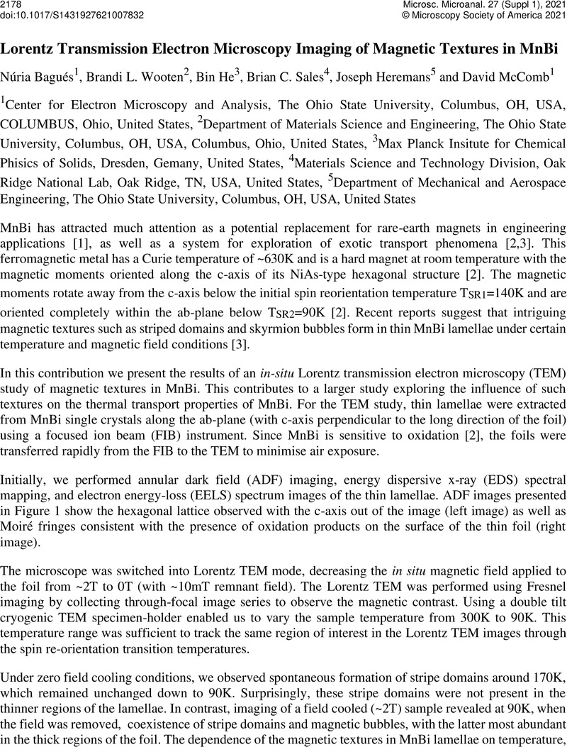
Image of the first page of this content. For PDF version, please use the ‘Save PDF’ preceeding this image.'
Type
Investigating Phase Transitions in Functional Materials and Devices by In Situ/Operando TEM
Copyright
Copyright © The Author(s), 2021. Published by Cambridge University Press on behalf of the Microscopy Society of America

References

Rama Rao, N. V. et al. , J. Phys. D: Appl. Phys. 46 (2013), 062001CrossRefGoogle Scholar
He, B. et al. , arXiv:2009.02211 (2020)Google Scholar
He, Y. et al. , arXiv:2011.06340 (2020)Google Scholar
McGuire, M. A. et al. , Physical Review B 90, (2014), 174425CrossRefGoogle Scholar
The authors acknowledge funding from Defense Advanced Research Projects Agency (DARPA) under Grant No. D18AP00008 and the Center for Emergent Materials at The Ohio State University, an NSF MRSEC (DMR-2011876). B.C.S. was supported by the DOE, Office of Science, Basic Energy Sciences, Materials Sciences and Engineering Division. The electron microscopy was performed at the Center for Electron Microscopy and Analysis (CEMAS) at The Ohio State University.Google Scholar