Hostname: page-component-8448b6f56d-jr42d Total loading time: 0 Render date: 2024-04-19T03:13:17.727Z Has data issue: false hasContentIssue false

Introduction to Special Issue on Electron Microscopy of Specimens in Liquid

Published online by Cambridge University Press:  19 March 2014

Niels de Jonge*
Affiliation:
Leibniz Institute for New Materials (INM), 66123 Saarbr̈cken, Germany
Get access

Abstract

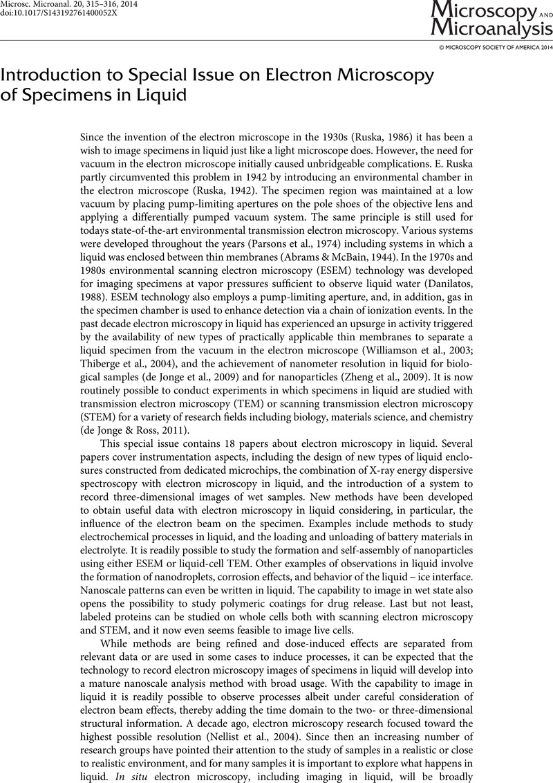
Image of the first page of this content. For PDF version, please use the ‘Save PDF’ preceeding this image.'
Type
In Situ Special Section
Copyright
© Microscopy Society of America 2014 

Access options

Get access to the full version of this content by using one of the access options below. (Log in options will check for institutional or personal access. Content may require purchase if you do not have access.)

References

Abrams, I.M. & McBain, J.W. (1944). A closed cell for electron microscopy. J Appl Phys 15, 607609.CrossRefGoogle Scholar
Danilatos, G.D. (1988). Foundations of environmental scanning electron microscopy. Adv Electron El Phys 71, 109251.CrossRefGoogle Scholar
de Jonge, N., Peckys, D.B., Kremers, G.J. & Piston, D.W. (2009). Electron microscopy of whole cells in liquid with nanometer resolution. Proc Natl Acad Sci 106, 21592164.Google Scholar
de Jonge, N. & Ross, F.M. (2011). Electron microscopy of specimens in liquid. Nat Nanotechnol 6, 695704.Google Scholar
Nellist, P.D., Chisholm, M.F., Dellby, N., Krivanek, O.L., Murfitt, M.F., Szilagyi, Z.S., Lupini, A.R., Borisevich, A., Sides, W.H. & Pennycook, S.J. (2004). Direct sub-angstrom imaging of a crystal lattice. Science 305, 1741.Google Scholar
Parsons, D.F., Matricardi, V.R., Moretz, R.C. & Turner, J.N. (1974). Electron microscopy and diffraction of wet unstained and unfixed biological objects. Adv Biol Med Phys 15, 161270.CrossRefGoogle ScholarPubMed
Ruska, E. (1942). Beitrag zur uebermikroskopischen Abbildungen bei hoeheren Drucken. Kolloid Zeitschrift 100, 212219.Google Scholar
Ruska, E. (1986). The development of the electron microscope and of electron microscopy (nobel lecture).Google Scholar
Thiberge, S., Nechushtan, A., Sprinzak, D., Gileadi, O., Behar, V., Zik, O., Chowers, Y., Michaeli, S., Schlessinger, J. & Moses, E. (2004). Scanning electron microscopy of cells and tissues under fully hydrated conditions. Proc Natl Acad Sci 101, 33463351.Google Scholar
Williamson, M.J., Tromp, R.M., Vereecken, P.M., Hull, R. & Ross, F.M. (2003). Dynamic microscopy of nanoscale cluster growth at the solid-liquid interface. Nat Mater 2, 532536.Google Scholar
Zheng, H., Smith, R.K., Jun, Y.W., Kisielowski, C., Dahmen, U. & Alivisatos, A.P. (2009). Observation of single colloidal platinum nanocrystal growth trajectories. Science 324, 13091312.CrossRefGoogle ScholarPubMed