Hostname: page-component-848d4c4894-tn8tq Total loading time: 0 Render date: 2024-07-02T02:06:16.912Z Has data issue: false hasContentIssue false

Integrative Atom Probe Tomography Using Scanning Transmission Electron Microscopy-Centric Atom Placement as a Step Toward Atomic-Scale Tomography

Published online by Cambridge University Press:  20 January 2021

Anna V. Ceguerra
Affiliation:
Australian Centre for Microscopy & Microanalysis (ACMM), The University of Sydney, Sydney, NSW2006, Australia School of Aerospace, Mechanical and Mechatronic Engineering (AMME), The University of Sydney, Sydney, NSW2006, Australia
Andrew J. Breen
Affiliation:
Australian Centre for Microscopy & Microanalysis (ACMM), The University of Sydney, Sydney, NSW2006, Australia School of Aerospace, Mechanical and Mechatronic Engineering (AMME), The University of Sydney, Sydney, NSW2006, Australia
Julie M. Cairney
Affiliation:
Australian Centre for Microscopy & Microanalysis (ACMM), The University of Sydney, Sydney, NSW2006, Australia School of Aerospace, Mechanical and Mechatronic Engineering (AMME), The University of Sydney, Sydney, NSW2006, Australia
Simon P. Ringer
Affiliation:
Australian Centre for Microscopy & Microanalysis (ACMM), The University of Sydney, Sydney, NSW2006, Australia School of Aerospace, Mechanical and Mechatronic Engineering (AMME), The University of Sydney, Sydney, NSW2006, Australia
Brian P. Gorman*
Affiliation:
Department of Metallurgical and Materials Engineering, Colorado School of Mines, Golden, CO80401, USA
*
*Author for correspondence: Brian Gorman, E-mail: bgorman@mines.edu
Get access

Abstract

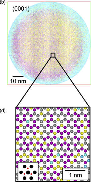
Current reconstruction methodologies for atom probe tomography (APT) contain serious geometric artifacts that are difficult to address due to their reliance on empirical factors to generate a reconstructed volume. To overcome this limitation, a reconstruction technique is demonstrated where the analyzed volume is instead defined by the specimen geometry and crystal structure as determined by transmission electron microscopy (TEM) and diffraction acquired before and after APT analysis. APT data are reconstructed using a bottom-up approach, where the post-APT TEM image is used to define the substrate upon which APT detection events are placed. Transmission electron diffraction enables the quantification of the relationship between atomic positions and the evaporated specimen volume. Using an example dataset of ZnMgO:Ga grown epitaxially on c-plane sapphire, a volume is reconstructed that has the correct geometry and atomic spacings in 3D. APT data are thus reconstructed in 3D without using empirical parameters for the reverse projection reconstruction algorithm.

Type
Software and Instrumentation
Copyright
Copyright © The Author(s), 2021. Published by Cambridge University Press on behalf of the Microscopy Society of America

Access options

Get access to the full version of this content by using one of the access options below. (Log in options will check for institutional or personal access. Content may require purchase if you do not have access.)

References

Araullo-Peters, VJ, Breen, A, Ceguerra, AV, Gault, B, Ringer, SP & Cairney, JM (2015). A new systematic framework for crystallographic analysis of atom probe data. Ultramicroscopy 154, 714.CrossRefGoogle ScholarPubMed
Arslan, I, Marquis, EA, Homer, M, Hekmaty, MA & Bartelt, NC (2008). Towards better 3-D reconstructions by combining electron tomography and atom-probe tomography. Ultramicroscopy 108(12), 15791585.CrossRefGoogle ScholarPubMed
Bas, P, Bostel, A, Deconihout, B & Blavette, D (1995). A general protocol for the reconstruction of 3D atom probe data. Appl Surf Sci 87–88(Suppl. C), 298304.CrossRefGoogle Scholar
Bunton, JH, Olson, JD, Lenz, DR & Kelly, TF (2007). Advances in pulsed-laser atom probe: Instrument and specimen design for optimum performance. Microsc Microanal 13(6), 418427.CrossRefGoogle ScholarPubMed
Burton, GL, Ricote, S, Foran, BJ, Diercks, DR & Gorman, BP (2020 a). Quantification of grain boundary defect chemistry in a mixed proton-electron conducting oxide composite. J Am Ceram Soc 103(5), 32173230.CrossRefGoogle Scholar
Burton, GL, Wright, S, Stokes, A, Diercks, DR, Clarke, A & Gorman, BP (2020 b). Orientation mapping with kikuchi patterns generated from a focused STEM probe and indexing with commercially available EDAX software. Ultramicroscopy 209, 112882.CrossRefGoogle ScholarPubMed
Ceguerra, AV, Day, AC & Ringer, SP (2019). Assessing the spatial accuracy of the reconstruction in atom probe tomography and a new calibratable adaptive reconstruction. Microsc Microanal 25(2), 309319.CrossRefGoogle Scholar
Ceguerra, AV, Moody, MP, Stephenson, LT, Marceau, RKW & Ringer, SP (2010). A three-dimensional Markov field approach for the analysis of atomic clustering in atom probe data. Philos Mag 90(12), 16571683.CrossRefGoogle Scholar
Diercks, DR & Gorman, BP (2018). Self-consistent atom probe tomography reconstructions utilizing electron microscopy. Ultramicroscopy 195, 3246.CrossRefGoogle ScholarPubMed
Downs, RT & Hall-Wallace, M (2003). The American Mineralogist crystal structure database. Am Mineral 88(1), 247250.Google Scholar
Du, S, Burgess, T, Gault, B, Gao, Q, Bao, P, Li, L, Cui, X, Kong Yeoh, W, Liu, H, Yao, L, Ceguerra, AV, Hoe Tan, H, Jagadish, C, Ringer, SP & Zheng, R (2013). Quantitative dopant distributions in GaAs nanowires using atom probe tomography. Ultramicroscopy 132, 186192.CrossRefGoogle ScholarPubMed
Gault, B, Moody, MP, Cairney, JM & Ringer, SP (2012). Atom Probe Microscopy. New York: Springer.CrossRefGoogle Scholar
Geiser, BP, Kelly, TF, Larson, DJ, Schneir, J & Roberts, JP (2007). Spatial distribution maps for atom probe tomography. Microsc Microanal 13(6), 437447.CrossRefGoogle ScholarPubMed
Gorman, B (2007). Atom probe reconstruction refinements by pre- and post- analysis TEM structure quantification. Microsc Microanal 13(S02), 16161617.CrossRefGoogle Scholar
Gorman, BP, Diercks, D, Salmon, N, Stach, E, Amador, G & Hartfield, C (2008 a). Hardware and techniques for cross- correlative TEM and atom probe analysis. Microsc Today 16(4), 4247.CrossRefGoogle Scholar
Gorman, BP, Diercks, DR & Jaeger, D (2008 b). 3-D Cross-correlation of atom probe and STEM tomography. Microsc Microanal 14(S2), 10421043.CrossRefGoogle Scholar
Gorman, BP, Diercks, DR & Kirchhofer, R (2013). Laser and material effects on laser pulsed atom probe analysis of oxide and nitride ceramics. Microsc Microanal 19(S2), 18821883.CrossRefGoogle Scholar
Haley, D, Petersen, T, Barton, G & Ringer, SP (2009). Influence of field evaporation on radial distribution functions in atom probe tomography. Philos Mag 89(11), 925943.CrossRefGoogle Scholar
Katnagallu, S, Stephenson, LT, Mouton, I, Freysoldt, C, Subramanyam, APA, Jenke, J, Ladines, AN, Neumeier, S, Hammerschmidt, T, Drautz, R, Neugebauer, J, Vurpillot, F, Raabe, D & Gault, B (2019). Imaging individual solute atoms at crystalline imperfections in metals. New J Phys 21(12), 123020.CrossRefGoogle Scholar
Kelly, T, Miller, M, Rajan, K, Ringer, S, Borisevich, A, Dellby, N & Krivanek, O (2011). Toward atomic-scale tomography: The ATOM project. Microsc Microanal 17(S2), 708709.CrossRefGoogle Scholar
Kelly, TF, Gribb, TT, Olson, JD, Martens, RL, Shepard, JD, Wiener, SA, Kunicki, TC, Ulfig, RM, Lenz, DR, Strennen, EM, Oltman, E, Bunton, JH & Strait, DR (2004). First data from a commercial local electrode atom probe (LEAP). Microsc Microanal 10(3), 373383.CrossRefGoogle Scholar
Kelly, TF, Miller, MK, Rajan, K & Ringer, SP (2013). Atomic-scale tomography: A 2020 vision. Microsc Microanal 19(3), 652664.CrossRefGoogle ScholarPubMed
Kirchhofer, R, Diercks, DR & Gorman, BP (2015). Near atomic scale quantification of a diffusive phase transformation in (Zn,Mg)O/Al2O3 using dynamic atom probe tomography. J Mater Res 30(8), 11371147.CrossRefGoogle Scholar
Kirchhofer, R, Diercks, DR & Gorman, BP (2018). Electron diffraction and imaging for atom probe tomography. Rev Sci Instrum 89(5), 053706.CrossRefGoogle ScholarPubMed
Kirchhofer, R, Teague, MC & Gorman, BP (2013). Thermal effects on mass and spatial resolution during laser pulse atom probe tomography of cerium oxide. J Nucl Mater 436(1), 2328.CrossRefGoogle Scholar
Larson, DJ, Gault, B, Geiser, BP, De Geuser, F & Vurpillot, F (2013 a). Atom probe tomography spatial reconstruction: Status and directions. Curr Opin Solid State Mater Sci 17(5), 236247.CrossRefGoogle Scholar
Larson, DJ, Prosa, TJ, Ulfig, RM, Geiser, BP, Kelly, TF & Humphreys, PSCJ (2013 b). Local Electrode Atom Probe Tomography: A User's Guide. New York: Springer.CrossRefGoogle Scholar
Lefebvre, W, Vurpillot, F & Sauvage, X (2016). Atom Probe Tomography: Put Theory Into Practice. London: Elsevier Science.Google Scholar
Maejima, K, Shibata, H, Tampo, H, Matsubara, K & Niki, S (2010). Characterization of Zn1−xMgxO transparent conducting thin films fabricated by multi-cathode RF-magnetron sputtering. Thin Solid Films 518(11), 29492952.CrossRefGoogle Scholar
Marceau, RKW, Ceguerra, AV, Breen, AJ, Raabe, D & Ringer, SP (2015). Quantitative chemical-structure evaluation using atom probe tomography: Short-range order analysis of Fe–Al. Ultramicroscopy 157, 1220.CrossRefGoogle ScholarPubMed
Miller, MK (2012). Atom Probe Tomography: Analysis at the Atomic Level. London: Springer US.Google Scholar
Miller, MK & Forbes, RG (2009). Atom probe tomography. Mater Charact 60(6), 461469.CrossRefGoogle Scholar
Moody, MP, Gault, B, Stephenson, LT, et al. (2011). Lattice rectification in atom probe tomography: Toward true three-dimensional atomic microscopy. Microsc Microanal 17, 226239.CrossRefGoogle ScholarPubMed
Ohtomo, A, Kawasaki, M, Ohkubo, I, Koinuma, H, Yasuda, T & Segawa, Y (1999). Structure and optical properties of ZnO/Mg0.2Zn0.8O superlattices. Appl Phys Lett 75(7), 980982.CrossRefGoogle Scholar
Ophus, C (2019). Four-Dimensional Scanning Transmission Electron Microscopy (4D-STEM): From Scanning Nanodiffraction to Ptychography and Beyond. Microscopy and Microanalysis 25(3), 563582. doi: http://dx.doi.org/10.1017/S1431927619000497.CrossRefGoogle ScholarPubMed
Wallace, ND, Ceguerra, AV, Breen, AJ & Ringer, SP (2018). On the retrieval of crystallographic information from atom probe microscopy data via signal mapping from the detector coordinate space. Ultramicroscopy 189, 6575.CrossRefGoogle ScholarPubMed
Weber, L (1923). XII. Ein einfacher Ausdruck für das Verhältnis der Netzdichten der Bravaisschen Baumgitter. Z. Kristallogr. 58, 220.Google Scholar