Hostname: page-component-848d4c4894-jbqgn Total loading time: 0 Render date: 2024-07-01T17:10:22.388Z Has data issue: false hasContentIssue false

In Situ Atomic Force Microscopy Depth-Corrected Three-Dimensional Focused Ion Beam Based Time-of-Flight Secondary Ion Mass Spectroscopy: Spatial Resolution, Surface Roughness, Oxidation

Published online by Cambridge University Press:  23 November 2020

Lex Pillatsch
Affiliation:
Empa, Swiss Federal Laboratories for Materials Science and Technology, Laboratory of Mechanics of Materials and Nanostructures, Thun CH-3602, Switzerland TOFWERK AG., Thun CH-3600, Switzerland
Szilvia Kalácska*
Affiliation:
Empa, Swiss Federal Laboratories for Materials Science and Technology, Laboratory of Mechanics of Materials and Nanostructures, Thun CH-3602, Switzerland
Xavier Maeder
Affiliation:
Empa, Swiss Federal Laboratories for Materials Science and Technology, Laboratory of Mechanics of Materials and Nanostructures, Thun CH-3602, Switzerland
Johann Michler
Affiliation:
Empa, Swiss Federal Laboratories for Materials Science and Technology, Laboratory of Mechanics of Materials and Nanostructures, Thun CH-3602, Switzerland
*
*Author for correspondence: Szilvia Kalácska, E-mail: szilvia.kalacska@empa.ch
Get access

Abstract

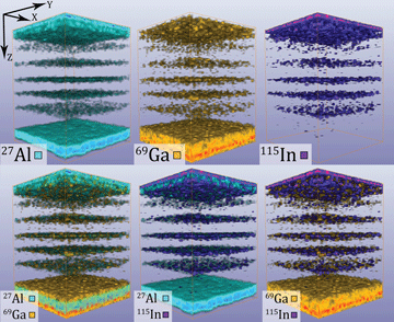
Atomic force microscopy (AFM) is a well-known tool for studying surface roughness and to collect depth information about features on the top atomic layers of samples. By combining secondary ion mass spectroscopy (SIMS) with focused ion beam (FIB) milling in a scanning electron microscope (SEM), chemical information of sputtered structures can be visualized and located with high lateral and depth resolution. In this paper, a high vacuum (HV) compatible AFM was installed in a TESCAN FIB-SEM instrument that was equipped with a time-of-flight SIMS (ToF-SIMS) detector. To calibrate the sputtering rate and measure the induced roughness caused by the ToF-SIMS analysis, subsequent AFM measurements were performed on an inorganic multilayer vertical cavity surface-emitting laser sample. Normalized sputtering rates were used to aid the accurate three-dimensional reconstruction of the sputtered volume's chemical composition. Achievable resolution, surface roughness during sputtering, and surface oxidation issues were analyzed. The integration of complementary detectors opens up the ability to determine the sample properties as well as to understand the influence of the Ga+ ion sputtering method on the sample surface during the analysis.

Type
Software and Instrumentation
Copyright
Copyright © The Author(s), 2020. Published by Cambridge University Press on behalf of the Microscopy Society of America

Access options

Get access to the full version of this content by using one of the access options below. (Log in options will check for institutional or personal access. Content may require purchase if you do not have access.)

References

Akiyama, T, de Rooij, NF, Staufer, U, Detterbeck, M, Braendlin, D, Waldmeier, S & Scheidiger, M (2010). Implementation and characterization of a quartz tuning fork based probe consisted of discrete resonators for dynamic mode atomic force microscopy. Rev Sci Instrum 81(6), 063706.CrossRefGoogle ScholarPubMed
Akiyama, T, Staufer, U & de Rooij, N (2003 a). Self-sensing and self-actuating probe based on quartz tuning fork combined with microfabricated cantilever for dynamic mode atomic force microscopy. Appl Surf Sci 210(1–2), 1821.CrossRefGoogle Scholar
Akiyama, T, Staufer, U, de Rooij, NF, Frederix, P & Engel, A (2003 b). Symmetrically arranged quartz tuning fork with soft cantilever for intermittent contact mode atomic force microscopy. Rev Sci Instrum 74(1), 112.CrossRefGoogle Scholar
Akiyama, T, Suter, K, de Rooij, NF, Baumgartner, A, Gildemeister, AE, Ihn, T, Ensslin, K & Staufer, U (2006). Scanning probe with tuning fork sensor, microfabricated silicon cantilever and conductive tip for microscopy at cryogenic temperature. Jpn J Appl Phys 45(3B), 19921995.CrossRefGoogle Scholar
Alberts, D, von Werra, L, Oestlund, F, Rohner, U, Hohl, M, Michler, J & Whitby, JA (2014). Design and performance of two orthogonal extraction time-of-flight secondary ion mass spectrometers for focused ion beam instruments. Instrum Sci Technol 42(4), 432445.CrossRefGoogle Scholar
Bayat, D, Akiyama, T, de Rooij, NF & Staufer, U (2008). Dynamic behavior of the tuning fork AFM probe. Microelectron Eng 85(5–6), 10181021.CrossRefGoogle Scholar
Busby, Y, Franquet, A, Spampinato, V, Casula, G, Bonfiglio, A, Cosseddu, P, Pireaux, JJ & Houssiau, L (2018). Combined AFM and ToF-SIMS analyses for the study of filaments in organic resistive switching memories. Proceedings SPIE 10738, Organic and Hybrid Sensors and Bioelectronics XI, 1073814.CrossRefGoogle Scholar
Casula, G, Busby, Y, Franquet, A, Spampinato, V, Houssiau, L, Bonfiglio, A & Cosseddu, P (2019). A flexible organic memory device with a clearly disclosed resistive switching mechanism. Org Electron 64, 209215. doi:10.1016/j.orgel.2018.10.018CrossRefGoogle Scholar
Dunn, DN & Hull, R (1999). Reconstruction of three-dimensional chemistry and geometry using focused ion beam microscopy. Appl Phys Lett 75(21), 34143416.CrossRefGoogle Scholar
Giannuzzi, LA & Stevie, FA (2005). Introduction to Focussed Ion Beams: Instrumentation, Theory, Techniques and Practice. New York: Springer.CrossRefGoogle Scholar
Giannuzzi, LA & Utlaut, M (2011). A review of Ga+ FIB/SIMS. Surf Interface Anal 43(1–2), 475478.CrossRefGoogle Scholar
Jeans, J (1982). Preface. In An Introduction to the Kinetic Theory of Gases, Cambridge Science Classics, pp. VVi. Cambridge: Cambridge University Press. doi:10.1017/CBO9780511628849.001CrossRefGoogle Scholar
Kushmerick, JG & Weiss, PS (2017). Scanning probe microscopes. In Encyclopedia of Spectroscopy and Spectrometry, Lindon, JC, Tranter, GE & Koppenaal, DW (Eds.), 3rd edn. pp. 714. Oxford: Academic Press.CrossRefGoogle Scholar
Lyytikäinen, J, Rautiainen, J, Toikkanen, L, Sirbu, A, Mereuta, A, Caliman, A, Kapon, E & Okhotnikov, OG (2009). 1.3-microm optically-pumped semiconductor disk laser by wafer fusion. Opt Express 17(11), 90479052.CrossRefGoogle ScholarPubMed
Nečas, D & Klapetek, P (2012). Gwyddion: An open-source software for SPM data analysis. Cent Eur J Phys 10(1), 181188.Google Scholar
Noel, C, Pescetelli, S, Agresti, A, Franquet, A, Spaminato, V, Felten, A, di Carlo, A, Houssiau, L & Busby, Y (2019). Hybrid perovskites depth profiling with variable-size argon clusters and monatonic ions beams. Materials 12(5), 726. doi:10.3390/ma12050726CrossRefGoogle ScholarPubMed
Perez-Cruz, A, Dominguez-Gonzalez, A, Stiharu, I & Osornio-Rios, RA (2012). Optimization of Q-factor of AFM cantilevers using genetic algorithms. Ultramicroscopy 115, 6167.CrossRefGoogle ScholarPubMed
Pillatsch, L, Östlund, F & Michler, J (2019). FIBSIMS: A review of secondary ion mass spectrometry for analytical dual beam focussed ion beam instruments. Prog Cryst Growth Charact Mater 65, 119.CrossRefGoogle Scholar
Priebe, A, Barnes, J-P, Edwards, TEJ, Pethö, L, Balogh, I & Michler, J (2019). 3D imaging of nanoparticles in an inorganic matrix using TOF-SIMS validated with STEM and EDX. Anal Chem 91(18), 1183411839.CrossRefGoogle Scholar
Rodríguez, TR & García, R (2003). Theory of Q control in atomic force microscopy. Appl Phys Lett 82(26), 48214823.CrossRefGoogle Scholar
Wei, Q, Lian, J, Lu, W & Wang, L (2008). Highly ordered Ga nanodroplets on a GaAs surface formed by a focused ion beam. Phys Rev Lett 100(7), 2023.CrossRefGoogle ScholarPubMed
Whitby, JA, Östlund, F, Horvath, P, Gabureac, M, Riesterer, JL, Utke, I, Hohl, M, Sedláček, L, Jiruše, J, Friedli, V, Bechelany, M & Michler, J (2012). High spatial resolution time-of-flight secondary ion mass spectrometry for the masses: A novel orthogonal ToF FIB-SIMS instrument with in situ AFM. Adv Mater Sci Eng 2012, 180437.CrossRefGoogle Scholar
Supplementary material: File

Pillatsch et al. Supplementary Materials

Pillatsch et al. Supplementary Materials

Download Pillatsch et al. Supplementary Materials(File)
File 14.6 KB