Hostname: page-component-848d4c4894-jbqgn Total loading time: 0 Render date: 2024-06-15T17:19:03.495Z Has data issue: false hasContentIssue false

Compositional Imaging of Cells and Bionanoparticles by EFTEM

Published online by Cambridge University Press:  27 August 2014

M.A. Aronova
Affiliation:
Laboratory of Cellular Imaging & Macromolecular Biophysics, NIBIB, NIH, Bethesda, MD 20892
R.D. Leapman
Affiliation:
Laboratory of Cellular Imaging & Macromolecular Biophysics, NIBIB, NIH, Bethesda, MD 20892

Abstract

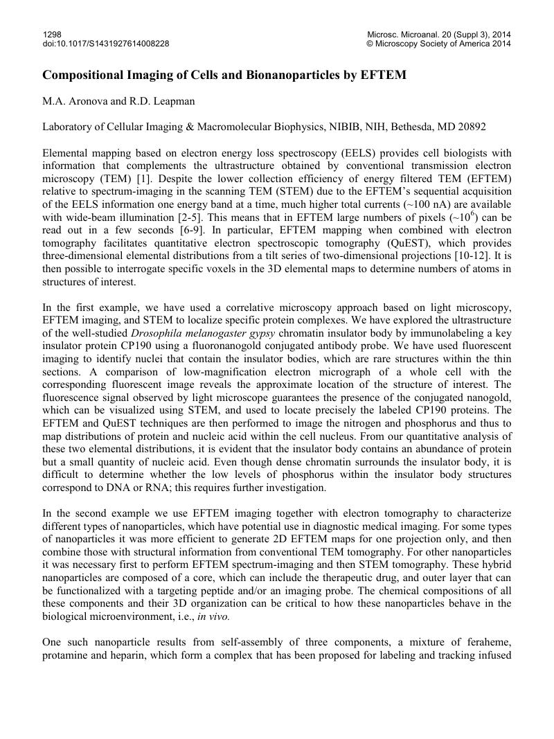
Image of the first page of this content. For PDF version, please use the ‘Save PDF’ preceeding this image.'
Type
Abstract
Copyright
Copyright © Microscopy Society of America 2014 

References

[1] Aronova, MA, Leapman, RD MRS Bulletin 37 (2012) p. 53.Google Scholar
[2] Shuman, H, Somlyo, AP Ultramicroscopy 21 (1987) p. 23.Google Scholar
[3] Jeanguillaume, C, Colliex, C Ultramicroscopy 28 (1989) p. 252.Google Scholar
[4] Hunt, JA, Williams, DB Ultramicroscopy 38 (1991) p. 47.Google Scholar
[5] Leapman, RD J Microsc 210 (2003) p. 5.Google Scholar
[6] Ottensmeyer, FP J Ultrastruct Res 88 (1984) p. 121.Google Scholar
[7] Bazett-Jones, DP, Hendzel, MJ, Kruhlak, MJ Micron 30 (1999) p. 151.Google Scholar
[8] Leapman, RD, Jarnik, M, Steven, AC J Struct Biol 120 (1997) p. 168.Google Scholar
[9] Goping, G, et al, Microsc Res Tech 61 (2003) p. 448.Google Scholar
[10] Leapman, RD, et al, Ultramicroscopy 100 (2004) p. 115.Google Scholar
[11] Boudier, T, et al, J Struct Biol 151 (2005 ) p. 151.Google Scholar
[12] Aronova, MA, et al, J Struct Biol 160 (2007) p. 35.Google Scholar
[13] Thu, MS, et al, Nature Med 18 (2012) p. 463.Google Scholar
[14] Xie, J, et al, ACS Nano 5 (2011) p. 3043.Google Scholar
[15] This research was supported by the intramural program of the National Institute of Biomedical Imaging and Bioengineering, NIH.Google Scholar