Hostname: page-component-8448b6f56d-xtgtn Total loading time: 0 Render date: 2024-04-19T08:11:22.841Z Has data issue: false hasContentIssue false

CDrift: An Algorithm to Correct Linear Drift From A Single High-Resolution STEM Image

Published online by Cambridge University Press:  24 July 2020

Guillermo Bárcena-González*
Affiliation:
Department of Computer Science and Engineering, University of Cádiz, Puerto Real, Cádiz, Spain
María de la Paz Guerrero-Lebrero
Affiliation:
Department of Computer Science and Engineering, University of Cádiz, Puerto Real, Cádiz, Spain
Elisa Guerrero
Affiliation:
Department of Computer Science and Engineering, University of Cádiz, Puerto Real, Cádiz, Spain
Andres Yañez
Affiliation:
Department of Computer Science and Engineering, University of Cádiz, Puerto Real, Cádiz, Spain
Bernardo Nuñez-Moraleda
Affiliation:
Department of Computer Science and Engineering, University of Cádiz, Puerto Real, Cádiz, Spain
Daniel Fernández-Reyes
Affiliation:
Department of Material Science and Metallurgy Engineering and Inorganic Chemistry, University of Cádiz, Puerto Real, Cádiz, Spain
Pedro Real
Affiliation:
Department of Applied Mathematics I, University of Seville, Seville, Spain
David González
Affiliation:
Department of Material Science and Metallurgy Engineering and Inorganic Chemistry, University of Cádiz, Puerto Real, Cádiz, Spain
Pedro L. Galindo
Affiliation:
Department of Computer Science and Engineering, University of Cádiz, Puerto Real, Cádiz, Spain
*
*Author for correspondence: Guillermo Bárcena-González, E-mail: guillermo.barcena@uca.es
Get access

Abstract

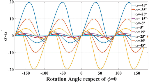
In this work, a new method to determine and correct the linear drift for any crystalline orientation in a single-column-resolved high-resolution scanning transmission electron microscopy (HR-STEM) image, which is based on angle measurements in the Fourier space, is presented. This proposal supposes a generalization and the improvement of a previous work that needs the presence of two symmetrical planes in the crystalline orientation to be applicable. Now, a mathematical derivation of the drift effect on two families of asymmetric planes in the reciprocal space is inferred. However, though it was not possible to find an analytical solution for all conditions, a simple formula was derived to calculate the drift effect that is exact for three specific rotation angles. Taking this into account, an iterative algorithm based on successive rotation/drift correction steps is devised to remove drift distortions in HR-STEM images. The procedure has been evaluated using a simulated micrograph of a monoclinic material in an orientation where all the reciprocal lattice vectors are different. The algorithm only needs four iterations to resolve a 15° drift angle in the image.

Type
Software and Instrumentation
Copyright
Copyright © Microscopy Society of America 2020

Access options

Get access to the full version of this content by using one of the access options below. (Log in options will check for institutional or personal access. Content may require purchase if you do not have access.)

References

Bárcena-González, G, Guerrero-Lebrero, MP, Guerrero, E, Fernández-Reyes, D, González, D, Mayoral, A, Utrilla, JM, Ulloa, JM & Galindo, PL (2016). Strain mapping accuracy improvement using super-resolution techniques. J Microsc 262(1), 5058. doi:10.1111/jmi.12341CrossRefGoogle ScholarPubMed
Bárcena-González, G, Guerrero-Lebrero, MP, Guerrero, E, Reyes, DF, Braza, V, Yañez, A, Nuñez-Moraleda, B, González, D & Galindo, PL (2018). Correcting sample drift using Fourier harmonics. Micron 110. doi:10.1016/j.micron.2018.04.004CrossRefGoogle ScholarPubMed
Bárcena-González, G, Guerrero-Lebrero, MP, Guerrero, E, Yañez, A, Fernández-Reyes, D, González, D & Galindo, PL (2017). Evaluation of high-quality image reconstruction techniques applied to high-resolution Z-contrast imaging. Ultramicroscopy 182, 283291. doi:10.1016/j.ultramic.2017.07.014CrossRefGoogle ScholarPubMed
Berkels, B & Liebscher, CH (2019). Ultramicroscopy Joint non-rigid image registration and reconstruction for quantitative atomic resolution scanning transmission electron microscopy. Ultramicroscopy 198, 4957. doi:10.1016/j.ultramic.2018.12.016CrossRefGoogle ScholarPubMed
Berkels, B, Sharpley, R, Binev, P, Yankovich, A, Shi, F, Voyles, P & Dahmen, W (2012 a). High precision STEM imaging by non-rigid alignment and averaging of a series of short exposures. Microsc Microanal 18(S2), 300301.CrossRefGoogle Scholar
Berkels, B, Yankovich, AB, Shi, F, Voyles, PM, Dahmen, W, Sharpley, R & Binev, P (2012 b). High precision STEM imaging by non-rigid alignment and averaging of a series of short exposures. Microsc Microanal 18(S2), 300301. doi:10.1017/S1431927612003352CrossRefGoogle Scholar
Binev, P, Blanco-Silva, F, Blom, D, Dahmen, W, Lamby, P, Sharpley, R & Vogt, T (2012). High-quality image formation by nonlocal means applied to high-angle annular dark-field scanning transmission electron microscopy (HAADF–STEM). Nanostruct Sci Technol. doi: doi:10.1007/978-1-4614-2191-7_5.CrossRefGoogle Scholar
Braidy, N, Le Bouar, Y, Lazar, S & Ricolleau, C (2012). Correcting scanning instabilities from images of periodic structures. Ultramicroscopy 118, 6776. doi:10.1016/j.ultramic.2012.04.001CrossRefGoogle ScholarPubMed
Chateigner, D, Chen, D, Ciriotti, D, Downs, RT, Gražulis, S, Kaminsky, W, Le Bail, A, Lutterotti, L, Matsushita, Y, Merkys, A, Moeck, P, Murray-Rust, P, Quirós Olozábal, M, Rajan, H & Yokochi, AFT (2020). Crystallography Open Database. Available at http://www.crystallography.net/cod/index.php.Google Scholar
Jones, L & Nellist, PD (2013). Identifying and correcting scan noise and drift in the scanning transmission electron microscope. Microsc Microanal 19(4), 10501060. doi:10.1017/S1431927613001402CrossRefGoogle ScholarPubMed
Muller, A & Grazul, J (2001). Optimizing the environment for sub-0.2 nm scanning transmission electron microscopy. J Electron Microsc 50(3), 219226. doi:10.1093/jmicro/50.3.219Google ScholarPubMed
Nakanishi, N, Yamazaki, T, Rečnik, A, Čeh, M, Kawasaki, M, Watanabe, K & Shiojiri, M (2002). Retrieval process of high-resolution HAADF-STEM images. J Electron Microsc 51(6), 383390. doi:10.1093/jmicro/51.6.383CrossRefGoogle ScholarPubMed
Ning, S, Fujita, T, Nie, A, Wang, Z, Xu, X, Chen, J, Chen, M, Yao, S & Zhang, T (2018). Scanning distortion correction in STEM images. Ultramicroscopy 184, 274283. doi:10.1016/j.ultramic.2017.09.003CrossRefGoogle ScholarPubMed
Ophus, C, Ciston, J & Nelson, CT (2016). Correcting nonlinear drift distortion of scanning probe and scanning transmission electron microscopies from image pairs with orthogonal scan directions. Ultramicroscopy 162, 19. doi:10.1016/j.ultramic.2015.12.002CrossRefGoogle ScholarPubMed
Pennycook, SJ & Jesson, DE (1991). High-resolution Z-contrast imaging of crystals. Ultramicroscopy 37(1–4), 1438. doi:10.1016/0304-3991(91)90004-PCrossRefGoogle Scholar
Rečnik, A, Möbus, G & Šturm, S (2005). IMAGE-WARP: A real-space restoration method for high-resolution STEM images using quantitative HRTEM analysis. Ultramicroscopy 103(4), 285301. doi:10.1016/j.ultramic.2005.01.003CrossRefGoogle ScholarPubMed
Saito, M, Kimoto, K, Nagai, T, Fukushima, S, Akahoshi, D, Kuwahara, H, Matsui, Y & Ishizuka, K (2009). Local crystal structure analysis with 10-pm accuracy using scanning transmission electron microscopy. J Electron Microsc 58(3), 131136. doi:10.1093/jmicro/dfn023CrossRefGoogle ScholarPubMed
Sang, X & LeBeau, JM (2014). Revolving scanning transmission electron microscopy: Correcting sample drift distortion without prior knowledge. Ultramicroscopy 138, 2835. doi:10.1016/j.ultramic.2013.12.004CrossRefGoogle ScholarPubMed
Sang, X, Lupini, AR, Ding, J, Kalinin, SV, Jesse, S & Unocic, RR (2017). Precision controlled atomic resolution scanning transmission electron microscopy using spiral scan pathways. Sci Rep 7, 43585. doi:10.1038/srep43585CrossRefGoogle ScholarPubMed
Von Harrach, HS (1995). Instrumental factors in high-resolution FEG STEM. Ultramicroscopy 58(1), 15. doi:10.1016/0304-3991(94)00172-JCrossRefGoogle Scholar
Zachariasen, WH & Ellinger, FH (1963). The crystal structure of alpha plutonium metal. Acta Crystallogr 16(8), 777783. doi:10.1107/S0365110X63002012CrossRefGoogle Scholar
Zuo, J-M, Shah, AB, Kim, H, Meng, Y, Gao, W & Rouviére, J-L (2014). Lattice and strain analysis of atomic resolution Z-contrast images based on template matching. Ultramicroscopy 136, 5060. doi:10.1016/j.ultramic.2013.07.018CrossRefGoogle ScholarPubMed