Hostname: page-component-7bb8b95d7b-495rp Total loading time: 0 Render date: 2024-10-04T02:30:44.873Z Has data issue: false hasContentIssue false

Ablation and Microstructure Imaging of Dentin-Enamel Junction Using Focused Electron Beam in an Environmental Scanning Electron Microscope

Published online by Cambridge University Press:  01 August 2018

Donggao Zhao
Affiliation:
School of Dentistry, University of Missouri-Kansas City, Kansas City, MO64108
Rasoul Seyedmahmoud
Affiliation:
School of Dentistry, University of Missouri-Kansas City, Kansas City, MO64108
Jacob D. McGuire
Affiliation:
School of Dentistry, University of Missouri-Kansas City, Kansas City, MO64108
Yong Wang
Affiliation:
School of Dentistry, University of Missouri-Kansas City, Kansas City, MO64108
Mary P. Walker
Affiliation:
School of Dentistry, University of Missouri-Kansas City, Kansas City, MO64108
Jeffrey P. Gorski
Affiliation:
School of Dentistry, University of Missouri-Kansas City, Kansas City, MO64108

Abstract

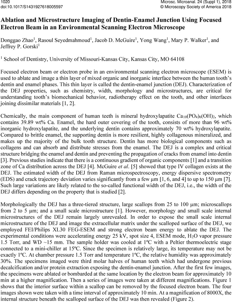
Image of the first page of this content. For PDF version, please use the ‘Save PDF’ preceeding this image.'
Type
Abstract
Copyright
© Microscopy Society of America 2018 

References

[1] Marshall, S.J., et al, J. European Ceramics Society 23 2003) p. 28972904.Google Scholar
[2] Walker, M.P. Fricke, B. A. Dentin-Enamel Junction of Human Teeth. In "Wiley Encyclopedia of Biomedical Engineering" (ed. M. Akay John Wiley & Sons Hoboken 2006) p 10611064.Google Scholar
[3] Nanci, A. Ten Cate's Oral histology: development, structure, and function, 7th ed 2008 Mosby Elsevier, St. Louis.Google Scholar
[4] Dusevich, V.M., et al., Microsc. Microanal. 18(Suppl 2 2012) p. 274275.Google Scholar
[5] McGuire, J.D., et al., J Dent Res 93(10 2014) p. 10281034.Google Scholar
[6] Xu, C., et al., Calcif. Tissue Int. 84(3 2009) p. 221228.Google Scholar
[7] Dong, X.D. Ruse, N.D. J. Biomed. Mater. Res. 66A 2003) p. 103109.Google Scholar
[8] The authors acknowledge funding from NIH/NIDCR Grant R01DE021462.Google Scholar