


Since the discovery of X-rays by Roentgen in 1895, human beings have embarked on an experiment of such massive proportions in which humans of all races and ages will be irradiated for years to come.
The proponents of whole body backscatter X-ray scanner use at the airport argue that the dose from a scan is low. The whole body X-ray unit is operated at 50 KV, with exposure time of 3 s and HVL of 1.2 mm Al. The X-ray output of these units is 5 uR per exposure (1.67uR/s). Comparing this with US natural background radiation is 300 mR/year or 30 uR/h (0.008uR/s). An air traveler at 30,000 feet receives radiation exposure on an average of 200–500 uR/h (0.055–0.138 uR/s). The exposure rate is higher in airport scanner than either with the background radiation or an air traveler. There is approximately 500 whole body backscatter X-ray scanners installed in the United States. It is estimated that more than 700 million people fly each year.
To date, no useful data regarding effects of radiation on human populations is available. The data we have is either derived from the atomic bomb explosions, radiation accidents or medical procedures. Only years later were effects of these determined to have radiobiological implications.
The question is how we test or assess whether the airport scanner is safe. On this note, a different approach is presented here to comprehend the effects of radiation on human populations. The data was abstracted from the literature on Hela cells S-3 in different phases of the cell cycles. Since mitotic phase is the most sensitive to radiation, it is therefore appropriate to assess the quality factor of X-rays in mitotic phase of the Hela cells S-3 at 220 KV, 280 KV and 1.25 MeV with their mean lethal radiation dose (Do).Reference Terasima and Tolmach1,Reference Sapozink2 In Figure 1, it is interesting to observe that as the energy decreases, the Do value decreases as well. Extrapolating the curve, it appears that at lower energy, Do decreases and becomes more potent. The relative biological effectiveness (RBE) value increases with decreasing energy, suggesting the harmful effects of low energy radiation.
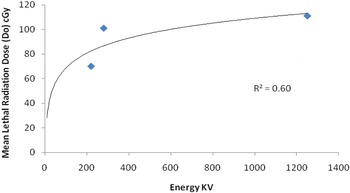
Figure 1. Correlation between mean lethal radiation dose (Do) and Energy (KV) in the mitotic phase of the Hela Cells S-3 yielding a correlation of 0.60.
Frankenberg et al.Reference Frankenberg, Kelnhofer, Bär and Frankenberg-Schwager3 observed from their study in human hybrid cell line (CGLI) that the RBE of mammography X-rays relative to 200 KV X-rays, was determined to be about four for doses of < 0.5 Gy.
The question is why low energy X-rays has a more potent effect compared to high energy. Chen and WattReference Chen and Watt4 showed that the optimum damage occurs when the mean free path between ionizations is equivalent to the strand separation in the double strand DNA. This criterion is met by low energy X-rays only.
SancheReference Sanche5 suggested that DNA lesions are induced by the lower energy secondary electrons generated by the primary (X-rays) ionizing radiation. SancheReference Sanche5 showed that the low energy electrons induce both single and double strand breaks at energy below the ionization limit of DNA (7.5 eV). Even the sun rays, at 10 eV, can cause skin cancer.
Today, in dental X-rays, chest X-rays and other radiological procedures, the genital organs are shielded with a lead shield. For chest X-rays, the kV used are 110 to 120 with 10 mR exposures. The scatter radiation is one-third of the peak energy (120) which is about 40 kV: well below the airport scanners kV. But these scatter radiation has the potential of radiation damage. Airport scanners have direct exposure to the genital organs, skin, breast and eyes and the implication of its harmful effects cannot be ruled out.
If only 1% of all humans scanned by the airport whole body X-ray scanners, and if the exposed organ has their cells in mitotic phase or high percentage of cells in mitotic phase in cell kinetics during irradiation: the damage to mitotic phase and duplication and progression of cells in those individual may cause cancer. This is too large a number to ignore. In fact, there is no such thing as safe dose of X-rays.

