


Introduction
Mycetomas are chronic subcutaneous infections caused by fungi or actinomycetes, known as eumycetomas or actinomycetomas respectively [Reference Fahal1, Reference Zijlstra2].
Mycetoma has a worldwide distribution, mainly confined to tropical regions in the area between the latitudes of 15°S and 30°N known as the ‘Mycetoma belt’ [Reference Desnos-Ollivier3]. India and Sudan have higher than average prevalence of the infection. Regional distribution of mycetoma includes Sri Lanka, India, Pakistan, Sudan, Senegal, Somalia, Mexico and South America [Reference Desnos-Ollivier3]. In India, Rajasthan (North-West India) and South India are mostly affected. Eumycetoma constitutes one-third of the total cases mainly reported from Uttar Pradesh (North India) and Central Rajasthan, while actinomycotic mycetoma is usually present in South India, South-East Rajasthan and Chandigarh [Reference Singh4, Reference Mathur5].
The organisms are chronic infections which are traumatically implanted into the subcutaneous tissue and deeper structures like fascia, joints and bones from the natural environment. They cause a subcutaneous infection characterised by progressive granulomatous lesions, with or without sinus tract formation with discharging grains, tumefaction and spreading into the adjacent tissue, bone, fascia and ligaments (Fig. 1) [Reference Rippon6–Reference Reddy9]. The main sites affected are lower limb and upper limb. Uncommon sites include trunks, buttocks, eyelids, lacrimal glands, paranasal sinuses, nails, mandible, scalp, neck, perineum and testes [Reference Rippon6].
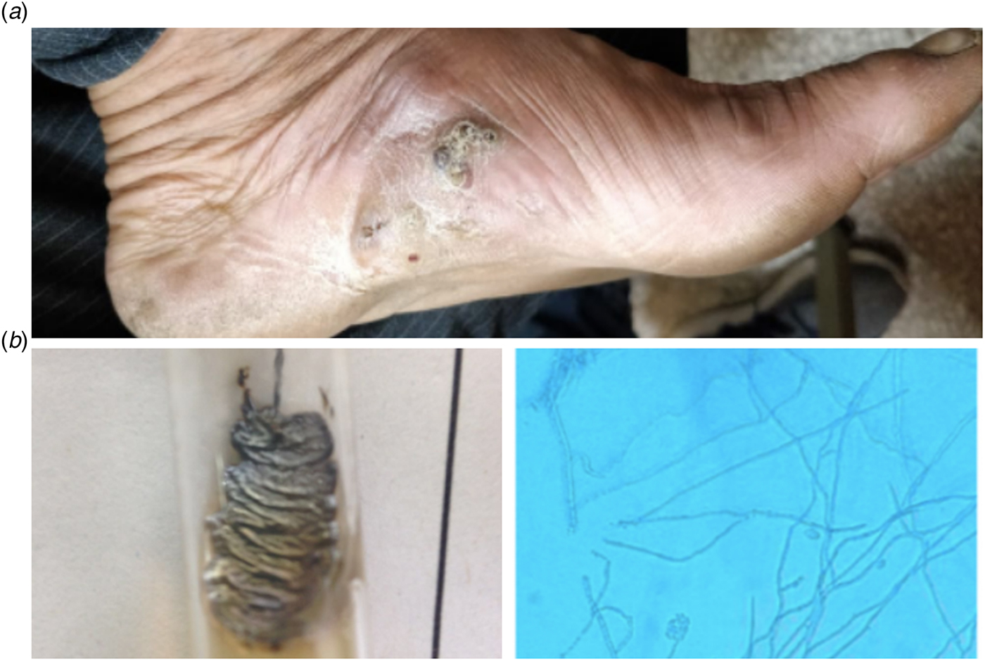
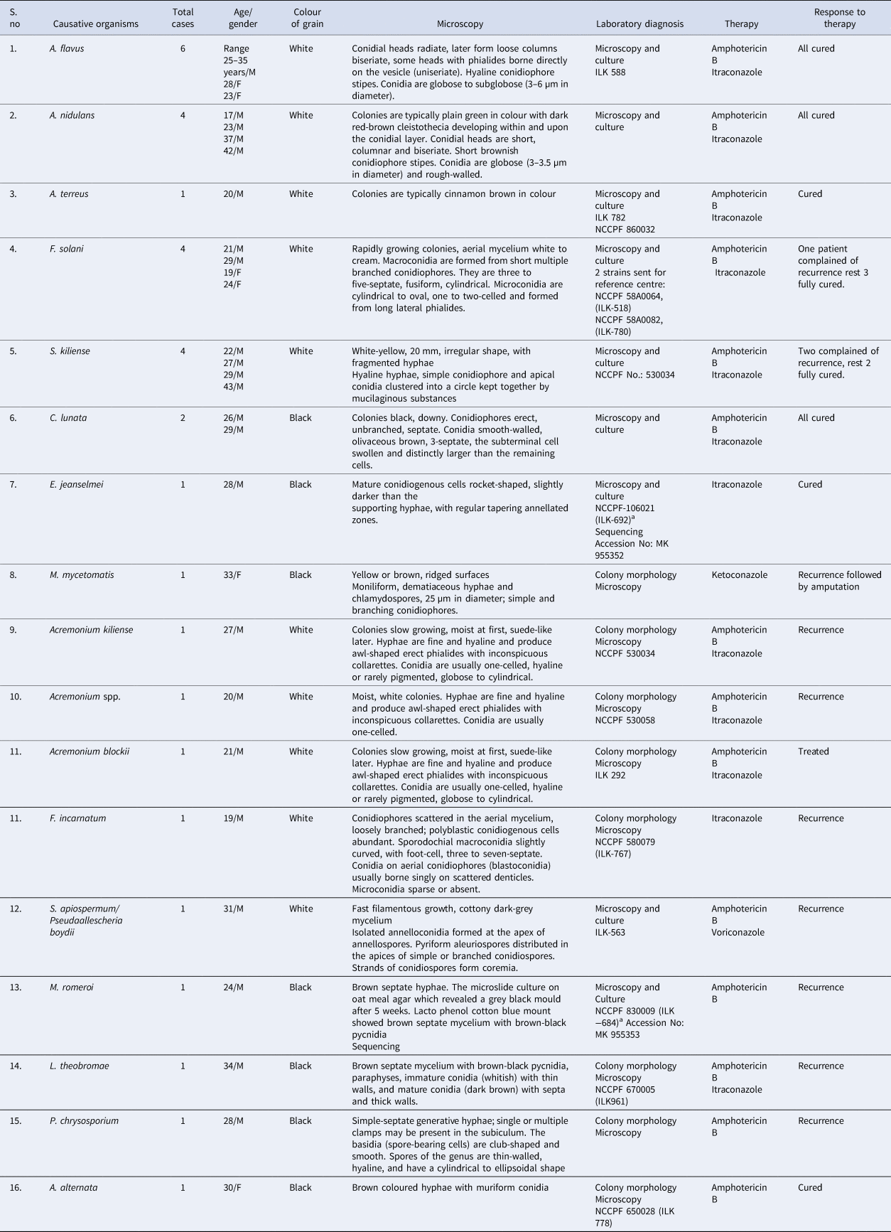
Fig. 1. (a) Granulomatous lesion with sinus tract formation and discharging black grains, tumefaction and (b) Colony morphology and microscopic picture of E. jeanselmei.
Black grain eumycetoma includes Madurella mycetomatis, Trematosphaeria grisea, Exophiala jeanselmei and Curvularia geniculata. White grain eumycetoma includes Scedosporium apiospermum complex, Aspergillus nidulans, Acremonium falciforme and Fusarium species [Reference Fahal10].
Prompt diagnosis and rapid treatment is required in the cases of eumycetoma. It is a chronic disease and the relapse rate is very high due to this it has become one of the most neglected disease and important public health problem in Africa and south Asia [11]. Anti-fungal susceptibility testing is still in its evolving stage. That is why correct identification of the species is of paramount importance [Reference Fahal1, Reference Zijlstra2, Reference Fahal10].
The current study was performed to know the epidemiological profile and spectrum of eumycetoma in Delhi, North India.
Methods
Thirty patients with eumycetoma seen in the last 13 years (January 2004–June 2018) were evaluated retrospectively at the Department of Microbiology, VMMC and Safdarjung Hospital. All the cases of actinomycetoma were excluded from the present study.
A diagnosis was made clinically on the basis of the classical triad of tumefaction, discharging sinuses and the presence of grains in these sinuses (Fig. 1a). A total of 59 samples were received.
The age, gender, occupation, site of involvement, duration of disease and underlying bony involvement detected by X-ray examination of patients were recorded. The size, shape, colour and consistency of granules were examined macroscopically. Direct examination of the granules was done by washing in normal saline followed by making a 10% KOH mount. The size of hyphae, septations and pigment formation in the hyphal walls was examined under a microscope. In cases of non-sporulating mould, slide culture was put up. The eumycotic grains appeared as 2–6 µm wide hyphae, have large, globose swollen cells (up to 15 µm) at margins. Gram's stain preparation was made by crushing the grain or tissue sample between two slides, heat fixing and staining. Gram's staining and Kinyoun's modified acid fast staining using 1% sulphuric acid were done [Reference Mencarini12, Reference Welsh13].
When no growth was obtained, the etiologic description was restricted to grain colour and/or actinomycotic/eumycotic aetiology [Reference van de Sande14]. The organisms were identified on the basis of their cultural characteristics and morphology and conidiation pattern on lacto phenol cotton blue (LPCB) mount. On LPCB mount the varied appearances were identified as follows:
(1) Sub spherical cells with annellidic butt producing long chains of conidia. The conidiogenous cells of hyphae were seen as intercalary or rocket shaped and these were identified as E. jeanselmei (Fig. 1b).
(2) M. mycetomatis showed small conidia that are ovoid to globose, on corn meal agar pointed conidiophores bearing conidia at the tips of flask-shaped phialide was seen.
(3) Fusarium solani appeared as single or grouped conidiophores, conidia were seen in conidial balls with hyaline transverse septae.
(4) Sarocladium kiliense had slimy colonies with pinkish appearance on SDA, microscopically hyaline hyphae with scanty sporulation were seen.
(5) A. nidulans had globose conidia surrounded by mass of hulle cells.
(6) Fusarium incarnatum showed hyaline hyphae with clustered and chain of verrucose chlamydospores and coiled sterile hyphae and characteristic curved slender septate macroconidia. Sporodochia were produced on a piece of carnation leaf.
(7) S. apiospermum complex had broad hyphae of up to 2–5 µm width, cleistothecia formation was seen, conidia were single celled pale brown and oval in shape (Fig. 1c).
(8) Curvularia lunata had simple brown coloured conidiophores bent at points showing sympodial geniculate growth, conidia had 3–4 septae at third cell from base.
(9) Medicopsis romeroi: Lacto phenol cotton blue mount of the colonies revealed brown septate hyphae. The microslide culture on oat meal agar which revealed a grey black mould after 5 weeks. Lacto phenol cotton blue mount showed brown septate mycelium with brown-black pycnidia.
(10) Lasiodiplodia theobromae: Lactophenol cotton blue mount showed brown septate mycelium with brown-black pycnidia, paraphyses and immature conidia (whitish) with thin walls, and mature conidia (dark brown) with septa and thick walls.
(11) Alternaria alternata: simple brown coloured conidiophores with muriform conidia.
Few nonsporulating isolates and rare fungal isolates were further sent for reconfirmation and sequencing of the internal transcribed spacer (ITS) ITS1, ITS2, ITS4 and D1, D2 sequencing regions of ribosomal DNA (rDNA) at National Culture Collection for Pathogenic Fungi (NCCPF), Post Graduate Institute of Medical Research and Education (PGIMER), Chandigarh, India. Out of these M. romeroi (Accession No: MK 955353, NCCPF 830008) and E. jeanselmei (Accession No: MK955352, NCCPF 106021) were sequenced and were then alloted NCCPF (National Culture Collection for Pathogenic Fungi) collection numbers. Strains of F. incarnatum (1), F. solani (2), S. kiliense (1), E. jeanselmei (1), Acremonium species (1), S. apiospermum complex (1), L. theobromae (1), Aspergillus flavus (1), A. alternata (1) and Aspergillus terreus (1) were reconfirmed in reference centre on culture and then given NCCPF (National Culture Collection for Pathogenic Fungi) collection numbers for future reference. Rest strains could not be revived (Refer to Table 1).
Table 1. Distribution of the patients according to etiological agent, laboratory diagnosis, treatment and outcome of therapy
M, male; F, female; NCCPF, National Culture Collection for Pathogenic Fungi.
a Collection Numbers.
All patients with eumycetomas were managed with surgical debridement and either of oral antifungal drugs such as ketoconazole, voriconazole, itraconazole and intravenous amphotericin B and periodic surgical debridement.
Results
A total of 59 skin tissue samples were received from the Department of Surgery and Department of Dermatology, out of which 30 cases of eumycetoma, three cases of actinomycetoma were diagnosed and 26 cases showed no growth. Thirty cases of eumycetoma were analysed. The age group included young adults (15–45 years), males were affected more than females. The microscopic examination of thirty samples (10% KOH) revealed fungal elements and fungal hyphae and yielded fungal growth. Thirty cases yielded eumycotic aetiology, three cases yielded actinomycotic aetiology and rest 26 samples were showing no growth on prolonged incubation. In the present study, only the species of eumycetoma are taken into consideration. The age group included young adults (15–45 years), males (24) were affected more than females (6). All of them were agricultural workers or labourers involved in construction work. The patients were from Rajasthan (12), Bihar (7), Haryana, (4), Uttar Pradesh (4), Madhya Pradesh (1), West Bengal (1) and Delhi (1). The spectrum of Mycetoma obtained was A. flavus (6), A. nidulans (4), F. solani (4), S. kiliense (4), C. lunata (3), E. jeanselmei (1), M. mycetomatis (1), Acremonium blochii (1), F. incarnatum (1), S. apiospermum complex (1), M. romeroi (1), L. theobromae (1), A. alternata (1), A. terreus (1) and Phanerochaete chrysosporium.
A total of 22 isolates of white grain mycetoma and eight isolates of black grain mycetoma were diagnosed.
Table 1 shows distribution of the patients according to etiological agent, laboratory diagnosis, treatment and outcome of therapy. Grains of eumycetoma cases were characterised by the presence of parallel running hyphae with or without chlamydospores and were better delineated on intense positivity in periodic acid Schiff (PAS) stain. The grains of M. mycetomatis were larger and had foreign body type of giant cell reaction around them.
In the histology section of the grain of M. mycetomatis, the filamentous grain consists of brown septate and branched hyphae that were slightly more swollen towards the periphery. In the black grains of Falciformispora senegalensis and T. grisea the centre was non-pigmented and cement was absent, whereas at the peripheries the grains were dark coloured and brown cement was present. It was difficult to distinguish between F. senegalensis and T. grisea based on histology alone.
The criteria for cure includes disappearance of the subcutaneous mass, healing of the sinuses and the skin return to normal, the bone regains its normal radiological appearance with remodelling, the absence of hyper reflective echoes and cavities on ultrasonic examination and no grains seen in fine needle aspiration.
The wound was surgically debrided and antifungal therapy was initiated. Eleven patients in the present study showed response to treatment by antifungal therapy. The antifungal therapy ranged from amphotericin B, ketoconazole, itraconazole and voriconazole. We observed a high drop-out rate in the patients. Ten out of 30 patients (33.33%) had multiple surgical excisions and had recurrence. Out of the 30 cases, one patient's lower limb had to be amputated and two patients could not be followed up. This patient developed a nodule in the anterior aspect of shin after the trauma and had tibial involvement on X-ray after the disease progression. M. mycetomatis was isolated in this patient. However, despite antifungal therapy, the patient underwent limb amputation.
Discussion
Mycetoma is defined as a chronic subcutaneous granulomatous reaction caused by traumatic implantation of either true fungi or aerobic bacteria present in the soil [Reference Fahal1]. It is also known as ‘Madura foot’ because it was first described in Madurai (South India) in 1842 [Reference Desnos-Ollivier3]. Presently, more than 70 species are proven as agents of mycetoma [Reference Zijlstra15].
Mycetoma predominantly affects men as compared to women especially in rural areas, and it is mostly seen in patients who work barefoot on land such as farmers and daily labourers [Reference Fahal1]. In the present study, 24 of 30 patients were men and this is in accordance with previous reports from the Sudan [Reference Fahal1]. Males were mostly affected in our series and this is in agreement with preceding reports from the Sudan and globally, however, the sex ratio reported in this series is smaller [Reference Zijlstra15–Reference Zein18]. The explanation for this is unclear and suggested that sex hormones might play a role in this predominance [Reference Fahal10]. All of them were farmers and labourers in our case series. This is an important finding as the nature of their work puts them in direct contact with the soil on a daily basis and it has been postulated that the soil harbours the causative organisms and these patients are constantly exposed to minor injuries which facilitate the traumatic subcutaneous inoculation of the organisms [Reference Fahal1]. The patients were mainly from north Indian states closer to Delhi like Rajasthan, Uttar Pradesh, Haryana, Bihar, Madhya Pradesh. In India, eumycetoma constitutes one-third of the total cases mainly reported from Uttar Pradesh and Central Rajasthan in North India and few states in South India [Reference Singh4, Reference Mathur5].
The majority of the reported patients were young adults with a mean age of 25 ± 15 years and this is a typical age in mycetoma patients [Reference Desnos-Ollivier3]. In endemic regions, any age group can be affected, although it mostly affects young individuals of age group 20–40 years. The young adults were most frequent affected cohort in the present study which is in agreement with other series [Reference Zijlstra15–Reference Zein18]. In developing country, the young adults are often the working force and therefore mycetoma in these patients leads to serious socio-economic consequences [Reference Zein18]. Risk factors include low-socioeconomic status, insufficient nutrition and poor hygiene [Reference Rippon6]. The entry of the causative agent into the subcutaneous tissue is through abrasion of the skin or through traumatic implantation [Reference Rippon6]. The most common site of involvement was the lower extremities especially the foot (22 of 30, 73.3%) [Reference Fahal1]. The extra-pedal sites of involvement in this study were hands and trunk. The mean disease duration at presentation among the affected study population is quite long. This may be explained by the painless nature of the disease, the lack of health education, low socio-economic status of the affected patients and lack of medical and health facilities in the endemic regions [Reference Castro19]. The clinical presentation of patients in this series was typical and in agreement with other reports [Reference Desnos-Ollivier3]. It started gradually at the subcutaneous tissue and progressed to affect the deep structures. It was painless in the majority of patients and that may be an important contributory factor for the late presentation in most patients.
The study showed that 10 out of 30 patients (33.33%) had multiple surgical excisions and recurrence and had surgery. The treatment dropouts were high and they were likely related to delayed clinical responses and the prolonged treatment times. Recurrence was also frequent [Reference Venkatswami, Sankarasubramanian and Subramanyam20] and prevailed in patients that had undergone surgery [Reference Sampaio21]. The reasons are unknown, but may be likely due to the existence of undiagnosed subclinical lesions fungal defence mechanisms against antifungal drugs or incomplete surgical procedures. It is a well-known fact that incomplete surgical excision performed under local anaesthesia is the major factor leading in recurrence [Reference Fahal10]. At presentation majority of the patients had massive ulcers which is caused by their late presentation, and also eumycetoma are known to be aggressive and can invade the deep structures and bone at an early disease stage. Sequence based identification of black granule mycetoma agents facilitated identification because many black grain producing fungi do not sporulate or require prolonged incubation to do so. Species specific sequences allowed differentiation of M. mycetomatis and other eumycetoma agents: C. lunata, F. senegalensis, T. grisea and M. romeroi [Reference Desnos-Ollivier3] as was also done of nonsporulating and rare fungi in this study [Reference Desnos-Ollivier3].
Indian studies by Desai et al. highlight the most common site as lower limb with majority of cases of actinomycetoma diagnosed [Reference Desai8]. The other Indian study by Mathur et al., diagnosed most of the cases as eumycotic mycetoma with lower extremities affecting in most of the patients [Reference Bakshi and Mathur22]. This is in accordance with the present study. Maiti et al., studied between 1981 and 2000, 264 cases of mycetoma were diagnosed clinically and microbiologically at Calcutta School of Tropical Medicine, Kolkata, East India [Reference Maiti23]. Retrospective analysis of the records revealed that the ratio of actinomycetomas and eumycetomas was 197 : 67; the male to female ratio was 183 : 81. Ninety-four cases occurred in the 1980s and 170 in 1990s, with significantly more infections of Actinomadura spp. (P < 0.01) and fewer with Nocardia caviae (P < 0.01) during the last decade. Pricking was the most common injury associated with eumycetomas (P < 0.01). A total of 196 infections were in exposed body parts and 68 in covered areas. The localisation of mycetomas differed significantly (P < 0.01) according to sex, incidence of actinomycetomas or eumycetomas, and obvious history of trauma. Exposed area cases were more common among agricultural workers (P < 0.01), while covered area mycetomas were almost always actinomycetomas with a remarkably lower incidence of N. caviae, A. madurae and Madurella grisea infections. The peak age of onset was between 16 and 25 years. In the present study also, the mean age for presentation is in accordance with other Indian studies [Reference Singh4, Reference Mathur5, Reference Desai8, Reference Reddy9].
Dieng et al., in their analysis of 130 cases of mycetoma from Senegal, had 76 cases of actinomycetoma and 54 cases of eumycetomas [Reference Dieng24]. The commonest isolates among actinomycetomas were Actinomadura pelletieri and Actinomadura madurae whereas among eumycetomas were M. mycetomatis and S. apiospermum complex. The predominant etiologic agents in Mexico differed from those in Sudan. Nocardia brasiliensis (86%) and Actinomadura madurae (10%) were the most common etiologic agent in Mexico [Reference Bonifaz25].
Table 2 shows the comparative data between this study and the other studies.
Table 2. Comparative data between this study and the other Indian studies
The present study showed poor treatment outcome, only 20 patients were cured and this is in line with previous reports [Reference Dieng24]. In the present study, amphotericin B and itraconazole combination was given for A. flavus, A. nidulans, F. solani, S. kiliense, C. lunata, A. blochii and L. theobromae. The isolates of E. jeanselmei and F. incarnatum were treated with itraconazole alone, M. mycetomatis was treated with ketoconazole alone. The cases of M. romeroi and Phanerochaete chrysosporium were treated with amphotericin B alone. Eumycetoma has no acceptable treatment at present due to the presence of entangled hyphae making the blood grain barrier making the penetration of the drug very difficult [Reference Dieng24]. Reports on medical treatment in eumycetoma are scarce and disappointing. Over the years the treatment of eumycetoma was based on personal clinical experience and on the results of sporadic case reports, rather than controlled clinical trials. Still, in many centres, massive surgical excisions or amputation of the affected part are the treatment of choice [Reference Fahal16, Reference Hassan, Fahal, Kamil and Lumby26]. Amphotericin B has been used with limited success, and it is no longer popular due to its serious toxic side effects [Reference Lupi, Tyring and McGinnis27]. The most popular treatment regimens nowadays for eumycetoma are ketoconazole 400–800 mg/day or itraconazole 400 mg/day for extended periods of time with a mean duration of 9–12 months [Reference Lupi, Tyring and McGinnis27]. Both of these drugs alone are not curative in most eumycetoma patients, but they help in localising the disease. In vitro susceptibilities of M. mycetomatis, the most common eumycetoma causative organism, to amphotericin B, fluconazole, itraconazole, ketoconazole, 5-flucytosine and voriconazole were determined [Reference Fahal28]. The organism appeared to be most susceptible to the azole group; ketoconazole, itraconazole and voriconazole, with minimum inhibitory concentrations (MICs) of 0.125, 0.064 and 0.125 µg/ml, respectively [Reference Fahal28]. Amphotericin B appeared to be less effective than ketoconazole, itraconazole and voriconazole (MIC 2 µg/ml) [Reference Ahmed29, Reference van de Sande30]. These susceptibility tests indicate that M. mycetomatis is extremely susceptible to the azole group – ketoconazole and itraconazole, which are currently used in the medical treatment of eumycetoma caused by this organism [Reference Ahmed29, Reference van de Sande30]. The black compound in the M. mycetomatis grain is melanin produced by the organism. It was thought to protect the fungus from the host immune system and antifungal agents; a fact that was proved experimentally [Reference Ahmed29, Reference van de Sande31]. This may explain the poor response to ketoconazole and itraconazole in clinical practice [Reference van de Sande31]. The reasons for the high dropout rate in case of treatment are multifactorial. It can be due to and to the dissatisfaction of the patient due to the high cost and the prolonged treatment duration which is commonly more than one year duration, the drug side effects and complications, low socio-economic status of the patient and the lack of health education. Therefore, early diagnosis and prompt treatment is required. The long treatment duration, poor therapy response and high rate of relapse have prompted trials of novel antifungals like posaconazole, voriconazole and terbinafine. But access to drug therapies in the mycetoma belt countries remains limited due to poor availability and high cost.
Emmanuel et al. concluded the gruesome complications due to delay in visiting the health facility and initiation of appropriate choice of regimen [Reference Emmanuel32]. Abbas et al., evaluated the disabling consequences of mycetoma and clear areas for intervention and further research were assessed [Reference Abbas33]. Inclusion of mycetoma in 2016 to WHO's official list of neglected tropical disease, is a crucial step for national and global responses for addressing mycetoma, although strategic control and preventive measures are yet to be outlined [Reference Emmanuel32].
In conclusion, mycetoma is a serious medical and health problem, and is associated with serious complications, low cure rate and high follow-up dropout rate. The route of infection, susceptibility and resistance in mycetoma remains poorly understood. Its low reporting and lack of familiarity may predispose patients from misdiagnosis and consequently delayed treatment. Furthermore, this is compounded by the lack of preventive and control measures. Hence health education and awareness campaign on the national and international level in the mycetoma belt is crucial. All the more, improvement in the existing and the newer modalities for early diagnosis and management in the population at risk is warranted to improve to reduce the disease morbidity and mortality.
Acknowledgements
The authors acknowledge the help of Late Mrs Kamlawati and Mr Satya Prakash, Senior Technician, Department of Microbiology, VMMC and Safdarjung Hospital, New Delhi, India, for their technical expertise in the case.
Conflict of interest
The authors declare that they have no conflict of interest. The authors alone are responsible for the content and writing of the paper.
Ethical standards
The manuscript is in compliance with ethical standards of the journal. Informed consent of the patients was not required as it was a retrospective study. This article does not contain any studies with human participants or animals performed by any of the authors.


 Open access