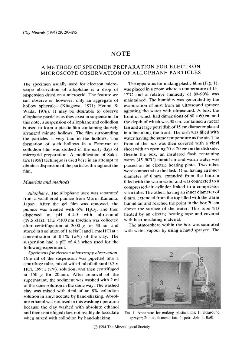
Hostname: page-component-848d4c4894-tn8tq Total loading time: 0 Render date: 2024-07-04T12:57:27.660Z Has data issue: false hasContentIssue false

A Method of Specimen Preparation for Electron Microscope Observation of Allophane Particles

Published online by Cambridge University Press:  09 July 2018

M. Hagiwara*
Affiliation:
Faculty of Agriculture, Hirosaki University, Hirosaki 036, Japan

Abstract

Image of the first page of this content. For PDF version, please use the ‘Save PDF’ preceeding this image.'
Type
Note
Copyright
Copyright © The Mineralogical Society of Great Britain and Ireland 1994

Access options

Get access to the full version of this content by using one of the access options below. (Log in options will check for institutional or personal access. Content may require purchase if you do not have access.)

References

Henmi, T. & Wada, K. (1976) Morphology and composition of allophane. Am. Miner, 61, 379390.Google Scholar
Kitagawa, Y. (1971) The ‘unit particle’ of allophane. Am. Miner, 56, 465475.Google Scholar
Sakata, S. (1958) Preparation of micro-grid for the electron microscopic observation with high magnification. J. Electron Microscop, 6, 7576.Google Scholar
Sjöstrand, F.S. (1956) A method to improve contrast in high resolution electron microscopy of ultrathin tissue sections. Exp. Cell Res, 10, 657664.Google Scholar