


No CrossRef data available.
Article contents
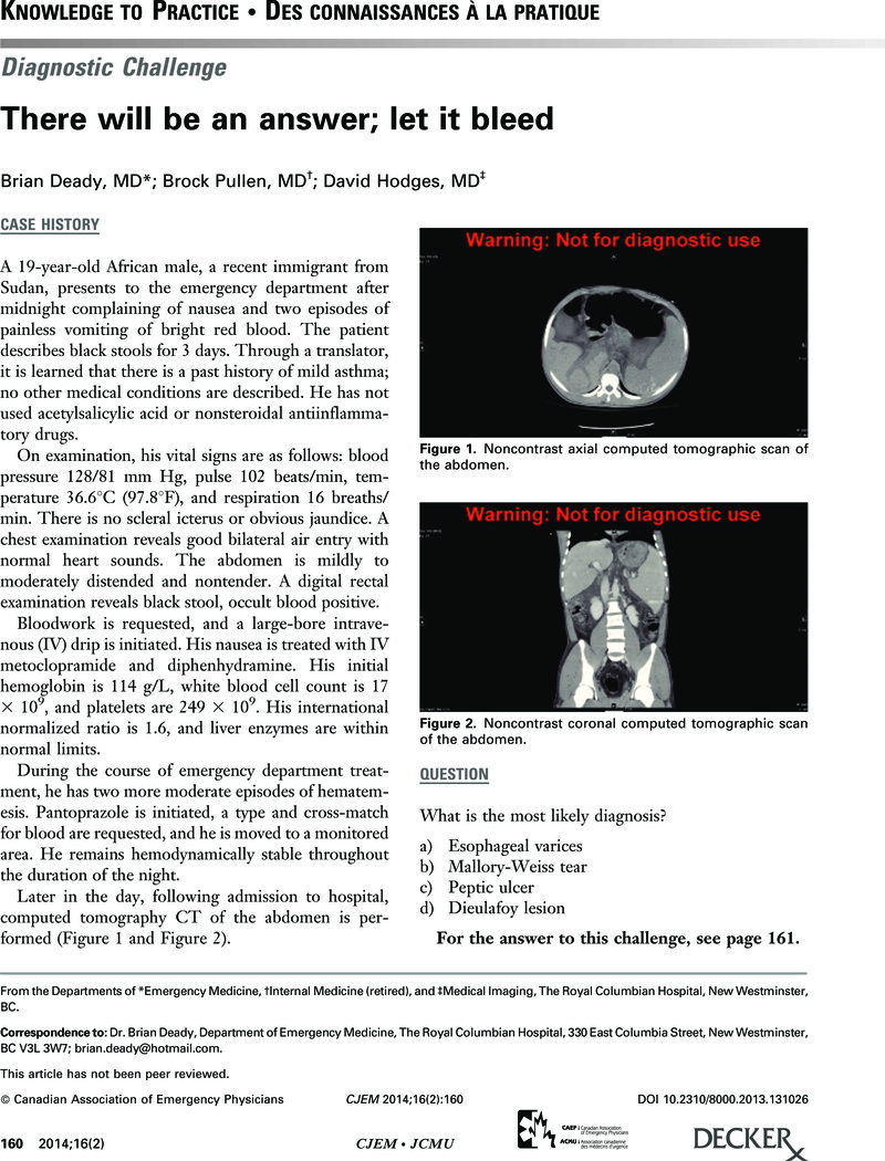
There will be an answer; let it bleed
Published online by Cambridge University Press: 04 March 2015
Abstract
An abstract is not available for this content so a preview has been provided. As you have access to this content, a full PDF is available via the ‘Save PDF’ action button.
- Type
- Knowledge To Practice • Des connaissances à la pratique
- Information
- Copyright
- Copyright © Canadian Association of Emergency Physicians 2014
References
REFERENCES
1.
Weaver, DH, Maxwell, JG, Castleton, KB.
Mallory-Weiss syndrome. Am J Surg
1969;118:887–92, doi:10.1016/0002-9610(69)90252-9.Google Scholar
2.
Watts, HD, Admirand, WH.
Mallory-Weiss syndrome. A reappraisal. JAMA
1974;230:1674–5, doi:10.1001/jama.1974.03240120042018.Google Scholar
3.
Michel, L, Serrano, A, Malt, RA.
Mallory-Weiss syndrome. Evolution of diagnostic and therapeutic patterns over two decades. Ann Surg
1980;192:716–21, doi:10.1097/00000658-198012000-00004.Google Scholar
4.
Sugawa, C, Benishek, D, Walt, AJ.
Mallory-Weiss syndrome. A study of 224 patients. Am J Surg
1983;145:30–3, doi:10.1016/0002-9610(83)90162-9.Google Scholar
5.
Harris, JM, DiPalma, JA.
Clinical significance of Mallory-Weiss tears. Am J Gastroenterol
1993;88:2056–8.Google ScholarPubMed
6.
Skok, P.
Fatal hemorrhage from a giant Mallory-Weiss tear. Endoscopy
2003;35:635, doi:10.1055/s-2003-40214.Google Scholar
7.
Knauer, CM, Mallory-Weiss syndrome. Characterization of 75 Mallory-Weiss lacerations in 528 patients with upper gastrointestinal hemorrhage. Gastroenterology
1976;71:5–8.Google ScholarPubMed
8.
Kovacs, TO, Jensen, DM.
Endoscopic diagnosis and treatment of bleeding Mallory-Weiss tears. Gastrointest Endosc Clin North Am
1991;1:387.CrossRefGoogle Scholar
9.
Bharucha, AE, Gostout, CJ, Balm, RK.
Clinical and endoscopic risk factors in the Mallory-Weiss syndrome. Am J Gastroenterol
1997;92:805–8.Google Scholar
10.
Laine, L, Peterson, WL.
Bleeding peptic ulcer. N Engl J Med
1994;331:717–27, doi:10.1056/NEJM199409153311107.Google Scholar
11.
Wara, P, Stodkilde, H.
Bleeding pattern before admission as guideline for emergency endoscopy. Scand J Gastroenterol
1985;20:72–8, doi:10.3109/00365528509089635.Google Scholar
12.
Jensen, DM, Machicado, GA.
Diagnosis and treatment of severe hematochezia: the role of urgent colonoscopy after purge. Gastroenterology
1988;95:1569–74.Google Scholar
13.
Mumtaz, R, Shaukat, M, Ramirez, FC.
Outcomes of endoscopic treatment of gastroduodenal Dieulafoy’s lesion with rubber band ligation and thermal/injection therapy. J Clin Gastroenterol
2003;36:310–4, doi:10.1097/00004836-200304000-00006.Google Scholar
14.
Lee, YT, Walmsley, RS, Leong, RW, et al. Dieulafoy’s lesion. Gastrointest Endosc
1993;58:236–43, doi:10.1067/mge.2003.328.Google Scholar
15.
Pollack, R, Lipsky, H, Goldberg, RI.
Duodenal Dieulafoy’s lesion. Gastrointest Endosc
1993;39:820–2, doi:10.1016/S0016-5107(93)70276-X.Google Scholar
16.
Anireddy, D, Timberlake, G, Seibert, D.
Dieulafoy’s lesion of the esophagus. Gastrointest Endosc
1993;39:604, doi:10.1016/S0016-5107(93)70198-4.Google Scholar
17.
Linhares, MM, Filho, BH, Schraibman, V, et al. Dieulafoy lesion: endoscopic and surgical management. Surg Laparosc Endosc Percutan Tech
2006;16:1–3, doi:10.1097/01.sle.0000202191.59322.5f.Google Scholar
18.
Gryseels, B.
Schistosomiasis. Infect Dis Clin North Am
2012;26:383–97, doi:10.1016/j.idc.2012.03.004.Google Scholar
19.
Gryseels, B, Polman, K, Clerinx, J, et al. Human schistosomiasis. Lancet
2006;368:1106–18, doi:10.1016/S0140-6736(06)69440-3.Google Scholar
20.
Chitsulo, L, Engels, D, Montresor, A, et al. The global status of schistosomiasis and its control. Acta Trop
2000;77:41–51, doi:10.1016/S0001-706X(00)00122-4.CrossRefGoogle ScholarPubMed
21.
Bottieau, E, Clerinx, J, De Vega, MR, et al. Imported Katayama fever: clinical and biological features at presentation and during treatment. J Infect
2006;52:339–45, doi:10.1016/j.jinf.2005.07.022.Google Scholar
22.
Lambertucci, JR.
Acute schistosomiasis: clinical, diagnostic and therapeutic features. Rev Inst Med Trop Sao Paulo
1993;35:399–404, doi:10.1590/S0036-4665199300 0500003.Google Scholar
23.
Cheever, AW, Hoffmann, KF, Wynn, TA.
Immunopathology of schistosomiasis mansoni in mice and men. Immunol Today
2000;21:46566, doi:10.1016/S0167-5699(00)01626-1.Google Scholar
24.
Thoeni, RF.
The role of imaging in patients with ascites. AJR Am J Roentgenol
1995;165:16–8.CrossRefGoogle ScholarPubMed
25.
Cattau, EL, Benjamin, SB, Knuff, TE, et al. The accuracy of the physical examination in the diagnosis of suspected ascites. JAMA
1982;247:1164–6, doi:10.1001/jama.1982.03320330060027.Google Scholar
26.
Amin, HM, Omran, SA, el-Bassuoni, NE, et al. Assessment of factors II, VII, IX, X and protein C in hepatosplenic schistosomiasis. Haemostasis
1994;24:22–6.Google Scholar
27.
Bottieau, E, Clerinx, J.
Imported Katayama fever: clinical and biological features at presentation and during treatment. J Infect
2006;52:339–45, doi:10.1016/j.jinf.2005.07.022.Google Scholar
28.
Ross, AG, Vickers, D.
Katayama syndrome. Lancet Infect Dis
2007;7:218–24, doi:10.1016/S1473-3099(07)70053-1.Google Scholar
29.
Jauréguiberry, S, Paris, L, Caumes, E.
Acute schistosomiasis, a diagnostic and therapeutic challenge. Clin Microbiol Infect
2010;16:225–31, doi:10.1111/j.1469-0691.2009.03131.x.Google Scholar
 You have
Access
You have
Access