


A 51-year-old female with esophageal cancer presented with a 1-day history of retrosternal chest pain with hypotension. She had no previous cardiac disease but was a chronic smoker. Her electrocardiogram (ECG) revealed inferior ST elevation. Given her well-looking appearance and active malignancy, etiologies other than ST elevation myocardial infarction (STEMI) were considered. Point-of-care ultrasound (POCUS) was performed to evaluate for a pericardial effusion (PCE). It revealed a small PCE and a mass in the inferior wall of the right ventricle (RV) (Figure 1).
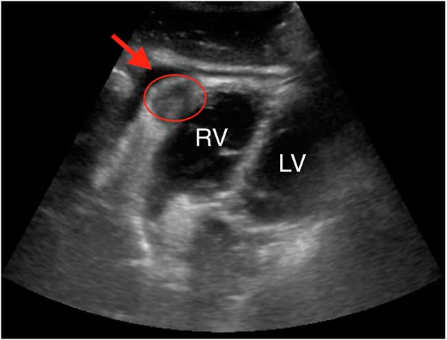
Figure 1 Subxiphoid cardiac view revealing a cardiac mass (red oval) in the inferior wall of the right ventricle (RV), adjacent to the left ventricle (LV), and a small pericardial effusion (red arrow).
Additional cardiac ultrasound views demonstrated appropriate fractional shortening and no regional wall motion abnormalities. Troponin levels were elevated but stable. Cardiology reviewed the case and POCUS images and agreed that a STEMI was unlikely. Therefore, the cath lab was not activated. Pulmonary embolus investigations were negative. Cardiac magnetic resonance imaging confirmed a mass in the RV wall believed to be metastatic esophageal cancer and the cause of her symptoms.
Case reports exist on secondary cardiac cancer causing ECG changes mimicking STEMI, particularly metastatic esophageal cancer.Reference Suga, Akuzawa and Hatori 1 - Reference Reynen, Köckeritz and Strasser 4 However, we believe that this is the first published case of the diagnosis being made using POCUS in the emergency department. This case demonstrates the utility of POCUS in clarifying the clinical presentation when there is diagnostic uncertainty, providing physicians with rapid information at the bedside that can significantly alter patient management.
ACKNOWLEDGEMENT
Thank you to Dr. Sheila Peters for her assistance in the management of this case.
Competing interests: None declared.
Keywords: point-of-care ultrasound, POCUS, cardiac cancer, STEMI

