


A 42-year-old woman with uncontrolled diabetes mellitus presented to the emergency department with fever, nausea, dysuria, and left flank pain. Physical examination revealed knocking tenderness on the left costovertebral angle. Laboratory results revealed leukocytosis, diabetic ketoacidosis, and pyuria. An abdominal radiograph showed air in the left renal fossa and ureter (Figure 1). Computed tomography (CT) demonstrated left emphysematous pyelonephritis (EPN) with air collections in the left kidney, perinephric space and ureter, and a calculus at the ureterovesical junction (Figures 2 and 3). The patient was treated with resuscitation, glycemic control, antibiotic therapy, and a percutaneous drainage. After stabilization of the general condition, subsequent nephroureterectomy was performed. The cultures of pus and urine yielded Escherichia coli.
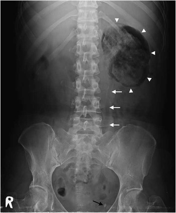
Figure 1 Abdominal radiography showing abnormal gas shadows in the left renal fossa (white arrowheads) and ureter (white arrows), and a radiopaque stone (black arrow).

Figure 2 Air collections in the left renal parenchyma, perinephric space, and ureter (arrow).

Figure 3 A distal ureteral calculus at the left ureterovesical junction (arrow).
EPN is a life-threatening, necrotizing infection of the renal parenchyma, characterized by the presence of air within the kidney, collecting system, and perinephric tissue.Reference Ubee, McGlynn and Fordham 1 , Reference Lin, Chen and Lin 2 Diabetes mellitus is the single most common associated factor.Reference Ubee, McGlynn and Fordham 1 Once high levels of tissue glucose, presence of glucose-fermenting organisms (gas-forming coliform bacteria such as E. coli or Klebsiella pneumoniae), and ureteral obstruction coexist, a urinary tract infection may progress to EPN.Reference Ubee, McGlynn and Fordham 1 - Reference Pontin and Barnes 3
Plain abdominal radiography may show abnormal gas in the renal bed or collecting system.Reference Ubee, McGlynn and Fordham 1 CT is the preferred diagnostic modality because of its high sensitivity and ability to assess the extent of gas patterns.Reference Ubee, McGlynn and Fordham 1 , Reference Pontin and Barnes 3 Treatment of EPN comprises fluid resuscitation, correction of electrolytes and glucose abnormalities, and aggressive antibiotics with Gram-negative bacteria coverage. Definitive surgical management includes percutaneous drainage or nephrectomy for overwhelming sepsis with extensive gas and renal destruction.Reference Ubee, McGlynn and Fordham 1 - Reference Pontin and Barnes 3
Keywords: emphysematous pyelonephritis, diabetes mellitus, percutaneous drainage, nephrectomy
Competing interests: None declared.


