


No CrossRef data available.
Article contents
Use of Primary Filters in X-Ray Spectrography: A New Method for Trace Analysis*
Published online by Cambridge University Press: 06 March 2019
Abstract
A method to improve the detectability of trace elements by X-ray fluorescent spectrography is described. The method consists of using appropriate filters in the primary, or exciting, beam. The effects of using filters in the primary beam on the peak-to-background ratio R of a fluorescent line have been analyzed on theoretical grounds. In fact,
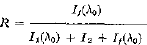
where I1(λ0) is the intensity of the analytic fluorescent line, I1(λ0) is the background intensity due to coherent and Compton scattering of the primary radiation by the specimen, and I2 is the background intensity due to scattering of the fluorescent radiation by the analyzing crystal. Analytical expressions were derived for I1(λ0), I2, and I1(λ0), from which it has been concluded:
1. The ratio I1(λ0)/I1(λ0) decreases when the filter used has its absorption edges at wavelengths longer than λ0.
2. The ratio I2/I1(λ0) can be separated into two parts which vary in opposite ways. The influence of these two parts on the value of R is discussed in the text.
It is then shown that the method should work well at short wavelengths and less well at longer wavelengths. The method was tested in the difficult case where overlapping of the analytical line with a characteristic line of the tube occurred, i.e., in the determination of traces of selenium by using tungsten radiation. The analytic line Se Kα. has a wavelength of 1.106 Å, while W Lγ1occurs at 1.098 Å. There is a marked effect of the filter thickness on the detectability; an optimum thickness appears to exist for each case. In the analysis of selenium, the best filter thickness (which can be selected by mere inspection of the diagrams reproduced in the text) increased the detectability of selenium traces by an order of magnitude. Finally, from statistical considerations, the quantity tσ2 is proposed as an index of the effectiveness of the filter: the smaller tσ2 is, the better the filter is. Here σ is the standard deviation of the intensity of the analytic line and t is the total counting time spent on the measure of the analytic line arid background. In order to study the dependence of the index tσ2 on the filter thickness, measurements were made on samples of sugar containing known concentrations of strontium. Then tσ2 was plotted against the thickness of the filter for each concentration; these curves do show a minimum. Thus, an optimum filter thickness exists in each case.
- Type
- Research Article
- Information
- Copyright
- Copyright © International Centre for Diffraction Data 1967
Footnotes
†
Present address: School of Physics, Georgia Institute of Technology, Atlanta, Georgia.
‡
Present address: Department of Physics, Northwestern University, Evanston, Illinois.
*
The section “Theory” was written by S. Caticha-Ellis; the section “Experimental” by Ariel Ramos, Luis Saravia, and S. Caticha-Ellis.
References
1.
Comptonand, A. H., Allison, S. K., X-Rays in Theory and Experiment,
D. Van Nostrand Co., Inc., Princeton, 1935.Google Scholar
2.
Birks, L. S., X-Ray Spectroehemkal Analysis,
Interscience Publishers, New York, 1959.Google Scholar