Skip to main content Accessibility help
×
Hostname: page-component-78c5997874-lj6df Total loading time: 0 Render date: 2024-11-18T08:38:29.983Z Has data issue: false hasContentIssue false

7 - Immunophenotyping of Mature B-Cell Lymphomas

Published online by Cambridge University Press:  01 February 2018

Anna Porwit
Affiliation:
Lunds Universitet, Sweden
Marie Christine Béné
Affiliation:
Université de Nantes, France
Get access

Summary

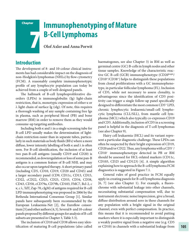
Image of the first page of this content. For PDF version, please use the ‘Save PDF’ preceeding this image.'
Type
Chapter
Information
Publisher: Cambridge University Press
Print publication year: 2018

Access options

Get access to the full version of this content by using one of the access options below. (Log in options will check for institutional or personal access. Content may require purchase if you do not have access.)

References

Wood, B.L., Arroz, M., Barnett, D., et al. 2006 Bethesda International Consensus recommendations on the immunophenotypic analysis of hematolymphoid neoplasia by flow cytometry: optimal reagents and reporting for the flow cytometric diagnosis of hematopoietic neoplasia. Cytometry Part B, Clinical Cytometry; 72 Suppl 1 (2007):S1422.CrossRefGoogle ScholarPubMed
Bene, M.C., Nebe, T., Bettelheim, P., et al. Immunophenotyping of acute leukemia and lymphoproliferative disorders: a consensus proposal of the European LeukemiaNet Work Package 10. Leukemia; 25 (2011):567–74.CrossRefGoogle ScholarPubMed
van Dongen, J.J., Lhermitte, L., Bottcher, S., et al. EuroFlow antibody panels for standardized n-dimensional flow cytometric immunophenotyping of normal, reactive and malignant leukocytes. Leukemia; 26 (2012):1908–75.CrossRefGoogle ScholarPubMed
Craig, F.E., and Foon, K.A.. Flow cytometric immunophenotyping for hematologic neoplasms. Blood; 111 (2008):3941–67.CrossRefGoogle ScholarPubMed
Calvo, K.R., McCoy, C.S., and Stetler-Stevenson, M.. Flow cytometry immunophenotyping of hematolymphoid neoplasia. Methods in Molecular Biology; 699 (2011):295316.CrossRefGoogle ScholarPubMed
Tanqri, S., Vall, H., Kaplan, D., et al. Validation of cell-based fluorescence assays: practice guidelines from the ICSH and ICCS - part III - analytical issues. Cytometry Part B, Clinical Cytometry; 84 (2013):291308.CrossRefGoogle Scholar
Johansson, U., Bloxham, D., Couzens, S., et al. Guidelines on the use of multicolour flow cytometry in the diagnosis of haematological neoplasms. British Committee for Standards in Haematology. British Journal of Haematology; 165 (2014):455–88.CrossRefGoogle ScholarPubMed
Rajab, A., and Porwit, A.. Screening bone marrow samples for abnormal lymphoid populations and myelodysplasia-related features with one 10-color 14-antibody screening tube. Cytometry Part B, Clinical Cytometry; 88 (2015):253–60.CrossRefGoogle ScholarPubMed
Preijers, F.W., Huys, E., and Moshaver, B.. OMIP-010: a new 10-color monoclonal antibody panel for polychromatic immunophenotyping of small hematopoietic cell samples. Cytometry Part A: The Journal of the International Society for Analytical Cytology; 81 (2012):453–5.Google ScholarPubMed
Hedley, B.D., Keeney, M., Popma, J., et al. Novel lymphocyte screening tube using dried monoclonal antibody reagents. Cytometry Part B, Clinical Cytometry; 88 (2015):361–70.CrossRefGoogle Scholar
Rajab, A, Axler, O, Leung, J, Wozniak, M, and Porwit, A. Ten-color 15-antibody flow cytometry panel for immunophenotyping of lymphocyte population. International Journal of Laboratory Hematology 2017 May; 39 Suppl 1:7685.CrossRefGoogle ScholarPubMed
Campo, S.H. Swerdlow, Harris, E., et al. WHO Classification of Tumours of Haematopoietic and Lymphoid Tissues. Lyon: IARC; 2008.Google Scholar
Swerdlow, S.H., Campo, E., Pileri, S.A., et al. The 2016 revision of the World Health Organization classification of lymphoid neoplasms. Blood; 127 (2016):2375–90.CrossRefGoogle Scholar
Stilgenbauer, S., Bullinger, L., Lichter, P., et al. Genetics of chronic lymphocytic leukemia: genomic aberrations and V(H) gene mutation status in pathogenesis and clinical course. Leukemia; 16 (2002):9931007.CrossRefGoogle ScholarPubMed
Juliusson, G., Oscier, D.G., Fitchett, M., et al. Prognostic subgroups in B-cell chronic lymphocytic leukemia defined by specific chromosomal abnormalities. The New England Journal of Medicine; 323 (1990):720–4.CrossRefGoogle ScholarPubMed
Landgren, O., Albitar, M., Ma, W., et al. B-cell clones as early markers for chronic lymphocytic leukemia. The New England Journal of Medicine; 360 (2009):659–67.CrossRefGoogle ScholarPubMed
Strati, P., and Shanafelt, T.D.. Monoclonal B-cell lymphocytosis and early-stage chronic lymphocytic leukemia: diagnosis, natural history, and risk stratification. Blood; 126 (2015):454–62.CrossRefGoogle ScholarPubMed
Mahdi, T., Rajab, A., Padmore, R., et al. Characteristics of lymphoproliferative disorders with more than one aberrant cell population as detected by 10-color flow cytometry. Cytometry Part B, Clinical Cytometry (2016). 20 July, E-pub ahead of print.Google ScholarPubMed
Rawstron, A.C., Fazi, C., Agathangelidis, A., et al. A complementary role of multiparameter flow cytometry and high-throughput sequencing for minimal residual disease detection in chronic lymphocytic leukemia: an European Research Initiative on CLL study. Leukemia; 30 (2016):929–36.CrossRefGoogle ScholarPubMed
Rawstron, A.C., Bottcher, S., Letestu, R., et al. Improving efficiency and sensitivity: European Research Initiative in CLL (ERIC) update on the international harmonised approach for flow cytometric residual disease monitoring in CLL. Leukemia; 27 (2013):142–9.CrossRefGoogle ScholarPubMed
Matutes, E., Owusu-Ankomah, K., Morilla, R., et al. The immunological profile of B-cell disorders and proposal of a scoring system for the diagnosis of CLL. Leukemia; 8 (1994):1640–5.Google ScholarPubMed
Ginaldi, L., De Martinis, M., Matutes, E., et al. Levels of expression of CD19 and CD20 in chronic B cell leukaemias. Journal of Clinical Pathology; 51 (1998):364–9.CrossRefGoogle ScholarPubMed
Molica, S., Levato, D., Dattilo, A., et al. Clinico-prognostic relevance of quantitative immunophenotyping in B-cell chronic lymphocytic leukemia with emphasis on the expression of CD20 antigen and surface immunoglobulins. European Journal of Haematology; 60 (1998):4752.CrossRefGoogle ScholarPubMed
Tam, C.S., Otero-Palacios, J., Abruzzo, L.V., et al. Chronic lymphocytic leukaemia CD20 expression is dependent on the genetic subtype: a study of quantitative flow cytometry and fluorescent in-situ hybridization in 510 patients. British Journal of Haematology; 141 (2008):3640.CrossRefGoogle ScholarPubMed
Quijano, S., Lopez, A., Rasillo, A., et al. Impact of trisomy 12, del(13q), del(17p), and del(11q) on the immunophenotype, DNA ploidy status, and proliferative rate of leukemic B-cells in chronic lymphocytic leukemia. Cytometry Part B, Clinical Cytometry; 74 (2008):139–49.Google Scholar
Lewis, R.E., Cruse, J.M., Pierce, S., et al. Surface and cytoplasmic immunoglobulin expression in B-cell chronic lymphocytic leukemia (CLL). Experimental and Molecular Pathology; 79 (2005):146–50.CrossRefGoogle ScholarPubMed
Baldini, L., Mozzana, R., Cortelezzi, A., et al. Prognostic significance of immunoglobulin phenotype in B cell chronic lymphocytic leukemia. Blood; 65 (1985):340–4.CrossRefGoogle ScholarPubMed
Potter, K.N., Mockridge, C.I., Neville, L., et al. Structural and functional features of the B-cell receptor in IgG-positive chronic lymphocytic leukemia. Clinical Cancer Research: an Official Journal of the American Association for Cancer Research; 12 (2006):1672–9.CrossRefGoogle ScholarPubMed
Tomomatsu, J., Isobe, Y., Oshimi, K., et al. Chronic lymphocytic leukemia in a Japanese population: varied immunophenotypic profile, distinctive usage of frequently mutated IGH gene, and indolent clinical behavior. Leukemia & Lymphoma; 51 (2010):2230–9.CrossRefGoogle Scholar
D'Avola, A., Drennan, S., Tracy, I., et al. Surface IgM expression and function are associated with clinical behavior, genetic abnormalities, and DNA methylation in CLL. Blood; 128 (2016):816–26.Google ScholarPubMed
Challagundla, P., Jorgensen, J.L., Kanagal-Shamanna, R., et al. Utility of quantitative flow cytometry immunophenotypic analysis of CD5 expression in small B-cell neoplasms. Archives of Pathology & Laboratory Medicine; 138 (2014):903–9.CrossRefGoogle ScholarPubMed
Durrieu, F., Genevieve, F., Arnoulet, C., et al. Normal levels of peripheral CD19(+) CD5(+) CLL-like cells: toward a defined threshold for CLL follow-up -- a GEIL-GOELAMS study. Cytometry Part B, Clinical Cytometry; 80 (2011):346–53.Google Scholar
Kriston, C., Bodor, C., Szenthe, K., et al. Low CD23 expression correlates with high CD38 expression and the presence of trisomy 12 in CLL. Hematological Oncology; 35 (2015):5863.CrossRefGoogle ScholarPubMed
Chen, C.C., Raikow, R.B., Sonmez-Alpan, E., et al. Classification of small B-cell lymphoid neoplasms using a paraffin section immunohistochemical panel. Applied Immunohistochemistry & Molecular Morphology: AIMM; 8 (2000):111.CrossRefGoogle ScholarPubMed
Zomas, A.P., Matutes, E., Morilla, R., et al. Expression of the immunoglobulin-associated protein B29 in B cell disorders with the monoclonal antibody SN8 (CD79b). Leukemia; 10 (1996):1966–70.Google Scholar
Alfarano, A., Indraccolo, S., Circosta, P., et al. An alternatively spliced form of CD79b gene may account for altered B-cell receptor expression in B-chronic lymphocytic leukemia. Blood; 93 (1999):2327–35.CrossRefGoogle ScholarPubMed
Marotta, G., Raspadori, D., Sestigiani, C., et al. Expression of the CD11c antigen in B-cell chronic lymphoproliferative disorders. Leukemia & Lymphoma; 37 (2000):145–9.CrossRefGoogle ScholarPubMed
Barrena, S., Almeida, J., Yunta, M., et al. Discrimination of biclonal B-cell chronic lymphoproliferative neoplasias by tetraspanin antigen expression. Leukemia; 19 (2005):1708–9.CrossRefGoogle Scholar
Gong, S., Osei, E.S., Kaplan, D., et al. CD317 is over-expressed in B-cell chronic lymphocytic leukemia, but not B-cell acute lymphoblastic leukemia. International Journal of Clinical and Experimental Pathology; 8 (2015):1613–21.Google Scholar
Porwit, A., Borgonovo, L., Osby, E., et al. B-cell chronic lymphocytic leukaemia with aberrant expression of CD8 antigen. European Journal of Haematology; 39 (1987):311–17.CrossRefGoogle ScholarPubMed
Carulli, G., Stacchini, A., Marini, A., et al. Aberrant expression of CD8 in B-cell non-Hodgkin lymphoma: a multicenter study of 951 bone marrow samples with lymphomatous infiltration. American Journal of Clinical Pathology; 132 (2009):186–90; quiz 306.CrossRefGoogle ScholarPubMed
Dorwal, P., Mehra, S., Pande, A., et al. Aberrant NK cell associated marker (CD56 and CD57) expression in chronic lymphocytic leukemia. Cytometry Part B, Clinical Cytometry; 88 (2015):348–51.CrossRefGoogle ScholarPubMed
Malavasi, F., Deaglio, S., Damle, R., et al. CD38 and chronic lymphocytic leukemia: a decade later. Blood; 118 (2011):3470–8.CrossRefGoogle ScholarPubMed
Hamblin, T.J., Orchard, J.A., Ibbotson, R.E., et al. CD38 expression and immunoglobulin variable region mutations are independent prognostic variables in chronic lymphocytic leukemia, but CD38 expression may vary during the course of the disease. Blood; 99 (2002):1023–9.CrossRefGoogle ScholarPubMed
Wiestner, A., Rosenwald, A., Barry, T.S., et al. ZAP-70 expression identifies a chronic lymphocytic leukemia subtype with unmutated immunoglobulin genes, inferior clinical outcome, and distinct gene expression profile. Blood; 101 (2003):4944–51.CrossRefGoogle ScholarPubMed
Wang, Y.H., Fan, L., Xu, W., et al. Detection methods of ZAP-70 in chronic lymphocytic leukemia. Clinical and Experimental Medicine; 12 (2012):6977.CrossRefGoogle ScholarPubMed
Rossi, F.M., Del Principe, M.I., Rossi, D., et al. Prognostic impact of ZAP-70 expression in chronic lymphocytic leukemia: mean fluorescence intensity T/B ratio versus percentage of positive cells. Journal of Translational Medicine; 8 (2010):23.CrossRefGoogle ScholarPubMed
Wiggers, T.G., Westra, G., Westers, T.M., et al. ZAP70 in B-CLL cells related to the expression in NK cells is a surrogate marker for mutational status. Cytometry Part B, Clinical Cytometry; 86 (2014):280–7.CrossRefGoogle Scholar
Gattei, V., Bulian, P., Principe, M.I. Del, et al. Relevance of CD49d protein expression as overall survival and progressive disease prognosticator in chronic lymphocytic leukemia. Blood; 111 (2008):865–73.CrossRefGoogle ScholarPubMed
Dal Bo, M., Bulian, P., Bomben, R., et al. CD49d prevails over the novel recurrent mutations as independent prognosticator of overall survival in chronic lymphocytic leukemia. Leukemia; 30 (2016):2011–18.CrossRefGoogle ScholarPubMed
Kwok, M., Rawstron, A.C., Varghese, A., et al. Minimal residual disease is an independent predictor for 10-year survival in CLL. Blood; 128 (2016):2770–3.CrossRefGoogle ScholarPubMed
Dowling, A.K., Liptrot, S.D., O'Brien, D., et al. Optimization and validation of an 8-color single-tube assay for the sensitive detection of minimal residual disease in B-cell chronic lymphocytic leukemia detected via flow cytometry. Laboratory Medicine; 47 (2016):103–11.CrossRefGoogle ScholarPubMed
Ruchlemer, R., Parry-Jones, N., Brito-Babapulle, V., et al. B-prolymphocytic leukaemia with t(11;14) revisited: a splenomegalic form of mantle cell lymphoma evolving with leukaemia. British Journal of Haematology; 125 (2004):330–6.CrossRefGoogle Scholar
Put, N., Van Roosbroeck, K., Konings, P., et al. Chronic lymphocytic leukemia and prolymphocytic leukemia with MYC translocations: a subgroup with an aggressive disease course. Annals of Hematology; 91 (2012):863–73.CrossRefGoogle ScholarPubMed
Hoehn, D., Miranda, R.N., Kanagal-Shamanna, R., et al. Splenic B-cell lymphomas with more than 55% prolymphocytes in blood: evidence for prolymphocytoid transformation. Human Pathology; 43 (2012):1828–38.CrossRefGoogle ScholarPubMed
Porwit, A., Fend, F., Kremer, M., et al. Issues in diagnosis of small B cell lymphoid neoplasms involving the bone marrow and peripheral blood. Report on the Bone Marrow Workshop of the XVIIth meeting of the European Association for Haematopathology and the Society for Hematopathology. Histopathology; 69 (2016):349–73.CrossRefGoogle Scholar
Baseggio, L., Traverse-Glehen, A., Petinataud, F., et al. CD5 expression identifies a subset of splenic marginal zone lymphomas with higher lymphocytosis: a clinico-pathological, cytogenetic and molecular study of 24 cases. Haematologica; 95 (2010):604–12.CrossRefGoogle ScholarPubMed
Demurtas, A., Stacchini, A., Aliberti, S., et al. Tissue flow cytometry immunophenotyping in the diagnosis and classification of non-Hodgkin's lymphomas: a retrospective evaluation of 1,792 cases. Cytometry Part B, Clinical Cytometry; 84 (2013):8295.CrossRefGoogle Scholar
Bahler, D.W., Pindzola, J.A., and Swerdlow, S.H.. Splenic marginal zone lymphomas appear to originate from different B cell types. The American Journal of Pathology; 161 (2002):81–8.CrossRefGoogle ScholarPubMed
Kost, C.B., Holden, J.T., and Mann, K.P.. Marginal zone B-cell lymphoma: a retrospective immunophenotypic analysis. Cytometry Part B, Clinical Cytometry; 74 (2008):282–6.Google ScholarPubMed
Franco, V., Florena, A.M., Ascani, S., et al. CD27 distinguishes two phases in bone marrow infiltration of splenic marginal zone lymphoma. Histopathology; 44 (2004):381–6.CrossRefGoogle ScholarPubMed
Miguet, L., Lennon, S., Baseggio, L., et al. Cell-surface expression of the TLR homolog CD180 in circulating cells from splenic and nodal marginal zone lymphomas. Leukemia; 27 (2013):1748–50.CrossRefGoogle ScholarPubMed
Mayeur-Rousse, C., Guy, J., Miguet, L., et al. CD180 expression in B-cell lymphomas: a multicenter GEIL study. Cytometry Part B, Clinical Cytometry; 90 (2016):462–6.CrossRefGoogle ScholarPubMed
Challagundla, P., Medeiros, L.J., Kanagal-Shamanna, R., et al. Differential expression of CD200 in B-cell neoplasms by flow cytometry can assist in diagnosis, subclassification, and bone marrow staging. American Journal of Clinical Pathology; 142 (2014):837–44.CrossRefGoogle ScholarPubMed
Venkataraman, G., Aguhar, C., Kreitman, R.J., et al. Characteristic CD103 and CD123 expression pattern defines hairy cell leukemia: usefulness of CD123 and CD103 in the diagnosis of mature B-cell lymphoproliferative disorders. American Journal of Clinical Pathology; 136 (2011):625–30.CrossRefGoogle ScholarPubMed
Julhakyan, H.L., Al-Radi, L.S., Moiseeva, T.N., et al. A single-center experience in splenic diffuse red pulp lymphoma siagnosis. Clinical Lymphoma, Myeloma & Leukemia; 16 Suppl (2016):S1669.CrossRefGoogle Scholar
Traverse-Glehen, A., Baseggio, L., Salles, G., et al. Splenic diffuse red pulp small-B cell lymphoma: toward the emergence of a new lymphoma entity. Discovery Medicine; 13 (2012):253–65.Google ScholarPubMed
Shao, H., Calvo, K.R., Gronborg, M., et al. Distinguishing hairy cell leukemia variant from hairy cell leukemia: development and validation of diagnostic criteria. Leukemia Research; 37 (2013):401–9.CrossRefGoogle ScholarPubMed
Jain, D., Dorwal, P., Gajendra, S., et al. CD5 positive hairy cell leukemia: a rare case report with brief review of literature. Cytometry Part B, Clinical Cytometry; 90 (2016):467–72.CrossRefGoogle ScholarPubMed
Chen, Y.H., Tallman, M.S., Goolsby, C., et al. Immunophenotypic variations in hairy cell leukemia. American Journal of Clinical Pathology; 125 (2006):251–9.CrossRefGoogle ScholarPubMed
Galani, K.S., Subramanian, P.G., Gadage, V.S., et al. Clinico-pathological profile of Hairy cell leukemia: critical insights gained at a tertiary care cancer hospital. Indian Journal of Pathology & Microbiology; 55 (2012):61–5.Google Scholar
Tallman, M.S.. Implications of minimal residual disease in hairy cell leukemia after cladribine using immunohistochemistry and immunophenotyping. Leukemia & Lymphoma; 52 Suppl 2 (2011):65–8.CrossRefGoogle ScholarPubMed
Garnache Ottou, F., Chandesris, M.O., Lhermitte, L., et al. Peripheral blood 8 colour flow cytometry monitoring of hairy cell leukaemia allows detection of high-risk patients. British Journal of Haematology; 166 (2014):50–9.CrossRefGoogle ScholarPubMed
Sainati, L., Matutes, E., Mulligan, S., et al. A variant form of hairy cell leukemia resistant to alpha-interferon: clinical and phenotypic characteristics of 17 patients. Blood; 76 (1990):157–62.CrossRefGoogle ScholarPubMed
Dong, H.Y., Weisberger, J., Liu, Z., et al. Immunophenotypic analysis of CD103+ B-lymphoproliferative disorders: hairy cell leukemia and its mimics. American Journal of Clinical Pathology; 131 (2009):586–95.CrossRefGoogle ScholarPubMed
Tiacci, E., Schiavoni, G., Forconi, F., et al. Simple genetic diagnosis of hairy cell leukemia by sensitive detection of the BRAF-V600E mutation. Blood; 119 (2012):192–5.CrossRefGoogle ScholarPubMed
Waterfall, J.J., Arons, E., Walker, R.L., et al. High prevalence of MAP2K1 mutations in variant and IGHV4-34-expressing hairy-cell leukemias. Nature genetics; 46 (2014):810.CrossRefGoogle ScholarPubMed
Kanellis, G., Mollejo, M., Montes-Moreno, S., et al. Splenic diffuse red pulp small B-cell lymphoma: revision of a series of cases reveals characteristic clinico-pathological features. Haematologica; 95 (2010):1122–9.CrossRefGoogle ScholarPubMed
Hunter, Z.R., Branagan, A.R., Manning, R., et al. CD5, CD10, and CD23 expression in Waldenstrom's macroglobulinemia. Clinical Lymphoma; 5 (2005):246–9.CrossRefGoogle ScholarPubMed
Konoplev, S., Medeiros, L.J., Bueso-Ramos, C.E., et al. Immunophenotypic profile of lymphoplasmacytic lymphoma/Waldenstrom macroglobulinemia. American Journal of Clinical Pathology; 124 (2005):414–20.CrossRefGoogle ScholarPubMed
Paiva, B., Montes, M.C., Garcia-Sanz, R., et al. Multiparameter flow cytometry for the identification of the Waldenstrom's clone in IgM-MGUS and Waldenstrom's Macroglobulinemia: new criteria for differential diagnosis and risk stratification. Leukemia; 28 (2014):166–73.CrossRefGoogle ScholarPubMed
San Miguel, J.F., Vidriales, M.B., Ocio, E., et al. Immunophenotypic analysis of Waldenstrom's macroglobulinemia. Seminars in Oncology; 30 (2003):187–95.CrossRefGoogle ScholarPubMed
Rosado, F.G., Morice, W.G., He, R., et al. Immunophenotypic features by multiparameter flow cytometry can help distinguish low grade B-cell lymphomas with plasmacytic differentiation from plasma cell proliferative disorders with an unrelated clonal B-cell process. British Journal of Haematology; 169 (2015):368–76.CrossRefGoogle ScholarPubMed
Owen, R.G., Kyle, R.A., Stone, M.J., et al. Response assessment in Waldenstrom macroglobulinaemia: update from the VIth International Workshop. British Journal of Haematology; 160 (2013):171–6.CrossRefGoogle ScholarPubMed
Garcia-Sanz, R., Ocio, E., Caballero, A., et al. Post-treatment bone marrow residual disease > 5% by flow cytometry is highly predictive of short progression-free and overall survival in patients with Waldenstrom's macroglobulinemia. Clinical Lymphoma, Myeloma & Leukemia; 11 (2011):168–71.CrossRefGoogle ScholarPubMed
Barakat, F.H., Medeiros, L.J., Wei, E.X., et al. Residual monotypic plasma cells in patients with Waldenstrom macroglobulinemia after therapy. American Journal of Clinical Pathology; 135 (2011):365–73.CrossRefGoogle ScholarPubMed
Pillai, R.K., Surti, U., and Swerdlow, S.H.. Follicular lymphoma-like B cells of uncertain significance (in situ follicular lymphoma) may infrequently progress, but precedes follicular lymphoma, is associated with other overt lymphomas and mimics follicular lymphoma in flow cytometric studies. Haematologica; 98 (2013):1571–80.CrossRefGoogle ScholarPubMed
Schmidt, B., Kremer, M., Gotze, K., et al. Bone marrow involvement in follicular lymphoma: comparison of histology and flow cytometry as staging procedures. Leukemia & Lymphoma; 47 (2006):1857–62.CrossRefGoogle ScholarPubMed
Iancu, D., Hao, S., Lin, P., et al. Follicular lymphoma in staging bone marrow specimens: correlation of histologic findings with the results of flow cytometry immunophenotypic analysis. Archives of Pathology & Laboratory Medicine; 131 (2007):282–7.CrossRefGoogle ScholarPubMed
Almasri, N.M., Iturraspe, J.A., and Braylan, R.C.. CD10 expression in follicular lymphoma and large cell lymphoma is different from that of reactive lymph node follicles. Archives of Pathology & Laboratory Medicine; 122 (1998):539–44.Google ScholarPubMed
Eshoa, C., Perkins, S., Kampalath, B., et al. Decreased CD10 expression in grade III and in interfollicular infiltrates of follicular lymphomas. American Journal of Clinical Pathology; 115 (2001):862–7.CrossRefGoogle ScholarPubMed
Li, Y., Hu, S., Zuo, Z., et al. CD5-positive follicular lymphoma: clinicopathologic correlations and outcome in 88 cases. Modern Pathology: an Official Journal of the United States and Canadian Academy of Pathology, Inc; 28 (2015):787–98.CrossRefGoogle ScholarPubMed
Miyoshi, H., Sato, K., Yoshida, M., et al. CD5-positive follicular lymphoma characterized by CD25, MUM1, low frequency of t(14;18) and poor prognosis. Pathology International; 64 (2014):95103.CrossRefGoogle ScholarPubMed
Olteanu, H., Fenske, T.S., Harrington, A.M., et al. CD23 expression in follicular lymphoma: clinicopathologic correlations. American Journal of Clinical Pathology; 135 (2011):4653.CrossRefGoogle ScholarPubMed
Fujiwara, S., Muroi, K., Tatara, R., et al. Clinical features of de novo CD25-positive follicular lymphoma. Leukemia & Lymphoma; 55 (2014):307–13.CrossRefGoogle ScholarPubMed
Mestrallet, F., Sujobert, P., Sarkozy, C., et al. CD180 overexpression in follicular lymphoma is restricted to the lymph node compartment. Cytometry Part B, Clinical Cytometry; 90 (2016):433–9.CrossRefGoogle Scholar
Carulli, G., Cannizzo, E., Zucca, A., et al. CD45 expression in low-grade B-cell non-Hodgkin's lymphomas. Leukemia Research; 32 (2008):263–7.CrossRefGoogle ScholarPubMed
Wang, F., Xu, D., and Cui, W.. Leukocyte common antigen (CD45) negative follicular lymphoma, a rare immunophenotypic presentation. Clinica Chimica Acta; International Journal of Clinical Chemistry; 442 (2015):46–8.CrossRefGoogle ScholarPubMed
Ray, S., Craig, F.E., and Swerdlow, S.H.. Abnormal patterns of antigenic expression in follicular lymphoma: a flow cytometric study. American Journal of Clinical Pathology; 124 (2005):576–83.CrossRefGoogle ScholarPubMed
Mantei, K., and Wood, B.L.. Flow cytometric evaluation of CD38 expression assists in distinguishing follicular hyperplasia from follicular lymphoma. Cytometry Part B, Clinical Cytometry; 76 (2009):315–20.Google ScholarPubMed
Cook, J.R., Craig, F.E., and Swerdlow, S.H.. Bcl-2 expression by multicolor flow cytometric analysis assists in the diagnosis of follicular lymphoma in lymph node and bone marrow. American Journal of Clinical Pathology; 119 (2003):145–51.CrossRefGoogle Scholar
Laane, E., Tani, E., Bjorklund, E., et al. Flow cytometric immunophenotyping including Bcl-2 detection on fine needle aspirates in the diagnosis of reactive lymphadenopathy and non-Hodgkin's lymphoma. Cytometry Part B, Clinical Cytometry; 64 (2005):3442.CrossRefGoogle ScholarPubMed
Wu, J.M., Borowitz, M.J., and Weir, E.G.. The usefulness of CD71 expression by flow cytometry for differentiating indolent from aggressive CD10+ B-cell lymphomas. American Journal of Clinical Pathology; 126 (2006):3946.CrossRefGoogle ScholarPubMed
Wahlin, B.E., Sander, B., Christensson, B., et al. CD8+ T-cell content in diagnostic lymph nodes measured by flow cytometry is a predictor of survival in follicular lymphoma. Clinical Cancer Research: an Official Journal of the American Association for Cancer Research; 13 (2007):388–97.CrossRefGoogle ScholarPubMed
Magnano, L., Martinez, A., Carreras, J., et al. T-cell subsets in lymph nodes identify a subgroup of follicular lymphoma patients with favorable outcome. Leukemia & Lymphoma (2016):19.Google ScholarPubMed
Marcheselli, L., Bari, A., Anastasia, A., et al. Prognostic roles of absolute monocyte and absolute lymphocyte counts in patients with advanced-stage follicular lymphoma in the rituximab era: an analysis from the FOLL05 trial of the Fondazione Italiana Linfomi. British Journal of Haematology; 169 (2015):544–51.CrossRefGoogle ScholarPubMed
He, L., Liang, J.H., Wu, J.Z., et al. Low absolute CD4+ T cell counts in peripheral blood are associated with inferior survival in follicular lymphoma. Tumour Biology: the Journal of the International Society for Oncodevelopmental Biology and Medicine; 37 (2016):12589–95.CrossRefGoogle ScholarPubMed
He, L., Zhu, H.Y., Qin, S.C., et al. Low natural killer (NK) cell counts in peripheral blood adversely affect clinical outcome of patients with follicular lymphoma. Blood Cancer Journal; 6 (2016):e457.CrossRefGoogle ScholarPubMed
Lobetti-Bodoni, C., Mantoan, B., Monitillo, L., et al. Clinical implications and prognostic role of minimal residual disease detection in follicular lymphoma. Therapeutic Advances in Hematology; 4 (2013):189–98.CrossRefGoogle ScholarPubMed
Drandi, D., Kubiczkova-Besse, L., Ferrero, S., et al. Minimal residual disease detection by droplet digital PCR in multiple myeloma, mantle cell lymphoma, and follicular lymphoma: a comparison with real-time PCR. The Journal of Molecular Diagnostics: JMD; 17 (2015):652–60.CrossRefGoogle ScholarPubMed
Kato, H., Yamamoto, K., Oki, Y., et al. Clinical value of flow cytometric immunophenotypic analysis for minimal residual disease detection in autologous stem-cell products of follicular and mantle cell lymphomas. Leukemia; 26 (2012):166–9.CrossRefGoogle ScholarPubMed
Fernandez, V., Salamero, O., Espinet, B., et al. Genomic and gene expression profiling defines indolent forms of mantle cell lymphoma. Cancer Research; 70 (2010):1408–18.CrossRefGoogle ScholarPubMed
Cohen, P.L., Kurtin, P.J., Donovan, K.A., et al. Bone marrow and peripheral blood involvement in mantle cell lymphoma. British Journal of Haematology; 101 (1998):302–10.CrossRefGoogle ScholarPubMed
Jares, P., Colomer, D., Campo, E.. Molecular pathogenesis of mantle cell lymphoma. The Journal of Clinical Investigation; 122 (2012):3416–23.CrossRefGoogle ScholarPubMed
Williams, M.E., Swerdlow, S.H.. Cyclin D1 overexpression in non-Hodgkin's lymphoma with chromosome 11 bcl-1 rearrangement. Annals of Oncology: Official Journal of the European Society for Medical Oncology; 5 Suppl 1 (1994):71–3.CrossRefGoogle ScholarPubMed
Elnenaei, M.O., Jadayel, D.M., Matutes, E., et al. Cyclin D1 by flow cytometry as a useful tool in the diagnosis of B-cell malignancies. Leukemia Research; 25 (2001):115–23.CrossRefGoogle ScholarPubMed
Jain, P., Giustolisi, G.M., Atkinson, S., et al. Detection of cyclin D1 in B cell lymphoproliferative disorders by flow cytometry. Journal of Clinical Pathology; 55 (2002):940–5.CrossRefGoogle Scholar
Wasik, A.M., Priebe, V., Lord, M., et al. Flow cytometric analysis of SOX11: a new diagnostic method for distinguishing B-cell chronic lymphocytic leukemia/small lymphocytic lymphoma from mantle cell lymphoma. Leukemia & Lymphoma; 56 (2015):1425–31.CrossRefGoogle ScholarPubMed
Gallo, M., Cacheux, V., Vincent, L., et al. Leukemic non-nodal mantle cell lymphomas have a distinct phenotype and are associated with deletion of PARP1 and 13q14. Virchows Archiv: an International Journal of Pathology; 469 (2016):697706.CrossRefGoogle ScholarPubMed
Gao, J., Peterson, L., Nelson, B., et al. Immunophenotypic variations in mantle cell lymphoma. American Journal of Clinical Pathology; 132 (2009):699706.CrossRefGoogle ScholarPubMed
Kelemen, K., Peterson, L.C., Helenowski, I., et al. CD23+ mantle cell lymphoma: a clinical pathologic entity associated with superior outcome compared with CD23- disease. American Journal of Clinical Pathology; 130 (2008):166–77.CrossRefGoogle ScholarPubMed
Kraus, T.S., Sillings, C.N., Saxe, D.F., et al. The role of CD11c expression in the diagnosis of mantle cell lymphoma. American Journal of Clinical Pathology; 134 (2010):271–7.CrossRefGoogle ScholarPubMed
Medd, P.G., Clark, N., Leyden, K., et al. A novel scoring system combining expression of CD23, CD20, and CD38 with platelet count predicts for the presence of the t(11;14) translocation of mantle cell lymphoma. Cytometry Part B, Clinical Cytometry; 80 (2011):230–7.Google Scholar
Sandes, A.F., Chauffaille, M. de Lourdes, Oliveira, C.R., et al. CD200 has an important role in the differential diagnosis of mature B-cell neoplasms by multiparameter flow cytometry. Cytometry Part B, Clinical Cytometry; 86 (2014):98105.CrossRefGoogle ScholarPubMed
Farren, T.W., Giustiniani, J., Liu, F.T., et al. Differential and tumor-specific expression of CD160 in B-cell malignancies. Blood; 118 (2011):2174–83.CrossRefGoogle ScholarPubMed
Miguet, L., Bechade, G., Fornecker, L., et al. Proteomic analysis of malignant B-cell derived microparticles reveals CD148 as a potentially useful antigenic biomarker for mantle cell lymphoma diagnosis. Journal of Proteome Research; 8 (2009):3346–54.CrossRefGoogle ScholarPubMed
Espinet, B., Ferrer, A., Bellosillo, B., et al. Distinction between asymptomatic monoclonal B-cell lymphocytosis with cyclin D1 overexpression and mantle cell lymphoma: from molecular profiling to flow cytometry. Clinical Cancer Research: an Official Journal of the American Association for Cancer Research; 20 (2014):1007–19.CrossRefGoogle ScholarPubMed
von Hohenstaufen, K.A., Conconi, A., de Campos, C.P., et al. Prognostic impact of monocyte count at presentation in mantle cell lymphoma. British Journal of Haematology; 162 (2013):465–73.Google ScholarPubMed
Zhang, X.Y., Xu, J., Zhu, H.Y., et al. Negative prognostic impact of low absolute CD4+ T cell counts in peripheral blood in mantle cell lymphoma. Cancer Science; 107 (2016):1471–6.CrossRefGoogle ScholarPubMed
Bottcher, S., Ritgen, M., Buske, S., et al. Minimal residual disease detection in mantle cell lymphoma: methods and significance of four-color flow cytometry compared to consensus IGH-polymerase chain reaction at initial staging and for follow-up examinations. Haematologica; 93 (2008):551–9.CrossRefGoogle ScholarPubMed
Cheminant, M., Derrieux, C., Touzart, A., et al. Minimal residual disease monitoring by 8-color flow cytometry in mantle cell lymphoma: an EU-MCL and LYSA study. Haematologica; 101 (2016):336–45.CrossRefGoogle ScholarPubMed
Rosenwald, A., Wright, G., Chan, W.C., et al. The use of molecular profiling to predict survival after chemotherapy for diffuse large-B-cell lymphoma. The New England Journal of Medicine; 346 (2002):1937–47.CrossRefGoogle ScholarPubMed
Alizadeh, A.A., Eisen, M.B., Davis, R.E., et al. Distinct types of diffuse large B-cell lymphoma identified by gene expression profiling. Nature; 403 (2000):503–11.CrossRefGoogle ScholarPubMed
Hans, C.P., Weisenburger, D.D., Greiner, T.C., et al. Confirmation of the molecular classification of diffuse large B-cell lymphoma by immunohistochemistry using a tissue microarray. Blood; 103 (2004):275–82.CrossRefGoogle ScholarPubMed
Chung, R., Lai, R., Wei, P., et al. Concordant but not discordant bone marrow involvement in diffuse large B-cell lymphoma predicts a poor clinical outcome independent of the International Prognostic Index. Blood; 110 (2007):1278–82.CrossRefGoogle Scholar
Kremer, M., Spitzer, M., Mandl-Weber, S., et al. Discordant bone marrow involvement in diffuse large B-cell lymphoma: comparative molecular analysis reveals a heterogeneous group of disorders. Laboratory Investigation; a Journal of Technical Methods and Pathology; 83 (2003):107–14.CrossRefGoogle ScholarPubMed
Tierens, A.M., Holte, H., Warsame, A., et al. Low levels of monoclonal small B cells in the bone marrow of patients with diffuse large B-cell lymphoma of activated B-cell type but not of germinal center B-cell type. Haematologica; 95 (2010):1334–41.CrossRefGoogle Scholar
Vallangeon, B.D., Tyer, C., Williams, B., et al. Improved detection of diffuse large B-cell lymphoma by flow cytometric immunophenotyping-Effect of tissue disaggregation method. Cytometry Part B, Clinical Cytometry; 90 (2016):455–61.CrossRefGoogle ScholarPubMed
Tokunaga, T., Tomita, A., Sugimoto, K., et al. De novo diffuse large B-cell lymphoma with a CD20 immunohistochemistry-positive and flow cytometry-negative phenotype: molecular mechanisms and correlation with rituximab sensitivity. Cancer Science; 105 (2014):3543.CrossRefGoogle ScholarPubMed
Hiraga, J., Tomita, A., Sugimoto, T., et al. Down-regulation of CD20 expression in B-cell lymphoma cells after treatment with rituximab-containing combination chemotherapies: its prevalence and clinical significance. Blood; 113 (2009):4885–93.CrossRefGoogle ScholarPubMed
Ohmoto, A., Maeshima, A.M., Taniguchi, H., et al. Histopathological analysis of B-cell non-Hodgkin lymphomas without light chain restriction by using flow cytometry. Leukemia & Lymphoma; 56 (2015):3301–5.CrossRefGoogle ScholarPubMed
Morice, W.G., Kurtin, P.J., Hodnefield, J.M., et al. Predictive value of blood and bone marrow flow cytometry in B-cell lymphoma classification: comparative analysis of flow cytometry and tissue biopsy in 252 patients. Mayo Clinic Proceedings; 83 (2008):776–85.CrossRefGoogle ScholarPubMed
Xu, Y., McKenna, R.W., and Kroft, S.H.. Comparison of multiparameter flow cytometry with cluster analysis and immunohistochemistry for the detection of CD10 in diffuse large B-Cell lymphomas. Modern Pathology: an Official Journal of the United States and Canadian Academy of Pathology, Inc; 15 (2002):413–19.CrossRefGoogle ScholarPubMed
Yamaguchi, M., Nakamura, N., Suzuki, R., et al. De novo CD5+ diffuse large B-cell lymphoma: results of a detailed clinicopathological review in 120 patients. Haematologica; 93 (2008):1195–202.CrossRefGoogle ScholarPubMed
Miyazaki, K., Yamaguchi, M., Imai, H., et al. Gene expression profiling of diffuse large B-cell lymphomas supervised by CD5 expression. International Journal of Hematology; 102 (2015):188–94.CrossRefGoogle ScholarPubMed
Calaminici, M., Piper, K., Lee, A.M., et al. CD23 expression in mediastinal large B-cell lymphomas. Histopathology; 45 (2004):619–24.CrossRefGoogle ScholarPubMed
Hashimoto, Y., Yokohama, A., Saitoh, A., et al. Prognostic importance of the soluble form of IL-2 receptoralpha (sIL-2Ralpha) and its relationship with surface expression of IL-2Ralpha (CD25) of lymphoma cells in diffuse large B-cell lymphoma treated with CHOP-like regimen with or without rituximab: a retrospective analysis of 338 cases. Journal of Clinical and Experimental Hematopathology: JCEH; 53 (2013):197205.CrossRefGoogle ScholarPubMed
Higashi, M., Tokuhira, M., Fujino, S., et al. Loss of HLA-DR expression is related to tumor microenvironment and predicts adverse outcome in diffuse large B-cell lymphoma. Leukemia & Lymphoma; 57 (2016):161–6.CrossRefGoogle ScholarPubMed
Sangle, N.A., Agarwal, A.M., Smock, K.J., et al. Diffuse large B-cell lymphoma with aberrant expression of the T-cell antigens CD2 and CD7. Applied Immunohistochemistry & Molecular Morphology: AIMM; 19 (2011):579–83.CrossRefGoogle ScholarPubMed
Stacchini, A., Barreca, A., Demurtas, A., et al. Flow cytometric detection and quantification of CD56 (neural cell adhesion molecule, NCAM) expression in diffuse large B cell lymphomas and review of the literature. Histopathology; 60 (2012):452–9.CrossRefGoogle Scholar
Suzuki, Y., Yoshida, T., Wang, G., et al. Incidence and clinical significance of aberrant T-cell marker expression on diffuse large B-cell lymphoma cells. Acta Haematologica; 130 (2013):230–7.CrossRefGoogle ScholarPubMed
Li, Z.M., Huang, J.J., Xia, Y., et al. Blood lymphocyte-to-monocyte ratio identifies high-risk patients in diffuse large B-cell lymphoma treated with R-CHOP. PloS one; 7 (2012):e41658.CrossRefGoogle ScholarPubMed
Li, Y.L., Pan, Y.Y., Jiao, Y., et al. Peripheral blood lymphocyte/monocyte ratio predicts outcome for patients with diffuse large B cell lymphoma after standard first-line regimens. Annals of Hematology; 93 (2014):617–26.CrossRefGoogle ScholarPubMed
Keane, C., Gill, D., Vari, F., et al. CD4(+) tumor infiltrating lymphocytes are prognostic and independent of R-IPI in patients with DLBCL receiving R-CHOP chemo-immunotherapy. American Journal of Hematology; 88 (2013):273–6.CrossRefGoogle ScholarPubMed
Wu, C., Wu, X., Liu, X., et al. Prognostic significance of monocytes and monocytic myeloid-derived suppressor cells in diffuse large B-cell lymphoma treated with R-CHOP. Cellular Physiology and Biochemistry: International Journal of Experimental Cellular Physiology, Biochemistry, and Pharmacology; 39 (2016):521–30.CrossRefGoogle ScholarPubMed
Bashashati, A., Johnson, N.A., Khodabakhshi, A.H., et al. B cells with high side scatter parameter by flow cytometry correlate with inferior survival in diffuse large B-cell lymphoma. American Journal of Clinical Pathology; 137 (2012):805–14.CrossRefGoogle ScholarPubMed
Johnson, N.A., Boyle, M., Bashashati, A., et al. Diffuse large B-cell lymphoma: reduced CD20 expression is associated with an inferior survival. Blood; 113 (2009):3773–80.CrossRefGoogle ScholarPubMed
Blum, K.A., Lozanski, G., and Byrd, J.C.. Adult Burkitt leukemia and lymphoma. Blood; 104 (2004):3009–20.CrossRefGoogle ScholarPubMed
McGowan, P., Nelles, N., Wimmer, J., et al. Differentiating between Burkitt lymphoma and CD10+ diffuse large B-cell lymphoma: the role of commonly used flow cytometry cell markers and the application of a multiparameter scoring system. American Journal of Clinical Pathology; 137 (2012):665–70.CrossRefGoogle ScholarPubMed
Schniederjan, S.D., Li, S., Saxe, D.F., et al. A novel flow cytometric antibody panel for distinguishing Burkitt lymphoma from CD10+ diffuse large B-cell lymphoma. American Journal of Clinical Pathology; 133 (2010):718–26.CrossRefGoogle ScholarPubMed
Mandelker, D.L., Dorfman, D.M., Li, B., et al. Antigen expression patterns of MYC-rearranged versus non-MYC-rearranged B-cell lymphomas by flow cytometry. Leukemia & Lymphoma; 55 (2014):2592–6.CrossRefGoogle ScholarPubMed
Sarkozy, C., Traverse-Glehen, A., and Coiffier, B.. Double-hit and double-protein-expression lymphomas: aggressive and refractory lymphomas. The Lancet Oncology; 16 (2015):e55567.CrossRefGoogle ScholarPubMed
Harrington, A.M., Olteanu, H., Kroft, S.H., et al. The unique immunophenotype of double-hit lymphomas. American Journal of Clinical Pathology; 135 (2011):649–50.CrossRefGoogle ScholarPubMed
Moench, L., Sachs, Z., Aasen, G., et al. Double- and triple-hit lymphomas can present with features suggestive of immaturity, including TdT expression, and create diagnostic challenges. Leukemia & Lymphoma; 57 (2016):2626–35.CrossRefGoogle ScholarPubMed
Kelemen, K., Holden, J., Johnson, L.J., et al. Immunophenotypic and cytogenetic findings of B-lymphoblastic leukemia/lymphoma associated with combined IGH/BCL2 and MYC rearrangement. Cytometry Part B, Clinical Cytometry; 92 (2017):310–4.CrossRefGoogle ScholarPubMed
Geyer, J.T., Subramaniyam, S., Jiang, Y., et al. Lymphoblastic transformation of follicular lymphoma: a clinicopathologic and molecular analysis of 7 patients. Human Pathology; 46 (2015):260–71.CrossRefGoogle Scholar

Save book to Kindle

To save this book to your Kindle, first ensure coreplatform@cambridge.org is added to your Approved Personal Document E-mail List under your Personal Document Settings on the Manage Your Content and Devices page of your Amazon account. Then enter the ‘name’ part of your Kindle email address below. Find out more about saving to your Kindle.

Note you can select to save to either the @free.kindle.com or @kindle.com variations. ‘@free.kindle.com’ emails are free but can only be saved to your device when it is connected to wi-fi. ‘@kindle.com’ emails can be delivered even when you are not connected to wi-fi, but note that service fees apply.

Find out more about the Kindle Personal Document Service.

Available formats
×

Save book to Dropbox

To save content items to your account, please confirm that you agree to abide by our usage policies. If this is the first time you use this feature, you will be asked to authorise Cambridge Core to connect with your account. Find out more about saving content to Dropbox.

Available formats
×

Save book to Google Drive

To save content items to your account, please confirm that you agree to abide by our usage policies. If this is the first time you use this feature, you will be asked to authorise Cambridge Core to connect with your account. Find out more about saving content to Google Drive.

Available formats
×