Skip to main content Accessibility help
×
Hostname: page-component-78c5997874-94fs2 Total loading time: 0 Render date: 2024-11-19T12:11:09.226Z Has data issue: false hasContentIssue false

Chapter 4 - Inflammatory Dermatoses Mimicking Lymphomas

Published online by Cambridge University Press:  04 November 2017

Mai P. Hoang
Affiliation:
Harvard Medical School, Boston
Get access

Summary

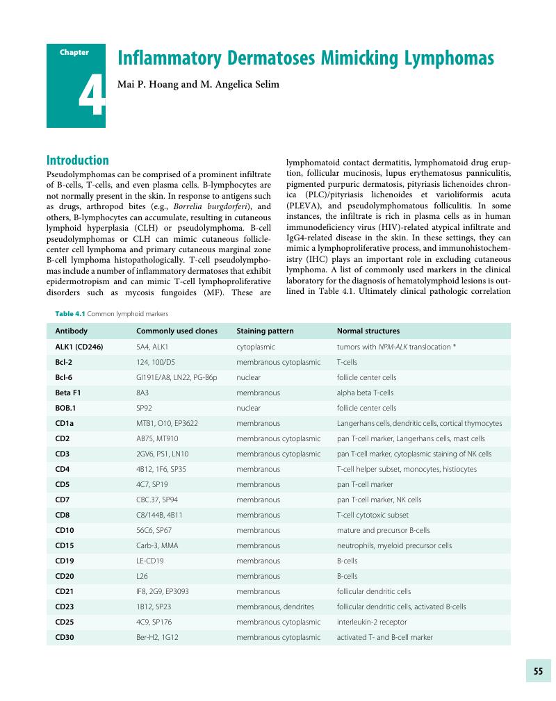
Image of the first page of this content. For PDF version, please use the ‘Save PDF’ preceeding this image.'
Type
Chapter
Information
Publisher: Cambridge University Press
Print publication year: 2017

Access options

Get access to the full version of this content by using one of the access options below. (Log in options will check for institutional or personal access. Content may require purchase if you do not have access.)

References

Cerroni, L, Arzberger, E, Putz, B, et al. Primary cutaneous follicular center cell lymphoma with follicular growth pattern. Blood 2000;95(12):3922–28.Google Scholar
Bergman, R, Khamaysi, K, Khamayi, Z, Ben Arie, Y. A study of histologic and immunophenotypical staining patterns in cutaneous lymphoid hyperplasia. J Am Acad Dermatol 2011;65(1):112–24.Google Scholar
Colli, C, Leinweber, B, Mullegger, R, et al. Borrelia burgdorferi-associated lymphocytoma cutis: Clinicopathologic, immunophenotypic, and molecular study of 106 cases. J Cutan Pathol 2004;31(3):232–40.Google Scholar
Arai, E, Shimizu, M, Hirose, T. A review of 55 cases of cutaneous lymphoid hyperplasia: Reassessment of the histopathologic findings leading to reclassification of 4 lesions as cutaneous marginal zone lymphoma and 19 as pseudolymphomatous folliculitis. Hum Pathol 2005;36(5):505–11.Google Scholar
Servitje, O, Gallardo, F, Estrach, T, et al. Primary cutaneous marginal zone B-cell lymphoma: A clinical, histopathological, immunophenotypic and molecular genetic study of 22 cases. Br J Dermatol 2002;147(6):1147–58.Google Scholar
de Leval, L, Harris, NL, Longtine, J, Ferry, JA, Duncan, LM. Cutaneous B-cell lymphomas of follicular and marginal zone types: Use of Bcl-6, CD10, Bcl-2, and CD21 in differential diagnosis and classification. Am J Surg Pathol 2001;25(6):732–41.Google Scholar
LeBoit, PE, McNutt, NS, Reed, JA, Jacobson, M, Weiss, LM. Primary cutaneous immunocytoma. A B-cell lymphoma that can easily be mistaken for cutaneous lymphoid hyperplasia. Am J Surg Pathol 1994;18(10):969–78.Google Scholar
Goodlad, JR, Krajewski, AS, Batstone, PJ, et al. Primary cutaneous follicular lymphoma: A clinicopathologic and molecular study of 16 cases in support of a distinct entity. Am J Surg Pathol 2002;26(6):733–41.Google Scholar
Orbaneja, JG, Diez, LI, Lozano, JL, Salazar, LC. Lymphomatoid contact dermatitis: A syndrome produced by epicutaneous hypersensitivity with clinical features and a histopathologic picture similar to that of mycosis fungoides. Contact Dermatitis 1976;2(3):139–43.Google Scholar
Smolle, J, Torne, R, Soyer, HP, Kerl, H. Immunohistochemical classification of cutaneous pseudolymphomas: Delineation of distinct patterns. J Cutan Pathol 1990;17(3):149–59.Google Scholar
Haynes, BF, Hensley, LL, Jegasothy, BV. Phenotypic characterization of skin-infiltrating T cells in cutaneous T-cell lymphoma: Comparison with benign cutaneous T-cell infiltrates. Blood 1982;60(2):463–73.Google Scholar
Souteyrand, P, d’Incan, M. Drug induced mycosis fungoides like lesions. Curr Probl Dermatol 1990;19:176–82.Google Scholar
Ploysangam, T, Breneman, DL, Mutasim, DF. Cutaneous pseudolymphomas. J Am Acad Dermatol 1998;38(6 Pt 1):877–95.Google Scholar
Magro, CM, Crowson, AN. Drug-induced immune dysregulation as a cause of atypical cutaneous lymphoid infiltrates: A hypothesis. Hum Pathol 1996;27(2):125–32.Google Scholar
Murphy, M, Fullen, D, Carlson, JA. Low CD7 expression in benign and malignant cutaneous lymphocytic infiltrates: Experience with an antibody reactive with paraffin-embedded tissue. Am J Dermatopathol 2002;24(1):616.Google Scholar
Florell, SR, Cessna, M, Lundell, RB, et al. Usefulness (or lack thereof) of immunophenotyping in atypical cutaneous T-cell infiltrates. Am J Clin Pathol 2006;125(5):727–36.Google Scholar
Michie, SA, Abel, EA, Hoppe, RT, Warnke, RA, Wood, GS. Expression of T-cell receptor antigens in mycosis fungoides and inflammatory skin lesions. J Invest Dermatol 1989;93(1):116–20.Google Scholar
Ormsby, A, Bergfeld, WF, Tubbs, RR, Hsi, ED. Evaluation of a new paraffin-reactive CD7 T-cell deletion marker and a polymerase chain reaction-based T-cell receptor gene rearrangement assay: Implications for diagnosis of mycosis fungoides in community clinical practice. J Am Acad Dermatol 2001;45(3):405–13.Google Scholar
Ortonne, N, Buyukbabani, N, Delfau-Larue, MH, Bago, M, Wechsler, J. Value of the CD8-CD3 ratio for the diagnosis of mycosis fungoides. Mod Pathol 2003;16(9):857–62.Google Scholar
Pulitzer, MP, Nolan, KA, Oshman, RG, Phelps, RG. CD30+ lymphomatoid drug reactions. Am J Dermatopathol 2013;35(3):343–50.Google Scholar
Cerroni, L, Fink-Puches, R, Back, B, Kerl, H. Follicular mucinosis: A critical reappraisal of clinicopathologic features and association with mycosis fungoides and Sezary syndrome. Arch Dermatol 2002;138(2):182–89.Google Scholar
Brown, HA, Gibson, LE, Pujol, RM, Lust, JA, Pittelkow, MR. Primary follicular mucinosis: Long-term follow-up of patients younger than 40 years with and without clonal T-cell receptor gene rearrangement. J Am Acad Dermatol 2002;47(6):856–62.Google Scholar
Gerami, P, Rosen, S, Kuzel, T, Boone, SL, Guitart, J. Folliculotropic mycosis fungoides: An aggressive variant of cutaneous T-cell lymphoma. Arch Dermatol 2008;144(6):738–46.Google Scholar
Burg, G, Kempf, W, Cozzio, A, et al. WHO/EORTC classification of cutaneous lymphomas 2005: Histological and molecular aspects. J Cutan Pathol 2005;32(10):647–74.Google Scholar
Rongioletti, F, De Lucchi, S, Meyes, D, et al. Follicular mucinosis: A clinicopathologic, histochemical, immunohistochemical and molecular study comparing the primary benign form and the mycosis fungoides-associated follicular mucinosis. J Cutan Pathol 2010;37(1):1519.Google Scholar
Demirkesen, C, Esirgen, G, Engin, B, Songur, A, Oquz, O. The clinical features and histopathologic patterns of folliculotropic mycosis fungoides in a series of 38 cases. J Cutan Pathol 2015;42(1):2231.Google Scholar
van Doorn, R, Scheffer, E, Willemze, R. Follicular mycosis fungoides, a distinct disease entity with or without associated follicular mucinosis: A clinicopathologic and follow-up study of 51 patients. Arch Dermatol 2002;138(2):191–98.Google Scholar
Smoller, BR, Kamel, OW. Pigmented purpuric eruptions: Immunopathologic studies supportive of a common immunophenotype. J Cutan Pathol 1991;18(6):423–27.Google Scholar
Lipsker, D. The pigmented and purpuric dermatitis and the many faces of mycosis fungoides. Dermatology 2003;207(3):246–47.Google Scholar
Geller, L, Antonov, NK, Lauren, CT, Morel, KD, Garzon, MC. Pityriasis lichenoides in childhood: Review of clinical presentation and treatment options. Pediatr Dermatol 2015;32(5):579–92.Google Scholar
Magro, C, Crowson, AN, Kovatich, A, Burns, F. Pityriasis lichenoides: A clonal T cell lymphoproliferative disorder. Hum Pathol 2002;33(8):788–95.Google Scholar
Kempf, W, Kazakov, DV, Palmedo, G, et al. Pityriasis lichenoides et varioliformis acuta with numerous CD30(+) cells: A variant mimicking lymphomatoid papulosis and other cutaneous lymphomas. A clinicopathologic, immunohistochemical, and molecular biological study of 13 cases. Am J Surg Pathol 2012;36(7):1021–29.Google Scholar
Martinez-Escala, ME, Sidiropoulos, M, Deonizio, J, et al. Gamma delta T-cell rich variants of lichenoides and lymphomatoid papulosis: Benign cutaneous disorders to be distinguished from aggressive cutaneous alpha beta T-cell lymphomas. Br J Dermatol 2015;172(2):372–79.Google Scholar
Magro, CM, Crowson, AN, Kovatich, AJ, Burns, F. Lupus profundus, indeterminate lymphocytic lobular panniculitis and subcutaneous T-cell lymphoma: A spectrum of subcuticular T-cell lymphoid dyscrasia. J Cutan Pathol 2001;28(5):235–47.Google Scholar
Aguilera, P, Mascaro, JM, Martinez, A, et al. Cutaneous gamma/delta T-cell lymphoma: A histopathologic mimicker of lupus erythematosus profundus (lupus panniculitis). J Am Acad Dermatol 2007;56(4):643–47.Google Scholar
Massone, C, Kodama, K, Salmhofer, W, et al. Lupus erythematosus panniculitis (lupus profundus): Clinical, histopathological, and molecular analysis of nine cases. J Cutan Pathol 2005;32(6):396404.Google Scholar
Park, HS, Choi, JW, Kim, BK, Cho, KH. Lupus erythematosus panniculitis: Clinicopathological, immunophenotypic, and molecular studies. Am J Dermatopathol 2010;32(1):2430.Google Scholar
LeBlanc, RE, Tavllaee, M, Kim, YH, Kim, J. Useful parameters for distinguishing subcutaneous panniculitis-like T-cell lymphoma from lupus erythematosus panniculitis. Am J Surg Pathol 2016;40(6):745–54.Google Scholar
Kumar, S, Krenacs, L, Medeiros, J, et al. Subcutaneous panniculitic T-cell lymphoma is a tumor of cytotoxic T lymphocytes. Hum Pathol 1998;29(4):397403.Google Scholar
Hoque, SR, Child, FJ, Whittaker, SJ, et al. Subcutaneous panniculitis-like T-cell lymphoma: A clinicopathological, immunophenotypic and molecular analysis of six patients. Br J Dermatol 2003;148(3):516–25.Google Scholar
Massone, C, Chott, A, Metze, D, et al. Subcutaneous, blastic natural killer (NK), NK/T-cell, and other cytotoxic lymphomas of the skin: A morphologic, immunophenotypic, and molecular study of 50 patients. Am J Surg Pathol 2004;28(6):719–35.Google Scholar
Willemze, R, Jansen, PM, Cerroni, L, et al. Subcutaneous panniculitis-like T-cell lymphoma: Definition, classification, and prognostic factors: An EORTC Cutaneous Lymphoma Group Study of 83 cases. Blood 2008;111(2):838–45.Google Scholar
Go, RS, Wester, SM. Immunophenotypic and molecular features, clinical outcomes, treatments, and prognostic factors associated with subcutaneous panniculitis-like T-cell lymphoma. Cancer 2004;101(6):1404–13.Google Scholar
Willemze, R, Jaffe, ES, Burg, G, et al. WHO-EORTC classification for cutaneous lymphomas. Blood 2005;105(10):3768–85.Google Scholar
Bosisio, F, Boi, S, Caputo, V, et al. Lobular panniculitic infiltrates with overlapping histopathologic features of lupus panniculitis (lupus profundus) and subcutaneous T-cell lymphoma: A conceptual and practical dilemma. Am J Surg Pathol 2015;39(2):206–11.Google Scholar
Magro, CM, Schaefer, JT, Morrison, C, Porcu, P. Atypical lymphocytic lobular panniculitis: A clonal subcutaneous T-cell dyscrasia. J Cutan Pathol 2008;35(10):947–54.Google Scholar
Garcia-Herrera, A, Song, JY, Chuang, SS, et al. Nonhepatosplenic gamma delta T-cell lymphomas represent a spectrum of aggressive cytotoxic T-cell lymphomas with a mainly extranodal presentation. Am J Surg Pathol 2011;35(8): 1214–25.Google Scholar
Kong, YY, Dai, B, Kong, JC, et al. Subcutaneous panniculitis-like T-cell lymphoma: A clinicopathologic, immunophenotypic, and molecular study of 22 Asian cases according to WHO-EORTC classification. Am J Surg Pathol 2008;32(10):1495–502.Google Scholar
Arai, E, Okubo, H, Tetsuya, T, Kitamura, K, Katayama, I. Pseudolymphomatous folliculitis: A clinicopathologic study of 15 cases of cutaneous pseudolymphoma with follicular invasion. Am J Surg Pathol 1999;23(11):1313–19.Google Scholar
Kwon, EJ, Kristjansson, AK, Meyerson, HJ, et al. A case of recurrent pseudolymphomatous folliculitis: A mimic of cutaneous lymphoma. J Am Acad Dermatol 2009;60(6):9941000.Google Scholar
Kazakov, DV, Belousova, IE, Kacerovska, D, et al. Hyperplasia of hair follicles and other adnexal structures in cutaneous lymphoproliferative disorders: A study of 53 cases, including so-called pseudolymphomatous folliculitis and overt lymphomas. Am J Surg Pathol 2008;32(10):1468–78.Google Scholar
Goyal, A, Moore, JB, Gimbel, D, et al. PD-1, S-100 and CD1a expression in pseudolymphomatous folliculitis, primary cutaneous marginal zone B-cell lymphoma (MALT lymphoma) and cutaneous lymphoid hyperplasia. J Cutan Pathol 2015;42(1):615.Google Scholar
Kempf, W, Kazakov, DV, Baumgartner, HP, Kutzner, H. Follicular lymphomatoid papulosis revisited: A study of 11 cases, with new histopathological findings. J Am Acad Dermatol 2013;68(5):809–16.Google Scholar
Guitart, J, Variakojis, D, Kuzel, T, Rosen, S. Cutaneous CD8+ T-cell infiltrates in advanced HIV infection. J Am Acad Dermatol 1999;41(5 Pt 1):722–77.Google Scholar
Zhang, P, Chiriboga, L, Jacobson, M, et al. Mycosis fungoides like T-cell cutaneous lymphoid infiltrates in patients with HIV infection. Am J Dermatopathol 1995;17(1):2935.Google Scholar
Friedler, S, Parisi, MT, Waldo, E, et al. Atypical cutaneous lymphoproliferative disorder in patients with HIV infection. Int J Dermatol 1999;38(2):111–18.Google Scholar
Lin, W, Lu, S, Chen, H, et al. Clinical characteristics of immunoglobulin G4-related disease: A prospective study of 118 Chinese patients. Rheumatology 2015;54(11):1982–90.Google Scholar
Deshpande, V, Zen, Y, Chan, JK, et al. Consensus statement on the pathology of IgG4-related disease. Mod Pathol 2012;25(9):1181–92.Google Scholar
Deng, C, Li, W, Chen, S, et al. Histolopathologic diagnostic value of the IgG4+/IgG+ ratio of plasmacytic infiltration for IgG4-related diseases: A PRISMA-compliant systematic review and meta-analysis. Medicine 2015;94(9):e579. doi: 10.1097/MD.00000000000000579.Google Scholar
Sato, Y, Takeuchi, M, Takata, K, et al. Clinicopathologic analysis of IgG4-related skin disease. Mod Pathol 2013;26(4):523–32.Google Scholar
Cheuk, W, Lee, K-C, Chong, L-Y, Yuen, S-T, Chan, JKC. IgG4-related sclerosing disease: A potential new etiology of cutaneous pseudolymphoma. Am J Surg Pathol 2009;33(11):1713–19.Google Scholar
Hattori, T, Miyanaga, T, Tago, O, et al. Isolated cutaneous manifestation of IgG4-related disease. J Clin Pathol 2012;65(9):815–18.Google Scholar
Lehman, JS, Smyrk, TC, Pittelkow, MR. Increased immunoglobulin (Ig) G4-positive plasma cell density and IgG4/IgG ratio are not specific for IgG4-related disease in the skin. Am J Clin Pathol 2014;141(2):234–38.Google Scholar
Aggarwal, N, Parwani, AV, Ho, J, Cook, JR, Swerdlow, SH. Plasma cell (Zoon) balanitis. Another inflammatory disorder that can be rich in IgG4+ plasma cells. Am J Surg Pathol 2014;38(10):1437–43.Google Scholar
Pincus, LB, LeBoit, PE, McCalmont, TH, et al. Subcutaneous panniculitis-like T-cell lymphoma with overlapping clinicopathologic features of lupus erythematosus: Coexistence of 2 entities? Am J Dermatopathol 2009;31(6):520–26.Google Scholar
Shiau, CJ, Abi Daoud, MS, Wong, SM, Crawford, RI. Lymphocytic panniculitis: An algorithmic approach to lymphocytes in subcutaneous tissue. J Clin Pathol 2015;68(12):954–62.Google Scholar
Lozzi, GP, Massone, C, Citarella, L, Kerl, H, Cerroni, L. Rimming of adipocytes by neoplastic lymphocytes: A histopathologic feature not restricted to subcutaneous T-cell lymphoma. Am J Dermatopathol 2006;28(1):912.Google Scholar
Requena, L, Kutzner, H, Palmedo, G, et al. Histiocytoid Sweet syndrome: A dermal infiltration of immature neutrophilic granulocytes. Arch Dermatol 2005;141(7):834–42.Google Scholar
Ghoufi, L, Ortonne, N, Ingen-Housz-Ora, S, et al. Histiocytoid Sweet syndrome is more frequently associated with myelodysplastic syndromes than the classical neutrophilic variant: A comparative series of 62 patients. Medicine 2016;95(15):e3033.Google Scholar

Save book to Kindle

To save this book to your Kindle, first ensure coreplatform@cambridge.org is added to your Approved Personal Document E-mail List under your Personal Document Settings on the Manage Your Content and Devices page of your Amazon account. Then enter the ‘name’ part of your Kindle email address below. Find out more about saving to your Kindle.

Note you can select to save to either the @free.kindle.com or @kindle.com variations. ‘@free.kindle.com’ emails are free but can only be saved to your device when it is connected to wi-fi. ‘@kindle.com’ emails can be delivered even when you are not connected to wi-fi, but note that service fees apply.

Find out more about the Kindle Personal Document Service.

Available formats
×

Save book to Dropbox

To save content items to your account, please confirm that you agree to abide by our usage policies. If this is the first time you use this feature, you will be asked to authorise Cambridge Core to connect with your account. Find out more about saving content to Dropbox.

Available formats
×

Save book to Google Drive

To save content items to your account, please confirm that you agree to abide by our usage policies. If this is the first time you use this feature, you will be asked to authorise Cambridge Core to connect with your account. Find out more about saving content to Google Drive.

Available formats
×