Skip to main content Accessibility help
×
  • Cited by 6
Publisher:
Cambridge University Press
Online publication date:
February 2014
Print publication year:
2009
Online ISBN:
9781107784703

Book description

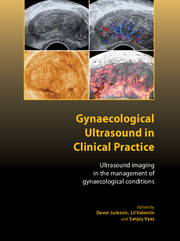
Ultrasound plays an integral part in the diagnosis and management of many gynaecological conditions; indeed, ultrasound forms part of the RCOG's mandatory training programme for doctors wishing to specialise in obstetrics and gynaecology. This book will be of use to both trainees and those already in clinical practice looking for a user-friendly reference guide. The use of ultrasound in gynaecology goes well beyond simple picture recognition: a skilful gynaecological sonographer will bring together scan findings and the clinical scenario to enhance patient care. This leads to targeted investigations and strategies for intervention. This book covers all aspects of the use of ultrasound in the fields of gynaecology and early pregnancy, with the contents including: postmenopausal bleeding; adnexal masses; pelvic pain; reproductive medicine; miscarriage; ectopic pregnancy; and ovarian cysts.

Refine List

Actions for selected content:

Select all | Deselect all
  • View selected items
  • Export citations
  • Download PDF (zip)
  • Save to Kindle
  • Save to Dropbox
  • Save to Google Drive

Save Search

You can save your searches here and later view and run them again in "My saved searches".

Please provide a title, maximum of 40 characters.
×

Contents

  • 1 - Ultrasound imaging in gynaecological practice
    pp 1-6
  • View abstract

    Summary

    This chapter provides an overview of the contribution of ultrasound examination to the evaluation of gynaecological conditions. Ultrasound imaging can be used to assess women with a history of acute or chronic pelvic pain. The imaging allows a quick non-invasive assessment of the pelvis and abdomen and it may be used as the first line investigation of patients with pelvic pain to confirm or exclude the provisional diagnosis based on clinical history. Ultrasound imaging determines the extent of ovarian and adnexal involvement in women with pelvic inflammatory disease. Ultrasound is helpful in assessing women with a history of post-menopausal bleeding and it can distinguish between women with post-menopausal bleeding who need to undergo invasive testing from those who do not require any intervention. Ultrasound is used to determine both the pregnancy location and viability. Transvaginal ultrasound has an important role in the study of female fertility.
  • 2 - Normal pelvic anatomy
    pp 7-16
  • View abstract

    Summary

    A gynaecological ultrasound examination can be performed transabdominally, transvaginally or, in exceptional cases, transrectally. Irrespective of the route of examination it is always important to optimise the image and to perform a systematic examination. There are two reasons to adopt a systematic scanning technique. First, it will ensure that a complete pelvic examination is performed. Second, if one always scans all the organs in the pelvis in a systematic way, he/she can build up a reference of what is normal, which increases confidence in detecting pelvic pathology. The uterus and ovaries are smaller in postmenopausal women than in women of fertile age. The endometrium has uniform ultrasound morphology because there are no cyclical hormonal changes. The ovaries contain no follicles but one or more inclusion cysts no larger than 10 mm are seen in healthy postmenopausal women.
  • 3 - The uterus
    pp 17-28
  • View abstract

    Summary

    The uterus is divided into four anatomical parts. The fundus is the uppermost part and extends into the interstitial portion of the fallopian tubes laterally. The corpus is the main body of the uterus, and the isthmus is the lowermost portion, which extends into the cervix. The lower uterine segment in the pregnant uterus forms at the isthmus, which is at the level of the reflection of the urinary bladder in the non-pregnant uterus. Uterine fibroids are the most common uterine abnormality encountered in women of reproductive age. Ultrasound has the advantages of being a widely available outpatient imaging modality, with the ability to measure and assess the depth of fibroid involvement into the myometrium more accurately than any other imaging technique. Uterine sarcoma is a rare tumour of the uterine myometrium that is usually diagnosed after hysterectomy. The clinical presentation is enlarging uterus in a post-menopausal woman.
  • 4 - Postmenopausal bleeding: presentation and investigation
    pp 29-42
  • View abstract

    Summary

    This chapter reviews evidence on various investigative modalities and management planning for women presenting to the gynaecology clinic with post-menopausal bleeding. It discusses some of the issues to be considered when planning a cost-effective, clinic-based service for these women. Dilatation and curettage (D&C) was for many years the investigation of choice in women presenting with post-menopausal bleeding. The Pipelle de Cornier is a widely used system. The system is a narrow plastic catheter, which is passed through the cervical canal into the uterine cavity. Transvaginal ultrasound (TVS) is an accurate, non-invasive diagnostic modality that enables examination of the uterine cavity and endometrium in the outpatient setting. With the uterus visualised in a longitudinal plane, the thickness of the endometrial echo can be measured. Endometrial polyps are a common finding in post-menopausal women and when they occur in association with post-menopausal bleeding they should be removed for histopathological diagnosis.
  • 5 - HRT, contraceptives and other drugs affecting the endometrium
    pp 43-54
  • View abstract

    Summary

    The endometrium undergoes cellular and structural changes that are essential for its function. These changes are cyclical and controlled by the production of estrogen and progesterone by the ovaries. Drugs with estrogenic or progestogenic modes of action also lead to alterations in the ultrasonographic appearances of the endometrium. This chapter helps the sonographer to interpret the appearances of the endometrium in women. The Committee on Safety of Medicines (CSM) has advised that hormone replacement therapy (HRT) is beneficial for the treatment of menopausal symptoms. Although unscheduled bleeding on HRT requires investigation, the underlying malignancy risk is low. In postmenopausal bleeding, there is a cut off of 4 mm for double-layer endometrial thickness on Transvaginal ultrasound (TVS) but for women on HRT there is no agreed cut-off point. Hormonal contraceptives and intrauterine contraceptive devices (IUCD) have multiple effects leading to their contraceptive action.
  • 6 - Diagnosis and management of adnexal masses
    pp 55-66
  • View abstract

    Summary

    Transvaginal ultrasound examination is an excellent tool for solving clinical problems in women with symptoms suggesting the presence of adnexal mass. An experienced ultrasound examiner can confidently discriminate between benign and malignant pelvic tumours in the adnexal region using pattern recognition. Some tumours - for example, endometriomas, dermoid cysts, hydropyo- and haematosalpinx, peritoneal pseudocysts, paraovarian cysts, haemorrhagic corpus luteum cysts, myomas, abscesses and ovarian fibromas, thecomas and Brenner tumours - may present with typical appearances at greyscale imaging. Doppler assessment of intratumoral blood flow contributes little to the correct specific diagnosis of adnexal mass. The ability to make a correct specific diagnosis in a series of pelvic tumours is highly dependent upon the types of tumour in the tumour series studied. The surgical removal of the adnexal mass with benign ultrasound morphology is incidentally detected at ultrasound examination in asymptomatic women.
  • 7 - Ultrasound assessment of women with pelvic pain
    pp 67-76
  • View abstract

    Summary

    Haemorrhagic corpus luteum cysts are common causes of acute pelvic pain in women of fertile age. At ultrasound examination, corpus luteum cysts are characterised by spiderweb-like contents but they may also contain bizarre-looking blood clots. This explains why corpus luteum cysts may sometimes be confused with malignancy. Endometriomas may cause pelvic pain but they are also quite common incidental ultrasound findings. A serious condition that may be more or less painful is torsion of the adnexa. Torsion occurs in adnexa containing a lesion, such as ovarian cyst or hydrosalpinx, but it may also occur in normal adnexa, especially in prepubertal girls. Uterine fibroids and adenomyosis may sometimes cause pelvic pain. Gynaecologists should be able to recognise the ultrasound image of non-gynaecological conditions that may cause pelvic pain. Ultrasound images of these conditions may be encountered when an ultrasound examination is performed because of suspicion of gynaecological disease.
  • 8 - Ultrasound of non-gynaecological pelvic lesions
    pp 77-90
  • View abstract

    Summary

    Transvaginal ultrasound has improved the ability of ultrasound to interrogate the pelvic organs with less interference from intervening structures such as gas or fat. The gynaecologist may need to distinguish between the symptoms caused by pelvic inflammatory disease and those of an inflamed pelvic appendix. Appendiceal mucocele occurs when there is accumulation of mucoid material within the lumen of the appendix distal to an obstruction. Mucoceles occur more commonly in women than men. Ultrasound can show different patterns, including a cystic structure with thin walls, a cyst with septations and, the most common appearance, layered rings of mucus of different echogenicity. Ultrasound is highly sensitive in detecting calculi in the kidneys. An ureterocele can be clinically silent and without upper tract dilation. The diagnosis may be made for the first time during sonography of the pelvis.
  • 9 - Ultrasound imaging in reproductive medicine
    pp 91-106
  • View abstract

    Summary

    Ultrasound examination is as effective a diagnostic test as hysteroscopy or laparoscopy for the diagnosis of uterine abnormalities. Three-dimensional ultrasound has been used to assess uterine anatomy and to detect congenital anomalies of the uterus. Ultrasound examination is used to monitor endometrial growth and ovarian response to the medical treatment. Appropriate endometrial growth indicates good endometrial receptivity. Appropriate ovarian response to gonadotrophin stimulation increases the chances that good quality eggs will be released in ovulation induction cycles and that good embryos will be available for embryo transfer in in vitro fertilisation (IVF) cycles. Ultrasound is also used to monitor embryo transfer. Both greyscale ultrasound examination of the endometrium and Doppler ultrasound examination of the uterine arteries have been used to assess 'endometrial receptivity' in IVF cycles. Ultrasound greyscale imaging is regarded as sufficiently accurate to be used alone for monitoring follicular growth during gonadotrophin therapy.
  • 10 - Ultrasound imaging of the lower urinary tract and uterovaginal prolapse
    pp 107-120
  • View abstract

    Summary

    Ultrasound has the advantage of being able to visualise fluid-filled structures without the need for contrast medium. It can demonstrate soft tissue structures such as the kidney, bladder wall, urethral and anal sphincters and surrounding pelvic floor musculature. Use of the transabdominal, transvaginal, transrectal and transperineal approaches for ultrasound scanning allows for easy visualisation of different aspects of the lower urinary tract. Bladder diverticula are easily visualised with transabdominal ultrasound. Transvaginal ultrasound allows clearer visualisation of the bladder base, bladder neck, inferior border of the pubic symphysis and periurethral structures. Translabial ultrasound is used to demonstrate uterovaginal prolapse. Three-dimensional ultrasound offers new insights into the pelvic floor as it allows imaging of both the levator ani muscle hiatus and paravaginal support structures in axial and transverse planes. This gives access to transverse planes similar to magnetic resonance imaging.
  • 11 - Ultrasound and diagnosis of obstetric anal sphincter injuries
    pp 121-132
  • View abstract

    Summary

    Anal endosonography is regarded as the gold standard investigation in patients presenting with faecal incontinence. The endosonography is also useful in the diagnosis of anal pain, anorectal tumours, fistulae, abscesses and anismus. The advent of anal endosonography has enabled considerable research into obstetric related anal sphincter trauma, the major aetiological factor in the development of anal incontinence. The internal anal sphincter is a thickened continuation of the circular smooth muscle layer of the bowel and appears homogeneously hypoechoic. The external anal sphincter usually appears hyperechoic, but has a heterogeneous appearance. Magnetic resonance imaging (MRI) defines the striated components of the sphincter with greater clarity. In 1994, Sultan et al. first described transvaginal endosonography to image the anal sphincters at rest with a rotating probe. The development of anal endosonography added a new dimension to understanding the pathogenesis of anal incontinence and the diagnosis of obstetric anal sphincter injuries.
  • 12 - Organisation of the early pregnancy unit
    pp 133-142
  • View abstract

    Summary

    Early pregnancy complications are one of the most common reasons for women of reproductive age seeking medical help. Many pregnancies are complicated by pain and/or bleeding, which often causes concern because of a perceived risk of miscarriage. A comprehensive service with easy and fast access needs to be provided for women in early pregnancy. An initial assessment to reach a correct diagnosis is carried out, and diagnostic tests are instigated. Follow-up is vital, and for those women who have suffered pregnancy loss, offering counselling and support is another important role for an early pregnancy unit. With the advent of computer databases, a paperless clinic can be a key element in the efficient running of an early pregnancy unit. It is important that the unit has multidisciplinary and trained staff, access to laboratory tests, a gynaecology ward and operating theatre, in case of direct admissions, and facilities for resuscitation.
  • 13 - Sonoembryology: ultrasound examination of early pregnancy
    pp 143-158
  • View abstract

    Summary

    Major improvement in the ultrasound assessment of early pregnancy came up with the introduction of transvaginal ultrasound at the end of 1980s. High-frequency transvaginal transducers improve the image quality to an extent that detailed description of the embryonic morphology became possible with in-depth anatomical studies of the brain compartments, the spine, the heart, the stomach, the midgut herniation and the limbs. There are three main characteristics that mark the early human conceptus: its small size, its rapidly changing anatomical appearance and its uniform development and constant growth. Embryologists use the Carnegie staging system to divide the human embryonic period into 23 developmental stages, commencing with fertilisation at stage 1, continuing into the fetal period with the onset of marrow formation in the humerus after stage 23, which takes place at 56-57 days post-ovulation, and the designation 'embryo' is replaced by 'fetus'.
  • 14 - Diagnosis and management of miscarriage
    pp 159-178
  • View abstract

    Summary

    Miscarriage occurs if there is a failure of embryonic growth or if a viable fetus dies. An incomplete miscarriage is diagnosed by history of bleeding, pain, passage of products of conception and an open internal cervical os on examination. Traditional clinical methods of diagnosing miscarriage have been largely replaced by ultrasound diagnosis. Early pregnancy units have been developed to streamline the diagnosis of abnormal early pregnancy. The Royal College of Radiologists and the Royal College of Obstetricians and Gynaecologists (RCR/RCOG) issued joint guidelines on the ultrasound diagnosis of early pregnancy loss. Low levels of progesterone have long been associated with early pregnancy failure. Treatment regimens include the use of the antiprogesterone, mifepristone and a prostaglandin analogue, the most commonly used of which is misoprostol. These regimens were initially devised for the management of first-trimester therapeutic abortion.
  • 15 - Tubal ectopic pregnancy
    pp 179-192
  • View abstract

    Summary

    Tubal ectopic pregnancy is an important cause of maternal morbidity and mortality worldwide. Clinical presentation of ectopic pregnancy varies from mild vaginal bleeding to sudden rupture and massive intra-abdominal haemorrhage. The diagnosis of the ectopic pregnancy was made at surgery and then confirmed on histological examination following salpingectomy. At laparoscopy, an unruptured ectopic pregnancy typically presents as a well-defined swelling in the fallopian tube. The diagnosis of intrauterine pregnancy becomes more difficult if the uterus is enlarged by fibroids. Fibroids often distort the shape of the endometrial cavity and prevent the operator from visualising in a single plane the continuity between the gestation sac and the cervical canal. Surgery remains the main therapeutic option for the treatment of tubal ectopic pregnancy. Medical management of ectopic pregnancy has grown in popularity following observational studies which reported success rates greater than 90% with single-dose systemic methotrexate.
  • 16 - Non-tubal ectopic pregnancies
    pp 193-210
  • View abstract

    Summary

    This chapter provides a summary of each type of non-tubal ectopic pregnancy, with particular emphasis on the ultrasound diagnosis and management options. Interstitial pregnancy is characterised by the implantation of the conceptus in the interstitial portion of the fallopian tube, which is surrounded by the muscular wall of the uterus. The advances in high-resolution transvaginal ultrasonography and the establishment of early pregnancy units have facilitated the early non-invasive diagnosis of interstitial pregnancy before complications occur. This has opened the door for more conservative management options such as medical treatment with methotrexate. The management of interstitial pregnancy was surgical, in the form of cornual resection or hysterectomy. The reason for this was the late detection of this condition, which used to be diagnosed at laparotomy following tubal rupture. Medical management using methotrexate, a folate antagonist, has been increasingly used in the treatment of women identified as having an unruptured interstitial pregnancy.
  • 17 - Ovarian cysts in pregnancy
    pp 211-228
  • View abstract

    Summary

    The presence of an ovarian cyst is traditionally considered to be an indication for operative intervention for fear of ovarian cancer and acute complications of ovarian cysts, such as torsion, rupture and obstruction of labour. Two studies described the prevalence of ovarian cysts in pregnancy before the routine use of ultrasound, when the diagnosis was based on clinical examination of women with symptoms suggestive of an adnexal mass. The vast majority of adnexal cystic masses detected in early pregnancy are functional cysts, such as corpus luteum cysts or follicular cysts. Dermoid cysts or mature cystic teratomas are the most common complex ovarian masses encountered in pregnancy, making up 24-40% of all ovarian tumours. Fimbrial cysts are usually seen on ultrasound examination as thin-walled, anechoic, unilocular adnexal masses. Ultrasound-guided cyst aspiration offers a less invasive alternative to the traditional techniques employed for surgical management of ovarian cysts in pregnancy.

Metrics

Full text views

Total number of HTML views: 0
Total number of PDF views: 0 *
Loading metrics...

Book summary page views

Total views: 0 *
Loading metrics...

* Views captured on Cambridge Core between #date#. This data will be updated every 24 hours.

Usage data cannot currently be displayed.