Skip to main content Accessibility help
×
Hostname: page-component-848d4c4894-4rdrl Total loading time: 0 Render date: 2024-06-28T22:26:42.232Z Has data issue: false hasContentIssue false

6 - Surgical Pathology of Non-neoplastic Conditions of the Pleura, Pericardium, and Peritoneum

Published online by Cambridge University Press:  16 March 2018

Alberto M. Marchevsky
Affiliation:
Cedars Sinai Medical Center, Los Angeles
Aliya N. Husain
Affiliation:
University of Chicago
Françoise Galateau-Sallé
Affiliation:
MESOPATH National Reference Center & Cancer Center Leon Berard
Get access

Summary

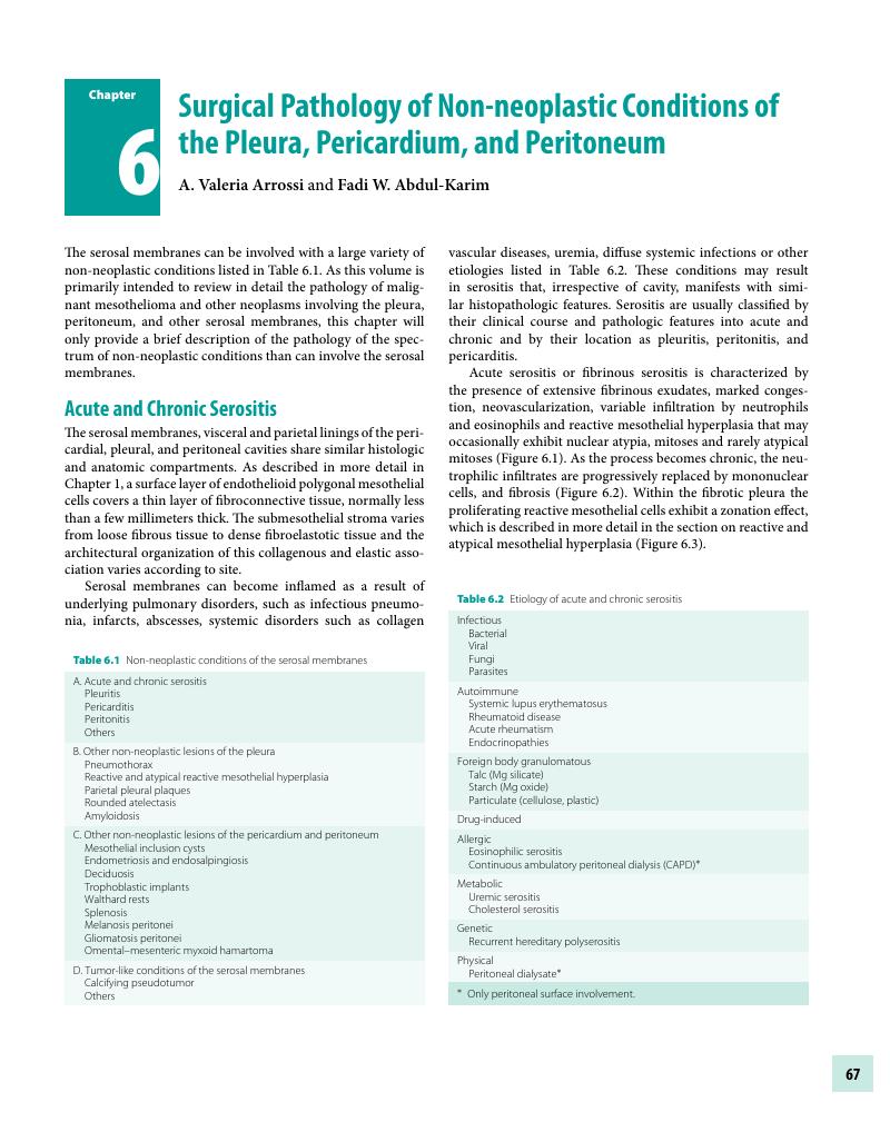
Image of the first page of this content. For PDF version, please use the ‘Save PDF’ preceeding this image.'
Type
Chapter
Information
Publisher: Cambridge University Press
Print publication year: 2018

Access options

Get access to the full version of this content by using one of the access options below. (Log in options will check for institutional or personal access. Content may require purchase if you do not have access.)

References

Ahmed, RA, Marrie, TJ, Huang, JQ. Thoracic empyema in patients with community-acquired pneumonia. Am J Med. 2006;119(10):877–83.CrossRefGoogle ScholarPubMed
Maskell, NA, Batt, S, Hedley, EL, Davies, CWH, Gillespie, SH, Davies, RJO. The bacteriology of pleural infection by genetic and standard methods and its mortality significance. Am J Respir Crit Care Med. 2006;174(7):817–23.CrossRefGoogle ScholarPubMed
Foster, S, Maskell, N. Bacteriology of complicated parapneumonic effusions. Curr Opin Pulm Med. 2007;13(4):319–23.CrossRefGoogle ScholarPubMed
Nestor, J, Huggins, T, Kummerfeldt, C, DiVietro, M, Walters, K, Sahn, S. Viral diseases affecting the pleura. J Clin Virol. 2013;58(2):367–73.CrossRefGoogle ScholarPubMed
Kummerfeldt, CE, Huggins, JT, Sahn, SA. Unusual bacterial infections and the pleura. Open Respir Med J. 2012;6:75.CrossRefGoogle ScholarPubMed
Dobbie, JW. Serositis: comparative analysis of histological findings and pathogenetic mechanisms in nonbacterial serosal inflammation. Perit Dial Int. 1993;13(4):256–69.CrossRefGoogle ScholarPubMed
Esther, CR, Barker, PM. Pulmonary lymphangiectasia: diagnosis and clinical course. Pediatr Pulmonol. 2004;38(4):308–13.CrossRefGoogle ScholarPubMed
Ryu, JH, Tomassetti, S, Maldonado, F. Update on uncommon pleural effusions. Respirology. 2011;16(2):238–43.CrossRefGoogle ScholarPubMed
Panchabhai, TS, Bandyopadhyay, D, Yadav, R, Arrossi, AV, Mehta, AC, Faress, JA. A 42-year-old woman with abnormal chest CT scan and chylous ascites. Chest. 2016;149(1):e2528.CrossRefGoogle ScholarPubMed
Lama, A, Ferreiro, L, Toubes, ME, Golpe, A, Gude, F, Álvarez-Dobaño, JM, et al. Characteristics of patients with pseudochylothorax – a systematic review. J Thorac Dis. 2016;8(8):2093.CrossRefGoogle ScholarPubMed
Padeh, S, Berkun, Y. Familial Mediterranean fever. Curr Opin Rheumatol. 2016;28(5):523–29.Google Scholar
Sarı, İ, Birlik, M, Kasifoğlu, T. Familial Mediterranean fever: an updated review. Eur J Rheumatol. 2014;1(1):2133.Google Scholar
Tschopp, J-M, Bintcliffe, O, Astoul, P, Canalis, E, Driesen, P, Janssen, JP, et al. ERS task force statement: diagnosis and treatment of primary spontaneous pneumothorax. Eur Respir J. 2015;46(2):321–35.CrossRefGoogle ScholarPubMed
Noppen, M. Spontaneous pneumothorax: epidemiology, pathophysiology and cause. Eur Respir Rev. 2010;19(117):217–19.Google Scholar
Ohata, M, Suzuki, H. Pathogenesis of spontaneous pneumothorax. With special reference to the ultrastructure of emphysematous bullae. Chest. 1980;77(6):771–76.Google Scholar
Noppen, M, Dekeukeleire, T, Hanon, S, Stratakos, G, Amjadi, K, Madsen, P, et al. Fluorescein-enhanced autofluorescence thoracoscopy in patients with primary spontaneous pneumothorax and normal subjects. Am J Respir Crit Care Med. 2006;174(1):2630.Google Scholar
Lichter, I, Gwynne, JF. Spontaneous pneumothorax in young subjects. A clinical and pathological study. Thorax. 1971;26(4):409–17.Google Scholar
Askin, FB, McCann, BG, Kuhn, C. Reactive eosinophilic pleuritis: a lesion to be distinguished from pulmonary eosinophilic granuloma. Arch Pathol Lab Med. 1977;101(4):187–91.Google ScholarPubMed
McDonnell, TJ, Crouch, EC, Gonzalez, JG. Reactive eosinophilic pleuritis. A sequela of pneumothorax in pulmonary eosinophilic granuloma. Am J Clin Pathol. 1989;91(1):107–11.CrossRefGoogle ScholarPubMed
Shekhel, TA, Ricciotti, RW, Blair, JE, Colby, TV, Sobonya, RE, Larsen, BT. Surgical pathology of pleural coccidioidomycosis: a clinicopathological study of 36 cases. Hum Pathol. 2014;45(5):961–69.Google Scholar
Evison, M, Holme, J, Alaloul, M, Doran, H, Bishop, P, Booton, R, et al. Olanzapine-induced eosinophilic pleuritis. Respir Med Case Rep. 2015;14:2426.Google ScholarPubMed
Sobonya, RE, Yanes, J, Klotz, SA. Cavitary pulmonary coccidioidomycosis: pathologic and clinical correlates of disease. Hum Pathol. 2014;45(1):153–59.Google Scholar
Cudzilo, C, Aragaki, A, Guitron, J, Benzaquen, S. Methotrexate-induced pleuropericarditis and eosinophilic pleural effusion. J Bronchology Interv Pulmonol. 2014;21(1):9092.Google Scholar
Middleton, KL, Santella, R, Couser, JI. Eosinophilic pleuritis due to propylthiouracil. Chest. 1993;103(3):955–56.Google Scholar
Sen, N, Ermis, H, Karatasli, M, Habesoglu, MA, Eyuboglu, FO. Propylthiouracil-associated eosinophilic pleural effusion: a case report. Respiration. 2007;74(6):703–05.CrossRefGoogle ScholarPubMed
Belchis, DA, Shekitka, K, Gocke, CD. A unique, histopathologic lesion in a subset of patients with spontaneous pneumothorax. Arch Pathol Lab Med. 2012;136(12):1522–27.CrossRefGoogle Scholar
Sauter, JL, Butnor, KJ. Pathological findings in spontaneous pneumothorax specimens: does the incidence of unexpected clinically significant findings justify routine histological examination? Histopathology. 2015;66(5):675–84.CrossRefGoogle ScholarPubMed
Butnor, KJ, Guinee, DG. Pleuropulmonary pathology of Birt–Hogg–Dubé syndrome. Am J Surg Pathol. 2006;30(3):395–99.Google ScholarPubMed
Joseph, J, Sahn, SA. Thoracic endometriosis syndrome: new observations from an analysis of 110 cases. Am J Med. 1996;100(2):164–70.Google Scholar
Ghigna, M-R, Mercier, O, Mussot, S, Fabre, D, Fadel, E, Dorfmuller, P, et al. Thoracic endometriosis: clinicopathologic updates and issues about 18 cases from a tertiary referring center. Ann Diagn Pathol. 2015;19(5):320–25.CrossRefGoogle ScholarPubMed
Flieder, DB, Moran, CA, Travis, WD, Koss, MN, Mark, EJ. Pleuro-pulmonary endometriosis and pulmonary ectopic deciduosis: a clinicopathologic and immunohistochemical study of 10 cases with emphasis on diagnostic pitfalls. Hum Pathol. 1998;29(12):1495–503.Google Scholar
Baker, PM, Clement, PB, Young, RH. Selected topics in peritoneal pathology. Int J Gynecol Pathol. 2014;33(4):393401.CrossRefGoogle ScholarPubMed
Oparka, R, McCluggage, WG, Herrington, CS. Peritoneal mesothelial hyperplasia associated with gynaecological disease: a potential diagnostic pitfall that is commonly associated with endometriosis. J Clin Pathol. 2011;64(4):313–18.CrossRefGoogle ScholarPubMed
Cheung, AN, Young, RH, Scully, RE. Pseudocarcinomatous hyperplasia of the fallopian tube associated with salpingitis. A report of 14 cases. Am J Surg Pathol. 1994;18(11):1125–30.Google Scholar
Clement, PB, Young, RH. Florid mesothelial hyperplasia associated with ovarian tumors: a potential source of error in tumor diagnosis and staging. Int J Gynecol Pathol. 1993;12(1):5158.Google Scholar
Chiosea, S, Krasinskas, A, Cagle, PT, Mitchell, KA, Zander, DS, Dacic, S. Diagnostic importance of 9p21 homozygous deletion in malignant mesotheliomas. Mod Pathol. 2008;21(6):742–47.Google Scholar
Ito, T, Hamasaki, M, Matsumoto, S, Hiroshima, K, Tsujimura, T, Kawai, T, et al. p16/CDKN2A FISH in differentiation of diffuse malignant peritoneal mesothelioma from mesothelial hyperplasia and epithelial ovarian cancer. Am J Clin Pathol. 2015;143(6):830–38.CrossRefGoogle ScholarPubMed
Hwang, HC, Pyott, S, Rodriguez, S, Cindric, A, Carr, A, Michelsen, C, et al. BAP1 immunohistochemistry and p16 FISH in the diagnosis of sarcomatous and desmoplastic mesotheliomas. Am J Surg Pathol. 2016;40(5):714–18.Google Scholar
Hida, T, Hamasaki, M, Matsumoto, S, Sato, A, Tsujimura, T, Kawahara, K, et al. BAP1 immunohistochemistry and p16 FISH results in combination provide higher confidence in malignant pleural mesothelioma diagnosis: ROC analysis of the two tests. Pathol Int. 2016;66(10):563–70.Google Scholar
Takeda, M, Kasai, T, Enomoto, Y, Takano, M, Morita, K, Kadota, E, et al. 9p21 deletion in the diagnosis of malignant mesothelioma, using fluorescence in situ hybridization analysis. Pathol Int. 2010;60(5):395–99.Google Scholar
Sheffield, BS, Hwang, HC, Lee, AF, Thompson, K, Rodriguez, S, Tse, CH, et al. BAP1 immunohistochemistry and p16 FISH to separate benign from malignant mesothelial proliferations. Am J Surg Pathol. 2015;39(7):977–82.Google Scholar
Monaco, SE, Shuai, Y, Bansal, M, Krasinskas, AM, Dacic, S. The diagnostic utility of p16 FISH and GLUT-1 immunohistochemical analysis in mesothelial proliferations. Am J Clin Pathol. 2011;135(4):619–27.Google Scholar
Walts, AE, Hiroshima, K, McGregor, SM, Wu, D, Husain, AN, Marchevsky, AM. BAP1 immunostain and CDKN2A (p16) FISH analysis: clinical applicability for the diagnosis of malignant mesothelioma in effusions. Diagn Cytopathol. 2016;44(7):599606.CrossRefGoogle ScholarPubMed
Husain, AN, Colby, T, Ordonez, N, Krausz, T, Attanoos, R, Beasley, MB, et al. Guidelines for pathologic diagnosis of malignant mesothelioma: 2012 update of the consensus statement from the International Mesothelioma Interest Group. Arch Pathol Lab Med. 2013;137(5):647–67.Google Scholar
Churg, A, Sheffield, BS, Galateau-Sallé, F. New markers for separating benign from malignant mesothelial proliferations: are we there yet? Arch Pathol Lab Med. 2016;140(4):318–21. PubMed PMID: 26288396Google Scholar
Hillerdal, G. The pathogenesis of pleural plaques and pulmonary asbestosis: possibilities and impossibilities. Eur J Respir Dis. 1980;61(3):129–38.Google Scholar
Ren, H, Lee, DR, Hruban, RH, Kuhlman, JE, Fishman, EK, Wheeler, PS, et al. Pleural plaques do not predict asbestosis: high-resolution computed tomography and pathology study. Mod Pathol. 1991;4(2):201–09.Google Scholar
Clarke, CC, Mowat, FS, Kelsh, MA, Roberts, MA. Pleural plaques: a review of diagnostic issues and possible nonasbestos factors. Arch Environ Occup Health. 2006;61(4):183–92.CrossRefGoogle ScholarPubMed
Hara, A, Kinoshita, Y, Hosoi, K, Okumura, Y, Song, M, Min, K. Pleural vasculitides of microscopic polyangiitis with asbestos-related plaques. Respirol Case Rep. 2015;3(4):148–50.CrossRefGoogle ScholarPubMed
Hillerdal, G. Rounded atelectasis. Clinical experience with 74 patients. Chest. 1989;95(4):836–41.Google ScholarPubMed
Dernevik, L, Gatzinsky, P. Pathogenesis of shrinking pleuritis with atelectasis – “rounded atelectasis.” Eur J Respir Dis. 1987;71(4):244–49.Google Scholar
Dernevik, L, Gatzinsky, P, Hultman, E, Selin, K, William-Olsson, G, Zettergren, L. Shrinking pleuritis with atelectasis. Thorax. 1982;37(4):252–58.CrossRefGoogle ScholarPubMed
Sinner, WN. Rounded atelectasis or pleuroma? Chest. 1985;88(2):312–13.Google Scholar
Onishi, Y, Nakahara, Y, Hirano, K, Sasaki, S, Kawamura, T, Mochiduki, Y. IgG4-related disease in asbestos-related pleural disease. Respirol Case Rep. 2016;4(1):2224.Google Scholar
Tetikkurt, C, Tetikkurt, S, Ozdemir, I, Bayar, N. Round atelectasis in sarcoidosis. Multidisc Respir Med. 2011;6(3):180.Google Scholar
Scala, R, Maccari, U, Madioni, C, Venezia, D, La Magra, LC. Amyloidosis involving the respiratory system: 5-year's experience of a multi-disciplinary group's activity. Ann Thorac Med. 2015;10(3):212–16.CrossRefGoogle ScholarPubMed
Bontemps, F, Tillie-Leblond, I, Coppin, MC, Frehart, P, Wallaert, B, Ramon, R, et al. Pleural amyloidosis: thoracoscopic aspects. Eur Respir J. 1995;8(6):1025–27.Google Scholar
Kavuru, MS, Adamo, JP, Ahmad, M, Mehta, AC, Gephardt, GN. Amyloidosis and pleural disease. Chest. 1990;98(1):2023.Google Scholar
Horger, M, Vogel, M, Brodoefel, H, Schimmel, H, Claussen, C. Omental and peritoneal involvement in systemic amyloidosis: CT with pathologic correlation. AJR Am J Roentgenol. 2006;186(4):1193–95.Google Scholar
Weinrauch, LA, Desautels, RE, Christlieb, AR, Kaldany, A, D'Elia, JA. Amyloid deposition in serosal membranes. Its occurrence with cardiac tamponade, bilateral ureteral obstruction, and gastrointestinal bleeding. Arch Intern Med. 1984;144(3):630–32.Google Scholar
Adams, AL, Castro, CY, Singh, SP, Moran, CA. Pleural amyloidosis mimicking mesothelioma: a clinicopathologic study of two cases. Ann Diagn Pathol. 2001;5(4):229–32.CrossRefGoogle ScholarPubMed
Coumbaras, M, Chopier, J, Massiani, MA, Antoine, M, Boudghène, F, Bazot, M. Diffuse mesenteric and omental infiltration by amyloidosis with omental calcification mimicking abdominal carcinomatosis. Clin Radiol. 2001;56(8):674–76.Google Scholar
Akl, MN, Kho, RM, McCullough, AE, Collins, JM, Lund, JT, Magtibay, PM. Mesenteric and omental amyloidosis mimicking intraperitoneal carcinomatosis. Surgery. 2008;144(3):473–75.Google Scholar
Weiss, SW, Tavassoli, FA. Multicystic mesothelioma. An analysis of pathologic findings and biologic behavior in 37 cases. Am J Surg Pathol. 1988;12(10):737–46.CrossRefGoogle ScholarPubMed
McFadden, DE, Clement, PB. Peritoneal inclusion cysts with mural mesothelial proliferation. A clinicopathological analysis of six cases. Am J Surg Pathol. 1986;10(12):844–54.Google Scholar
Figueira, PGM, Abrão, MS, Krikun, G, Taylor, HS, Taylor, H. Stem cells in endometrium and their role in the pathogenesis of endometriosis. Ann N Y Acad Sci. 2011;1221:1017.CrossRefGoogle ScholarPubMed
Signorile, PG, Baldi, A. Endometriosis: new concepts in the pathogenesis. Int J Biochem Cell Biol. 2010;42(6):778–80.Google Scholar
Rahmioglu, N, Nyholt, DR, Morris, AP, Missmer, SA, Montgomery, GW, Zondervan, KT. Genetic variants underlying risk of endometriosis: insights from meta-analysis of eight genome-wide association and replication datasets. Hum Reprod Update. 2014;20(5):702–16.Google Scholar
Clement, PB. The pathology of endometriosis: a survey of the many faces of a common disease emphasizing diagnostic pitfalls and unusual and newly appreciated aspects. Adv Anat Pathol. 2007;14(4):241–60.Google Scholar
Büttner, A, Bässler, R, Theele, C. Pregnancy-associated ectopic decidua (deciduosis) of the greater omentum. An analysis of 60 biopsies with cases of fibrosing deciduosis and leiomyomatosis peritonealis disseminata. Pathol Res Pract. 1993;189(3):352–59.CrossRefGoogle ScholarPubMed
Pal, L, Parkash, V, Rutherford, TJ. Omental trophoblastic implants and hemoperitoneum after laparoscopic salpingostomy for ectopic pregnancy. A case report. J Reprod Med. 2003;48(1):5759.Google Scholar
Fremont, RD, Rice, TW. Splenosis: a review. South Med J. 2007;100(6):589–93.Google Scholar
Liang, L, Zhang, Y, Malpica, A, Ramalingam, P, Euscher, ED, Fuller, GN, et al. Gliomatosis peritonei: a clinicopathologic and immunohistochemical study of 21 cases. Mod Pathol. 2015;28(12):1613–20.Google Scholar
Ludwig, K, Alaggio, R, Dall'Igna, P, Lazzari, E, d'Amore, ESG, Chou, PM. Omental mesenteric myxoid hamartoma, a subtype of inflammatory myofibroblastic tumor? Considerations based on the histopathological evaluation of four cases. Virchows Arch. 2015;467(6):741–47.Google Scholar
Coffin, CM, Hornick, JL, Fletcher, CDM. Inflammatory myofibroblastic tumor: comparison of clinicopathologic, histologic, and immunohistochemical features including ALK expression in atypical and aggressive cases. Am J Surg Pathol. 2007;31(4):509–20.CrossRefGoogle ScholarPubMed
Coffin, CM, Watterson, J, Priest, JR, Dehner, LP. Extrapulmonary inflammatory myofibroblastic tumor (inflammatory pseudotumor). A clinicopathologic and immunohistochemical study of 84 cases. Am J Surg Pathol. 1995;19(8):859–72.Google Scholar
IARC. WHO Classification of tumours of soft tissue and bone. 4th edition. Lyon: World Health Organization; 2013.Google Scholar
Gonzalez-Crussi, F, deMello, DE, Sotelo-Avila, C. Omental–mesenteric myxoid hamartomas. Infantile lesions simulating malignant tumors. Am J Surg Pathol. 1983;7(6):567–78.Google Scholar
Su, LD, Atayde-Perez, A, Sheldon, S, Fletcher, JA, Weiss, SW. Inflammatory myofibroblastic tumor: cytogenetic evidence supporting clonal origin. Mod Pathol. 1998;11(4):364–68.Google Scholar
Chun, YS, Wang, L, Nascimento, AG, Moir, CR, Rodeberg, DA. Pediatric inflammatory myofibroblastic tumor: anaplastic lymphoma kinase (ALK) expression and prognosis. Pediatr Blood Cancer. 2005;45(6):796801.Google Scholar
Griffin, CA, Hawkins, AL, Dvorak, C, Henkle, C, Ellingham, T, Perlman, EJ. Recurrent involvement of 2p23 in inflammatory myofibroblastic tumors. Cancer Res. 1999;59(12):2776–80.Google Scholar
Coffin, CM, Patel, A, Perkins, S, Elenitoba-Johnson, KS, Perlman, E, Griffin, CA. ALK1 and p80 expression and chromosomal rearrangements involving 2p23 in inflammatory myofibroblastic tumor. Mod Pathol. 2001;14(6):569–76.Google Scholar
Treissman, SP, Gillis, DA, Lee, CL, Giacomantonio, M, Resch, L. Omental–mesenteric inflammatory pseudotumor. Cytogenetic demonstration of genetic changes and monoclonality in one tumor. Cancer. 1994;73(5):1433–37.3.0.CO;2-F>CrossRefGoogle ScholarPubMed
Ma, Z, Hill, DA, Collins, MH, Morris, SW, Sumegi, J, Zhou, M, et al. Fusion of ALK to the Ran-binding protein 2 (RANBP2) gene in inflammatory myofibroblastic tumor. Genes Chromosomes Cancer. 2003;37(1):98105.Google Scholar
Mariño-Enríquez, A, Wang, W-L, Roy, A, Lopez-Terrado, D, Lazar, AJ, Fletcher, CD, et al. Epithelioid inflammatory myofibroblastic sarcoma: an aggressive intra-abdominal variant of inflammatory myofibroblastic tumor with nuclear membrane or perinuclear ALK. Am J Surg Pathol. 2011;35(1):135–44.CrossRefGoogle ScholarPubMed
Cook, JR, Dehner, LP, Collins, MH, Ma, Z, Morris, SW, Coffin, CM, et al. Anaplastic lymphoma kinase (ALK) expression in the inflammatory myofibroblastic tumor: a comparative immunohistochemical study. Am J Surg Pathol. 2001;25(11):1364–71.Google Scholar
Taheri, D, Zahavi, DJ, Del Carmen, Rodriguez M, Meliti, A, Rezaee, N, Yonescu, R, et al. For staining of ALK protein, the novel D5F3 antibody demonstrates superior overall performance in terms of intensity and extent of staining in comparison to the currently used ALK1 antibody. Virchows Arch. 2016;469(3):345–50.Google Scholar
Meis-Kindblom, JM, Kjellström, C, Kindblom, LG. Inflammatory fibrosarcoma: update, reappraisal, and perspective on its place in the spectrum of inflammatory myofibroblastic tumors. Semin Diagn Pathol. 1998;15(2):133–43.Google ScholarPubMed
Meis, JM, Enzinger, FM. Inflammatory fibrosarcoma of the mesentery and retroperitoneum. A tumor closely simulating inflammatory pseudotumor. Am J Surg Pathol. 1991;15(12):1146–56.CrossRefGoogle ScholarPubMed
Yu, L, Liu, J, Lao, IW, Luo, Z, Wang, J. Epithelioid inflammatory myofibroblastic sarcoma: a clinicopathological, immunohistochemical and molecular cytogenetic analysis of five additional cases and review of the literature. Diagn Pathol. 2016;11(1):67.CrossRefGoogle ScholarPubMed
Jain, A, Maheshwari, V, Alam, K, Jain, V. Calcifying fibrous pseudotumor of peritoneum. J Postgrad Med. 2007;53(3):189–90.Google Scholar
Minerowicz, C, Jagpal, S, Uppaluri, L, Deen, M, Langenfeld, J. Calcifying fibrous pseudotumor of the pleura. Am J Respir Crit Care Med. 2015;192(11):e5758.CrossRefGoogle ScholarPubMed
Nascimento, AF, Ruiz, R, Hornick, JL, Fletcher, CDM. Calcifying fibrous “pseudotumor”: clinicopathologic study of 15 cases and analysis of its relationship to inflammatory myofibroblastic tumor. Int J Surg Pathol. 2002;10(3):189–96.Google Scholar
Pinkard, NB, Wilson, RW, Lawless, N, Dodd, LG, McAdams, HP, Koss, MN, et al. Calcifying fibrous pseudotumor of pleura. A report of three cases of a newly described entity involving the pleura. Am J Clin Pathol. 1996;105(2):189–94.CrossRefGoogle ScholarPubMed
Medina, AM, Alexis, JB. A 27-year-old woman with incidental omental nodules. Calcifying fibrous pseudotumor of the omentum. Arch Pathol Lab Med. 2006;130(4):563–64.CrossRefGoogle ScholarPubMed
Sigel, JE, Smith, TA, Reith, JD, Goldblum, JR. Immunohistochemical analysis of anaplastic lymphoma kinase expression in deep soft tissue calcifying fibrous pseudotumor: evidence of a late sclerosing stage of inflammatory myofibroblastic tumor? Ann Diagn Pathol. 2001;5(1):1014.Google Scholar
Hill, KA, Gonzalez-Crussi, F, Chou, PM. Calcifying fibrous pseudotumor versus inflammatory myofibroblastic tumor: a histological and immunohistochemical comparison. Mod Pathol. 2001;14(8):784–90.Google Scholar

Save book to Kindle

To save this book to your Kindle, first ensure coreplatform@cambridge.org is added to your Approved Personal Document E-mail List under your Personal Document Settings on the Manage Your Content and Devices page of your Amazon account. Then enter the ‘name’ part of your Kindle email address below. Find out more about saving to your Kindle.

Note you can select to save to either the @free.kindle.com or @kindle.com variations. ‘@free.kindle.com’ emails are free but can only be saved to your device when it is connected to wi-fi. ‘@kindle.com’ emails can be delivered even when you are not connected to wi-fi, but note that service fees apply.

Find out more about the Kindle Personal Document Service.

Available formats
×

Save book to Dropbox

To save content items to your account, please confirm that you agree to abide by our usage policies. If this is the first time you use this feature, you will be asked to authorise Cambridge Core to connect with your account. Find out more about saving content to Dropbox.

Available formats
×

Save book to Google Drive

To save content items to your account, please confirm that you agree to abide by our usage policies. If this is the first time you use this feature, you will be asked to authorise Cambridge Core to connect with your account. Find out more about saving content to Google Drive.

Available formats
×