


Cerebral aneurysm is a rare neurological complication of atrial myxoma. We describe a case of recurrent embolic phenomena followed by a clinical presentation of right posterior communicating artery (Pcomm) aneurysm on a background of a diagnosis of left atrial myxoma.
A 37-year-old female with a 1-year history of complete left eye vision loss presented with three sudden and transient episodes of right eye vision loss, resolving within minutes. The vision loss was described as altitudinal, complete, and painless. There was no history of eye pain, redness, jaw claudication, headache, ulcers, or other neurological deficits. The patient was a non-smoker. Physical examination revealed a normal cardiovascular examination. On neurologic examination, there was a left relative afferent pupillary defect. Visual acuity was 20/20 in the right eye and with no light perception in the left eye. Funduscopic exam revealed a normal right fundus and left optic nerve atrophy consistent with remote central retinal artery occlusion. The remainder of the neurological exam was normal. Work-up included erythrocyte sedimentation rate, C-reactive protein, antinuclear antibodies, dsDNA antibodies, antineutrophil cytoplasmic antibodies, anticardiolipin (aCL), which were all negative. Lupus anticoagulant (LA) was weakly positive at 1.39. Twenty-four-hour Holter monitor demonstrated no cardiac rhythm abnormalities. Computed tomography arteriography (CTA) of the head and neck was reported as normal. Magnetic resonance imaging (MRI) of the brain demonstrated small foci of white matter T2 hyperintensity of unknown clinical significance. The patient left the hospital against medical advice before undergoing transthoracic echocardiogram (TTE) and was unfortunately lost to neurologic follow-up.
After 10 months, the patient presented again to hospital with a 1-week history of thunderclap headache followed by right-sided ptosis and inferolateral deviation of the right eye. Physical examination demonstrated a complete right cranial nerve III palsy. Cardiovascular examination demonstrated a 2/6 systolic ejection murmur at the left upper sternal border, new from previous, but otherwise normal findings. MRI of the brain demonstrated no change from previous or evidence of acute infarct. CTA of the head and neck demonstrated a right posterior communicating artery (Pcomm) aneurysm (Figure 1), which was very subtle in retrospective review of the CTA at initial presentation, 10 months prior. Neurosurgical consultation led to right Pcomm aneurysm clipping. TTE was subsequently performed to investigate potential causes of embolization and revealed a left atrial mass (Figure 2). CT of the chest was done to further characterize the mass, suggesting a diagnosis of atrial myxoma. After several weeks, the patient underwent open-heart surgery for mass resection. Pathology confirmed the diagnosis of atrial myxoma. Further history did not reveal personal or family history of aneurysms or disorders associated with cerebral aneurysm formation, such as Ehlers–Danlos syndrome or autosomal dominant polycystic kidney disease. The patient was discharged having experienced no recurrent embolic phenomena.
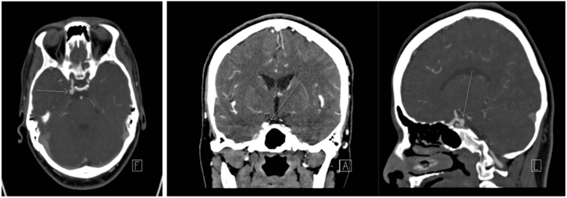
Figure 1: CTA for the head reveals right posterior communicating artery aneurysm.
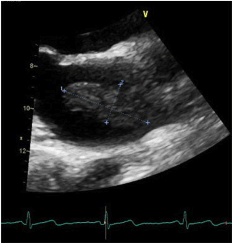
Figure 2: TTE discovered left atrial mass seized 1.9 cm x 4.9 cm.
Cardiac myxoma is a rare tumor of mesenchymal origin, but comprises 50% of all primary cardiac neoplasms. Left atrial myxoma represents about 75% of cardiac myxoma cases. Myxoma is associated with the broad clinical triad of cardiac presentation (60%), sequelae of cerebral or systemic embolization (30–40%), and constitutional symptoms (30%). Reference Chiang, Cheng, Chang, Chiu and Yen1 Transthoracic echocardiography is generally used initially, but transesophageal echocardiography is a superior method for diagnosing this condition. Reference Lee, Connolly and Brown2 The mainstay of treatment for myxoma is surgical resection, particularly when the risk of further tumor embolism or valve obstruction is very high. Reference Chiang, Cheng, Chang, Chiu and Yen1 The tumor can behave aggressively and tends to recur despite being a benign tumor. Reference Xu, Zhang, Wu, Wang, Zhou and Feng3 The mobility, but not the size of the mass, may influence embolic potential. Reference Lee, Connolly and Brown2 Neurological presentations of left atrial myxoma are varied. The most common presentation is of embolic stroke. Progressive vascular dementia is also described due to multiple cerebral infarctions. Meningeal and cerebral metastases may also occur. Although cardiac myxoma-related cerebral embolus is well documented, there are also other rare neurovascular complications described, including cerebral aneurysm, a possible complication of which is intracranial hemorrhage due to rupture. Reference Ashalatha, Moosa, Gupta, Krishna Manohar and Sandhyamani4
There have been approximately 50 cases of cerebral aneurysm related to myxoma reported in the literature since 1966. Reference Xu, Zhang, Wu, Wang, Zhou and Feng3 Aneurysms were detected prior to tumor resection 56% of the time. Reference Sabolek, Bachus-Banaschak, Bachus, Arnold and Storch5 Most commonly, aneurysms are fusiform (91%), but saccular aneurysms are also described. In our case, saccular aneurysm was observed. Our patient had no modifiable or syndromic risk factors for cerebral aneurysm formation. Furthermore, the aneurysm became clinically apparent in the context of clinical evidence of atrial myxoma. Taken together, this suggests an association between our patient’s PComm aneurysm and atrial myxoma. Unfortunately, we do not have a pathologic correlate for confirmation, and thus it is possible that the two disease processes are separate.
Atrial myxoma-associated aneurysms are most often localized to middle cerebral arteries (74.2%), followed by anterior cerebral arteries (13%), cerebellar arteries (7%), posterior cerebral arteries (5%), and finally the basilar artery (1%). Reference Sabolek, Bachus-Banaschak, Bachus, Arnold and Storch5 The pathophysiology of this condition remains unknown. Stoane et al. proposed a “vascular damage theory” in which temporary occlusion of cerebral vessels by tumor emboli leads to perivascular damage and endothelial scarring, and subsequent aneurysm formation. Reference Stoane, Allen and Collins6 Another theory is the “neoplastic theory” which postulates that hematogenous metastases of myxoma cells penetrate and damage the vessels with subsequent fibroblastic proliferation. Reference New, Price and Carter7 The latter was supported by histopathological findings in previous reports. Reference Furuya, Sasaki, Yoshimoto, Okada, Fujimaki and Kirino8 CTA, magnetic resonance angiography (MRA), and conventional cerebral angiography are all methods that can be used to diagnose myxomatous cerebral aneurysms.
There are no guidelines for the treatment of myxomatous cerebral aneurysms. Resection of the cardiac myxoma can help with preventing neurologic complications but does not eliminate completely the risk of delayed cerebral aneurysm formation. For aneurysms located in non-eloquent areas, surgical resection has been described. Reports on coil embolization are limited as most of the aneurysms are fusiform. Reference Chiang, Cheng, Chang, Chiu and Yen1 In patients with multiple cerebral aneurysms, physicians should consider cardiac myxoma. Likewise, patients with cardiac myxoma should be monitored for myxomatous cerebral aneurysms and embolism. Long-term follow-up is therefore recommended.
Conflict of Interest
None.


