


No CrossRef data available.
Article contents
Gray matter reduction in high-risk subjects, recently diagnosed and chronic patients with schizophrenia: A revised coordinate-based meta-analysis
Published online by Cambridge University Press: 13 August 2021
Abstract
Core share and HTML view are not available for this content. However, as you have access to this content, a full PDF is available via the ‘Save PDF’ action button.
Introduction
Characterizing neuroanatomical markers of different stages of schizophrenia (SZ) to assess of how the disorder develops is extremely important for the clinical practice. It still remains uncertain how abnormalities are formed as SZ progresses.
Objectives
We reviewed and analyzed 113 voxel based morphometry studies on people at risk of or with schizophrenia to assess GM alterations at different stages of the disorder and to functionally characterize these GM variations.
Methods
We performed a meta-analysis of voxel-based morphometry studies of genetic and clinical high-risk subjects (g-/c-HR), recently diagnosed (RDSZ) and chronic SZ patients (ChSZ). We quantified gray matter (GM) changes associated with these four conditions and compared them with contrast and conjunctional data. We performed the behavioral analysis and networks decomposition of alterations to obtain their functional characterization.
Results
Compared to previous investigations, results reveal a robust cortical-subcortical, left-to-right homotopic progression of GM loss. The right anterior cingulate is the only altered region in all conditions. Contrast analyses show left-lateralized insular, amygdalar and parahippocampal GM reduction in RDSZ, which appears bilateral in ChSZ. An overlap between RDSZ and ChSZ is observed in the left insula, amygdala, precentral and inferior frontal gyri. Functional decomposition shows involvement of the salience network, with an enlargement of the sensorimotor network in RDSZ and the thalamus-basal nuclei network in ChSZ.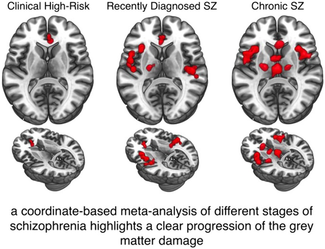
Conclusions
These results can help the research on diagnostic and neuroimaging biomarkers of SZ staging, as well as on the identification of new therapeutics neuroanotomic targets that could be addressed with focused magnetic or non-invasive electric stimulation.
Disclosure
No significant relationships.
- Type
- Abstract
- Information
- European Psychiatry , Volume 64 , Special Issue S1: Abstracts of the 29th European Congress of Psychiatry , April 2021 , pp. S129
- Creative Commons

 This is an Open Access article, distributed under the terms of the Creative Commons Attribution licence (http://creativecommons.org/licenses/by/4.0/), which permits unrestricted re-use, distribution, and reproduction in any medium, provided the original work is properly cited.
- Copyright
- © The Author(s), 2021. Published by Cambridge University Press on behalf of the European Psychiatric Association
 You have
Access
You have
Access
 Open access
Comments
No Comments have been published for this article.