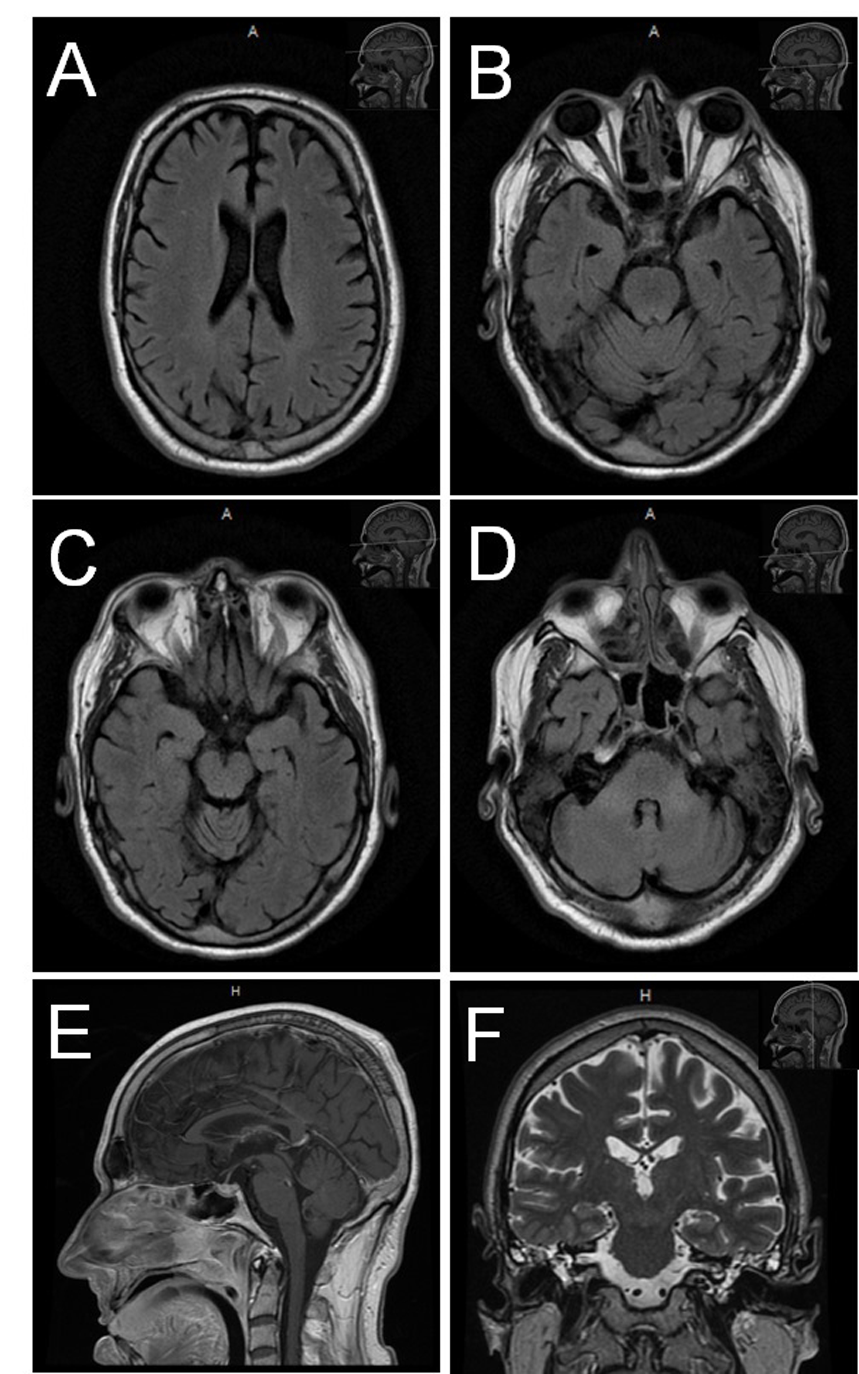
A 51-year-old Caucasian male with a past medical history of hypertension and retinitis pigmentosa (reads at ~6 inches) was admitted with acute nausea, vomiting, watery diarrhea, abdominal pain, lower extremity pain, and generalized weakness. Laboratory analysis revealed hypokalemia and hypoosmolar-hyponatremia (blood levels: Potassium: 3.1 mEq/L with magnesium: 0.5 mEq/L; Sodium: 131 mEq/L; Plasma osmolality: 252 milli-osmoles/kg). The electrolyte imbalance was corrected with intravenous normal saline and potassium supplements over the next few days. The following week he developed leukocytosis; subsequently he underwent an exploratory laparotomy which showed ileus. Extubation failed post-operatively. Patient developed weakness and loss of deep tendon reflexes in upper-limbs by day 7. By day 14, the patient developed lower-extremity weakness and right 6th nerve palsy. Facial diplegia, complete external ophthalmoplegia and sluggish pupillary reflexes were noted on day 18. Lumbar puncture showed albumino-cytologic dissociation. Tracheostomy tube for assisted ventilation and percutaneous endoscopic gastrostomy (PEG) tube for feeding was placed on day 28. By the 5th week, the patient was “locked-in” with complete bilateral ptosis, complete external ophthalmoplegia, complete flaccid quadriplegia, facial diplegia, and respiratory failure. Nerve conduction study (day 30) showed no recordable motor/sensory potentials and electromyography showed global, insertional positive sharp waves. Serology was significant for elevated anti-GM1 but not anti-Gq1b antibody titers. Magnetic resonance imaging (day 21: brain and whole spine; day 73: brain) were not significant for contributory intracranial etiologies (supplementary figure). Patient’s electroencephalogram was reported as mildly abnormal showing mild diffuse slowing of background without epileptiform or focal abnormalities.
The patient’s pupils (Figure) (day 41) were ~3.5 mm in diameter (room illumination and darkness) and light onset did not induce constriction at the expected latency (0.20 to 0.28 sec). Longer light exposure caused slow pupillary constriction for both direct and consensual light reflexes. Light offset after a two-minute exposure resulted in delayed but not immediate pupillary dilatation. Topically applied phenylephrine (2.5%, alpha-1 receptor agonist) and tropicamide (1%, non-selective muscarinic antagonist) caused pupillary dilatation. Titrating-up topical pilocarpine (non-selective muscarinic receptor agonist) to 2% did not constrict the pupils. Sector defects were visible in patient’s iris. The patient was not on any medication which could have affected the sympathetic or the parasympathetic system.

Figure Pupillary findings in anti-GM1 positive, anti-GQ1b negative inflammatory polyradiculoneuropathy. (A, B) Immediate pupillary direct light response (DLR) shows no change in pupil size. (C, D) DLR with extended light exposure showed a small constriction. (E, F) Consensual pupillary light response (CLR), recorded in the pupils contralateral to the eye exposed to light, showed constriction only with extended light exposure. (G, H, I) Pupillary response to an offset of a 120 second light exposure shows a slow dilatation of the pupils only at later time points (I) and not immediately after light offset (H). (J) Magnified (I) illustrating a sector defect (red-arrow). Patients pupils dilated after topical instillation of phenylephrine 2.5% (K) or Tropicamide 1% (L), but did not constrict to pilocarpine 2% (M,N). At baseline patient had complete ptosis (O) with divergent eyes (K, L). (P) Pupillary circumference (yellow-dashed circle) was normalized to the corneal circumference (red-dashed circle) measured at the limbus for comparing changes in pupil diameter. Images were captured from video recorded with a resolution of 1920x1080 at a frame rate of 30 frames per seconds.
Although this patient had sluggishly constricting pupils with sector defects, the lack of pupillary dilatation (observed over five months) and absence of cholinergic supersensitivity differentiates this case from Adies pupil.Reference Lee 1 Pupillary dilatation in response to tropicamide indicated muscarinic contribution to baseline pupillary aperture. Lack of pupillary constriction with 2% pilocarpine suggested muscarinic receptor saturation and/or desensitization in the constrictor-pupillae. The alpha-1 adrenergic receptor agonist caused pupillary dilation as expected. Absence of papillary dilation, responsiveness of pupils to muscarinic antagonists but unresponsiveness to muscarinic agonists are uncharacteristic of an oculomotor nuclear lesion. The post-ganglionic parasympathetic neuron innervates the sphincter muscles in a sector distribution. The presence of irideal sector defects similar to Adies pupils, indicates pathology in the post-ganglionic parasympathetic neuron.Reference Levin and Adler 2 Short-ciliary nerves that carry post-ganglionic parasympathetics to the iris are myelinated.Reference May 3 Ectopic activity from demyelinationReference Baker and Bostock 4 may augment post-ganglionic parasympathetic influence on the pupillo-constrictor fibers resulting in saturation and/or desensitization of muscarinic receptors.Reference el-Fakahany and Lee 5 Sluggish pupillary constriction and dilation in the presence of muscarinic receptor saturation/desensitization may indicate sympathetic involvement in the pupillary light reflex as with Parinaud’s syndrome.Reference Barbur 6 The sympathetic receptor sites on the dilator pupilae responded to phenylephrine, indicating that the sympathetic receptor sites on the dilator pupilae were not saturated/desensitized. Sympathetic compensation to parasympathetic overactivity or muscarinic receptor desensitization could both contribute to the normal sized pupils. Retinitis pigmentosa is unlikely to have caused acute pupillary changes. Pupillary abnormality in metabolic encephalopathy spares the light reflex hence cannot explain the observed pupillary changes.Reference Tokuda, Nakazato and Stein 7 The patient received three five-day courses of intravenous immunoglobulin and one three-day course of plasma exchange. By the 19th week, the patient was found to move his neck muscles, and showed some eye movement to lateral gaze bilaterally without any change in pupillary reactivity to light. Patient continued to be fed via PEG-tube and ventilated via the tracheostomy tube, and was subsequently moved to a long-term care facility. Whereas denervation of parasympathetic post-ganglionic neurons explains Adies pupil, demyelination of these fibers can produce normal-sized sluggish pupils with sector defects and sensitivity to muscarinic antagonism but not its agonism. As explained, other causes such as medication effect, nuclear oculomotor nerve lesions, and metabolic causes are unlikely to produce pupils with the described properties.
Disclosures
DM does not have anything to disclose. LL does not have anything to disclose. Source of support: Nil. Conflict of interest: None. Financial disclosures: None
Supplementary Materials
To view Supplementary Materials for this article, please visit http://dx.doi.org/10.1017/cjn.2015.47