


Alzheimer's disease, characterised by intracellular and extracellular deposits of filamentous proteins, progressive cognitive impairments and neuronal loss, is the most common form of dementia. Many biological factors appear to be involved in the development of Alzheimer's disease, such as β-amyloid (Aβ) peptide, oxidative stress and the inflammatory process, and the ApoE4 gene(Reference Almeida, Bolaños and Medina1–Reference Frey, Bonert and Kratzsch3). Aβ forms extracellular deposits in senile or diffusive plaques and in cerebral vasculatures in the Alzheimer's disease brain. Researchers have found that many Aβ peptides, such as Aβ1-42, Aβ1-40, Aβ25-35 and Aβ31-35 can induce neurons to undergo apoptosis in vitro, which is tightly associated with the process of Alzheimer's disease(Reference Chin, Hamid and Latiff4, Reference Clementi, Marini and Coletta5).
At present, Alzheimer's disease affects millions of people worldwide and cannot be diagnosed by a valid clinical method or a biomarker before the onset of disease, and there is no cure. Scientists have found that in addition to non-modifiable genetic risk factors, potentially modifiable factors including environmental exposure and diet-related chronic diseases have been identified as risk factors for Alzheimer's disease(Reference Patterson, Feightner and Garcia6). Epidemiological studies have suggested that the consumption of oestrogen is associated with a reduced risk of dementia in women(Reference Usui7). But oestrogen simultaneously contributes to the development of some oestrogen-dependent cancers, such as breast cancer and prostate cancer.
Genistein, a kind of phyto-oestrogen abundant in many plant-based diets, is the predominant isoflavone form in soya food and supplements. Many researches have showed that soya phyto-oestrogens influence cognitive function and behaviour, especially in postmenopausal women(Reference Lee, Lee and Sohn8). In an ovariectomised mouse experiment, the researchers found that escape latency was significantly shortened in a group orally administrated soyabean isoflavone continuously(Reference Zhu, Yang and Huang9). Recently, Azcoitia et al. (Reference Azcoitia, Moreno and Carrero10) reported that at high doses (10 mg/kg), genistein showed neuroprotective effects in a rat model.
Folic acid is not only essential for the maintenance of normal brain function, but may also be a potential source for brain therapeutics against excitotoxicity(Reference Das11). In general, cerebrospinal fluid (CSF) folic acid levels are three or four times higher than blood folic acid levels. Under normal conditions, CSF folic acid concentrations do not vary with age, while late-onset Alzheimer's disease patients have significantly lower CSF folic acid levels. Folic acid deficiency is the most common cause of hyperhomocysteinaemia that has been suggested as a risk factor of dementia or cognitive impairment(Reference Tassino, Campos and Guerra12). A randomised, double-blind, placebo-controlled study showed that daily supplementation of 800 μg oral folic acid for 3 years could improve cognitive function(Reference Durga, van Boxtel and Schouten13).
Genistein and folic acid prevent neuronal damage from many excitotoxicities; however, the mechanism of their neuroprotective actions has not been fully elucidated. Our previous study indicated that genistein and/or folic acid attenuated the toxic and apoptotic effects of cyclophosphamide in either rats with neural tube defects or cultured cortical neurons and we also found that the protective effects of genistein co-administered with folic acid were more pronounced(Reference Xiao, Yu and Zhao14). The present study is aimed to evaluate the effects of genistein and folic acid on Aβ31-35-induced apoptosis in cultured cortical neurons of rats and their joint functions.
Materials and methods
Materials
Specific pathogen-free newborn Wistar rats were provided by the Laboratory Animal Center (Capital Medical University, Beijing, China). Genistein and folic acid were purchased from Sigma (St Louis, MO, USA) and genistein was dissolved at 40 mm in dimethyl sulfoxide (DMSO), while folic acid was dissolved in neuron culture medium. The final concentrations of genistein and folic acid were 27 μg/ml (100 μm) and 40 μg/ml (90 μm), respectively. Aβ31-35 was purchased from Sigma Chemical. It was dissolved in sterile bi-distilled water at a concentration of 3 mg/ml and stored at − 20°C until use. Peptides were aggregated by incubation, at 3 mg/ml in sterile bi-distilled water, at 37°C for 4 d and then used as neurotoxin to cultured neurons.
Cortical neuron culture and neurotoxicity treatments to cultured neurons
The neuron culture protocol was adapted from the established method in our laboratory(Reference Xiao, Qiao and Zhao15) for primary cultured cortical neurons of the newborn (no more than 24 h) Wistar rat. Briefly, rats were killed by rapid decapitation and their brains quickly harvested. After cortical layers were minced and dissociated by mechanical trituration, cortical neurons were dissociated in Dulbecco's modified Eagle's medium supplemented with 10 % low-endotoxin horse serum, 10 % heat-inactivated fetal bovine serum (v/v), 26 mm-bicarbonate, 25 mm-d-glucose, 25 mm-HEPES, penicillin (10 mg/ml) and streptomycin (10 mg/ml). The single-cell suspension was plated into 35 mm diameter dishes (2 × 10⁵ cells/cm2) pre-coated with poly-l-lysine. Cultures were maintained in an incubator at 37°C under a 5 % CO2–95 % air atmosphere. Non-neuronal cell division was halted by exposure to 10 mm-cytosine arabinoside for 1 d and the cells were then transferred into a maintenance medium identical to the plating media.
After 48 h of culture, 2 h before the neurons were exposed to Aβ31-35 (25 μmol/l), genistein and/or folic acid were added. After another 24 h, cell viability assessment and other experiments were performed. Each experiment was repeated at least three times.
Cell viability
Cell viability was assessed using a modified 3-[4,5-dimethylthiazol-2]-2,5 diphenyltetrazolium bromide (MTT) assay. The MTT assay is based on the ability of a mitochondrial dehydrogenase enzyme from viable cells to cleave the tetrazolium rings of the pale yellow MTT and form a dark blue formazan crystal, which is accumulated within healthy cells, and the number of surviving cells is directly proportional to the level of the formazan product created. The colour can then be quantified using a simple colorimetric assay. Briefly, the cultured cortical neurons were pre-incubated with genistein and/or folic acid for 2 h before being exposed to 25 μm-Aβ31-35 for 24 h. Then 20 μl MTT stock solution (5 mg/ml) was added to the culture medium and the solution was incubated for another 4 h at 37°C. The resulted MTT formazan was extracted with 200 μl DMSO and the absorbance was recorded at 570 nm by microtiter plate reader (Tecan Sunrise Microplate Reader; Tecan Group Ltd, Männedorf, Switzerland). The sample solution was freshly prepared in which DMSO concentration was lower than 0·1 %.
Mitochondrial membrane potential
The fluorescent dye rhodamine 123 (Molecular Probes, Eugene, OR, USA) was used as a measure of mitochondrial membrane potential (MMP), as described by Almeida et al. (Reference Almeida, Bolaños and Medina1). Briefly, neurons were incubated for 30 min at 37°C in the presence of the dye (10 μg/ml) and then washed twice with PBS solution. Fluorescence signals were captured using a flow cytometer at 529 nm emission wavelength, corresponding to the fluorescence peak of the monomer and that of the aggregate.
Measurements of apoptosis
Hoechst 33342, a fluorescent stain for labelling DNA, was used to observe the apoptotic neurons. Briefly, to visualise nuclear morphology following Aβ31-35 treatment, cells were fixed with 4 % paraformaldehyde and stained in Hoechst 33342 DNA-binding dye (10 mg/l) for 15 min at 37°C in the dark, then washed with PBS three times and the nuclear morphology was observed under a fluorescence microscope (Olympus, Tokyo, Japan).
Single cell gel electrophoresis
The single cell gel electrophoresis assay was used to assess cells whose DNA was fragmented to migrate out of the cell under the influence of an electric field, whereas undamaged DNA migrates more slowly and remains within the confines of the nucleoid. Briefly, neurons were trypsinised, centrifuged (2 min; 1000 rpm) and cell pellets were suspended in a pre-warmed low-melting-point agarose (0·5 % in PBS) and deposited on conventional microscope slides (initially dipped in 1 % agarose and dried) precoated with normal agarose (0·8 % in PBS). The slides were then put in a lysis solution composed of 2·5 m-NaCl, 0·1 m-EDTA, 10 mm-2-amino-2-hydroxymethyl-propane-1,3-diol-HCl (Tris-HCl) at pH 10, extemporarily added with 10 % DMSO and 1 % Triton X-100, for 1 h at about 4°C. DNA was allowed to unwind for 20 min in fresh electrophoresis buffer (0·3 m-NaOH, 1 mm-EDTA, pH 13) and the electrophoretic migration was then performed at 4°C (24 min, 20 V, 300 mA, 25 min). The slides were bathed twice for 5 min in neutralising solution (0·4 m-Tris-HCl, pH 7·5) and dried for conservation with 95 % ethanol for 5 min. DNA was stained with propidium iodide (2·5 μg/ml in PBS) just before slide examination with a fluorescence microscope (TE2000-E; Nikon, Melville, NY, USA). At least 100 images per dose were analysed using the Comet Assay IV software (Perceptive Instruments, Haverhill, Suffolk, UK). Two parameters, the number of comet cells, defined as the number of neurons with a comet tail, and DNA migration length (μm), were used to evaluate the extent of DNA damage in individual cells.
Reverse transcriptase polymerase chain reaction
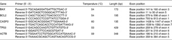
Total RNA was purified using Trizol (Invitrogen, Carlsbad, CA, USA) and reverse-transcribed using an RT kit (no. A3500; Applied Promega, Madison, WI, USA). mRNA encoding bax, bcl-2, caspase-3, p53 and β-actin (used as an invariant control) were analysed by RT-PCR. The primer sequences are listed in Table 1. PCR was carried out as follows: denature at 94°C for 5 min for the first cycle, and then cycles consisting of 94°C for 30 s, annealing at 54°C, 53°C or 59°C for 30 s, and extension at 72°C for 30 s. Thirty-five cycles were performed for all the genes. Amplification products underwent electrophoresis on a 2·0 % agarose gel and the relative quantity of mRNA was estimated by densitometry scanning with X-rays (Gel Doc XRTM; Bio-Rad, Hercules, CA, USA).
Table 1 Primers of BAX, BCL2,caspase-3 (CASP3), TP53 and β-actin (ACTB)
Statistical analysis
Data are shown as mean values and standard deviations. Statistical comparisons were performed by one-way ANOVA. The acceptable level of significance was set at P < 0·05.
Results
Cell viability
Viabilities of neurons in the control were 1·038 (sd 0·125) (vehicle). After treatment with Aβ31-35, neuron viability significantly decreased to 0·811 (sd 0·083). However, both folic acid and genistein increased neurons' viability when given 2 h before they were exposed to Aβ31-35. Though the neurons' viability was slightly increased, there were no statistically significant differences when genistein was co-administrated with folic acid, compared with genistein or folic acid singly administrated as indicated in Fig. 1.

Fig. 1 Effect of β-amyloid (Aβ) 31-35 on neuron viability, protection of genistein (Gen) and folic acid (FA) and the protective effect of the co-administration of FA and Gen. Neurons were exposed to vehicle (control), Aβ31-35, Aβ+FA, Aβ+Gen or Aβ+FA+Gen. FA and/or Gen was added 2 h before the neurons were exposed to Aβ31-35. Cell survival was quantified at 24 h by assaying 3-[4,5-dimethylthiazol-2]-2,5 diphenyltetrazolium bromide (MTT). Values are means, with standard deviations represented by vertical bars. * Mean value was significantly different from that of the Aβ31-35 group (P < 0·05).
Mitochondrial membrane potential
To analyse the alteration in MMP that follows Aβ apoptotic stimulation, we incubated cells treated with 10 μm-Aβ with the cationic dye rhodamine 123, and subsequently analysed the cells using a flow cytometer. Neurons treated with Aβ displayed significant decrease in mitochondrial potential. Both genistein and folic acid demonstrated potential protective activity. Though there were no significant differences between the genistein and folic acid singly treated and co-administrated groups, mitochondrial potential was much higher than that in the singly treated group (Fig. 2).

Fig. 2 Effect of β-amyloid (Aβ) 31-35 and protection of genistein (Gen) and folic acid (FA) on mitochondrial membrane potential (MMP) as assessed by fluorescence change of rhodamine 123 at 529 nm. Neurons were stained using the fluorescent probe rhodamine 123 to measure changes in MMP. Values are means, with standard deviations represented by vertical bars. * Mean value was significantly different from that of the Aβ31-35 group (P < 0·05).
DNA structure
Compared with the control, comet cells and DNA tail length were significantly increased in the Aβ31-35-treated group (Fig. 3). The comet cells and DNA migration length in three experiments were significantly decreased. It was exciting to find that the DNA migration length in the genistein co-administrated with folic acid group was much shorter than that in the genistein or folic acid solo-treated groups.

Fig. 3 Effect of β-amyloid (Aβ) 31-35 on DNA structure and protection of genistein (Gen) and folic acid (FA). The y axes represent the number of cells with a comet tail (%; ![]() ) and the DNA migration length (μm;
) and the DNA migration length (μm; ![]() ). Values are means, with standard deviations represented by vertical bars. * Mean value was significantly different from that of the Aβ31-35 group (P < 0·05). † Mean value was significantly different from that of the Aβ+FA+Gen group (P < 0·05).
). Values are means, with standard deviations represented by vertical bars. * Mean value was significantly different from that of the Aβ31-35 group (P < 0·05). † Mean value was significantly different from that of the Aβ+FA+Gen group (P < 0·05).
Apoptosis analysis
Neuron apoptosis was observed by Hoechst 33342 nuclear dyeing. Compared with the control group, Aβ31-35 induced neuronal shrinkage, nucleus pycnosis, chromatin margination or crescent-shaped and apoptotic body formation. The percentage of apoptotic neurons was significantly increased when treated with Aβ31-35 compared with that in the control group, while neurons pretreated with genistein and/or folic acid showed a significant but incomplete prevention of apoptosis (Fig. 4).

Fig. 4 Neurons were stained with the DNA-binding fluorochrome Hoechst 33342. Fluorescence micrographs are of cortical cell nuclei from untreated cells (control), cells exposed to 25 μm-β-amyloid (Aβ) 31-35, cells exposed to folic acid (40 μg/ml) 2 h before 25 μm-Aβ31-35 (FA), cells exposed to genistein (27 μg/ml) 2 h before 25 μm-Aβ31-35 (Gen), and cells co-incubated with genistein plus folic acid (Gen+FA). For explanation, see the Apoptosis analysis section.
mRNA levels of bax, bcl-2, caspase-3 and p53 genes
Compared with the control group, the expressions of bax, caspase-3 and p53 genes were all up-regulated and bcl-2 genes were down-regulated when the neurons were treated with Aβ31-35. The expression of bax was down-regulated when the neurons treated with Aβ31-35 were incubated with folic acid, genistein and genistein co-administrated with folic acid. The expressions of p53 and caspase-3 were down-regulated, while the expression of bcl-2 was up-regulated when the neurons treated with Aβ31-35 were incubated with genistein and genistein co-administered with folic acid (Table 2; Fig. 5).
Table 2 Effect of genistein (Gen) and/or folic acid (FA) on BAX, BCL2, caspase-3 (CASP3) and TP53 mRNA levels in the brain
(Mean values and standard deviations)

Aβ, β-amyloid.
* Mean value was significantly different from that of the Aβ31-35 group (P < 0·05).

Fig. 5 Effect of β-amyloid (Aβ) 31-35 and protection of genistein (Gen) and folic acid (FA) on the mRNA levels of β-actin (ACTB), BAX, BCL2, caspase-3 (CASP3) and TP53 genes in cortical neurons.
Discussion
Aβ31-35, the core sequence and active centre of the β-amyloid peptide, induces cell apoptosis in PC 12 cells(Reference Clementi, Marini and Coletta5) and rat cerebellar granule cells(Reference Matsui, Ramasamy and Ingelsson16). The mechanisms under the Aβ31-35-induced apoptosis include bax mRNA expression up-regulation(Reference Misiti, Clementi and Tringali17), caspase activation, DNA fragmentation(Reference Misiti, Sampaolese and Pezzotti18) and protein kinase A activation(Reference Zhao, Qian and Zhang19). We found that Aβ31-35 not only poisoned the neurons directly (cell viability decreased), but also decreased MMP, damaged the integrity of nuclear DNA, up-regulated the expression of bax, caspase-3 and p53 genes, down-regulated the expression of the bcl-2 genes, and finally led to neuronal apoptosis. The present results from the MTT assay (Fig. 1), MMP, comet assay and apoptotic analysis (Hoechst 33342 staining and determining expression of apoptosis-related genes) introduced the evidence that genistein and folic acid could protect cultured cortical neurons against Aβ31-35 toxicity.
Genistein, with potentially beneficial health effects such as anti-carcinogenic qualities, has been identified as having anti-prolific and pro-apoptotic effects on various malignant cell types derived from solid and non-solid tumours(Reference Schmidt, Knobbe and Frank20). However, the anti-apoptotic effects of genistein in pancreatic β-cells(Reference Kim, Kwon and Song21), primary neurons(Reference Kajta, Domin and Grynkiewicz22) and human mononuclear cells(Reference Wu and Chan23), induced by cytokines(Reference Kim, Kwon and Song21), glutamate(Reference Kajta, Domin and Grynkiewicz22) and methylglyoxal(Reference Wu and Chan23), have been reported widely. These studies always concluded that the anti-apoptotic effects of genistein were dose-dependent(Reference Kim, Kwon and Song21–Reference Wu and Chan23). Another study found that cells pre-treated with 50 μm-genistein could significantly prevent HCN1-A cells from cell death induced by 100 μm- and 1 mm-tertiary butylhydroperoxide(Reference Sonee, Sum and Wang24). In the present study, we did not find toxicity of 100 μm-genistein on the cultured cortical neurons treated with Aβ31-35. On the contrary, when administered 2 h before Aβ31-35 was added, genistein protected the neurons from the damage induced by Aβ31-35. These discrepancies may be due to differences in experimental conditions, such as the neural cell type and culture medium (for example, the toxic agent used)(Reference Jin, Wu and Cohen25). So, it is important to study the effective level of genistein, which is toxic or protective to primary cultured cortical neurons damaged by Aβ31-35.
Folic acid deficiency induces neurotoxicity by multiple routes. It was reported that folic acid deprivation increased cytosolic Ca and reactive oxygen species (ROS) and impaired mitochondrial function(Reference Tjiattas, Ortiz and Dhivant26). Moreover folic acid was remarkably neuroprotective against glutamate and N-methyl-d-aspartic acid cytotoxicity in a dose- and time-dependent manner(Reference Lin, Desbois and Jiang27). In the present study, folic acid supplementation decreased MMP and the percentage of comet cells that had been increased by Aβ31-35 in cultured cortical neurons. It has been reported that folic acid deprivation induced neurodegeneration changes typical of those observed in Alzheimer's disease, including increased cytosolic Ca, ROS, phospho-tau and the apoptotic process; an increase in glutathione and reduction in ROS levels were observed following supplementation of folic acid-deprived cultures(Reference Ho, Ashline and Dhitavat28). Substantial evidence has indicated that the neuronal damage caused by Aβ is mediated through oxidative stress(Reference Rottkamp, Raina and Zhu29, Reference Butterfield30). So, the mechanism of folic acid supplementation alleviating the damage of mitochondria and DNA induced by Aβ may be related to the alteration of oxidative stress conditions.
Mitochondria are integrated in a number of signalling pathways, including cell death cascades, thus controlling cellular homoeostasis in multiple ways(Reference McBride, Neuspiel and Wasiak31). Mitochondria undergo two major alterations during apoptosis. The first is the permeability of the outer mitochondrial membrane. This event is tightly regulated by members of the Bcl-2 family and involves the conformational change of pro-apoptotic family members such as Bax. Second, the electrochemical gradient that is normally present across the inner mitochondrial membrane is lost (membrane depolarisation)(Reference Knudson and Brown32). An array of evidence suggests that alteration of mitochondria function is critically involved in the apoptotic process. Dysfunctional mitochondria bear the risk of futile ATP hydrolysis and enhancement of oxidative stress. Furthermore, extensive mitochondrial damage may lead to the dissipation of the membrane potential across the inner membrane and induce cell death by the release of pro-apoptotic proteins(Reference Kroemer, Galluzzi and Brenner33). It is reported that in response to adverse stimuli, the mitochondrial permeability transition pore is actively opened, resulting in the collapse of MMP and release of multiple pro-apoptotic proteins such as Smac, Cyto C, AIF, Omi, etc, from the impaired mitochondria to cytosol(Reference Naderi, Somayajulu-Nitu and Mukerji34) and these proteins in turn trigger a cascade of events and eventually lead to cell apoptosis. We found that MMP markedly decreased after exposure to Aβ31-35. However, the reduction of MMP was attenuated after genistein and/or folic acid treatment, which indicated that the neuroprotective effect of genistein and/or folic acid might be related to maintaining the structure and function of mitochondria.
Bcl-2 family members are the arbiters of the mitochondrial apoptotic pathway, which is subdivided into two classes including anti-apoptotic members such as Bcl-2, Bcl-xl, Bcl-w and Mcl-1, which protect cells from apoptosis, and pro-apoptotic members such as Bax, Bak, Bad, Bid and Bim, which induce cell apoptosis. The stoichiometry of pro- v. anti-apoptotic Bcl-2 family members in the cell determines whether the cell lives or dies. This fine balance between anti-apoptosis and pro-apoptosis is regulated at the transcriptional or post-translational level in response to various cellular cues. Our earlier study found that the suppression of bcl-2 gene expression was the early signal in the neuron's programmed death when exposed to an apoptotic agent(Reference Xiao, Yu and Zhao14, Reference Yan, Qiao and Dou35). When the bax:bcl-2 ratio was >1, bax regulated mitochondrial cytochrome c release in vivo and in vitro (Reference Raisova, Hossini and Eberle36). In the present study, the expression of the bcl-2 gene was significantly down-regulated while the expression of the bax gene was significantly up-regulated and the ratio of bax:bcl-2 was greater than 1 in Aβ31-35-treated neurons, which indicated the ongoing of apoptosis. If pretreated with genistein or genistein co-administrated with folic acid, the situation could be reversed (Table 2).
It has been demonstrated that Aβ1-42 and Aβ25-35 may induce caspase-dependent apoptosis. Apoptotic cell death associated with the activation of caspases has been found in several neuronal cell types exposed to Aβ. Activation of caspase-3 is a key event in the execution of the apoptotic cascade in central nervous system disorders including Alzheimer's disease. Aβ31-35 can also induce apoptosis in the cortical and hippocampal neurons as Aβ25-35 does and further study showed that apoptosis was mediated by caspase-dependent pathways(Reference Misiti, Clementi and Tringali17–Reference Zhao, Qian and Zhang19, Reference Yan, Qiao and Dou35). In the present study, we gave evidence that genistein or genistein co-administrated with folic acid could alleviate the up-regulation of the caspase-3 gene induced by Aβ31-35, which indicated that the effect of anti-apoptosis by genistein was achieved possibly via caspase-dependent pathways.
Recently, involvement of p53 in neuronal death occurring in Alzheimer's disease(Reference Zhang, McLaughlin and Goodyer37) has been detected. Cell culture studies have established strong relationships between p53 expression and neuronal death induced by DNA-damaging agents and glutamate(Reference Inamura, Araki and Enokido38). From these results, we could suggest that the expression of p53, bax, bcl-2 and caspase3 were changed by Aβ31-35, which induced the neurons to undergo apoptosis and genistein or genistein co-administrated with folic acid could intervene this apoptotic progress.
In the study, the protective effects of genistein co-administrated with folic acid to DNA integrality were significantly different from that of genistein or folic acid, although there were no significant differences for cell viability and MMP. The reason may be related to the isolated protective effects of genistein and folic acid that were strong enough so that the combination was not prominent, while the protective effect was enhanced when the isolated effects were not sufficient. On the other hand, the antioxidative activity of genistein has been reported widely(Reference Wu and Chan23, Reference Sonee, Sum and Wang24, Reference Baluchnejadmojarad, Roghani and Nadoushan39–Reference Sánchez, Amrán and Fernández41). The anti-apoptotic effect of folic acid and genistein may be mainly related to their antioxidative activity; common physiological mechanisms may be affected, such that there was no enhanced protection in some variables examined.
The present results suggest that genistein and/or folic acid protected the neurons from the damage of Aβ31-35 by maintaining mitochondrial function and DNA integrality and regulating the apoptosis-related genes. The cooperation effects of genistein and folic acid were significantly displayed when the single effects were not strong enough. The molecular mechanisms of the anti-apoptosis function of genistein in cultured cortical neurons and if there is an effective genistein concentration spectrum that is beneficial to Aβ-treated neurons will be the next goals in further studies.
Acknowledgements
The present study was supported by grants from the National Natural Science Foundation of China (no. 30771802 and 30571560) and the Beijing Municipal Commission of Education Science and Technology Developmental Plan Foundation (no. KM200610025010 and KZ200710025011). H.-L. Y contributed to the drafting of the paper and the Hoechst 33342 nuclear dyeing; J.-F. F. did the data analysis; L. L., X.-H. Z, J. Z. and L. X. contributed to the cell culture, RT-PCR, single cell gel electrophoresis, MMP and MTT analyses; R. X. designed the study and was the final principal of the study.
There is no conflict of interest associated with the present study.


