Skip to main content Accessibility help
×
Hostname: page-component-78c5997874-t5tsf Total loading time: 0 Render date: 2024-11-19T12:02:16.723Z Has data issue: false hasContentIssue false

Part I - Non-Traumatic Conditions

Published online by Cambridge University Press:  05 April 2016

Bharti Khurana
Affiliation:
Brigham and Women's Hospital
Jacob Mandell
Affiliation:
Brigham and Women's Hospital, and Harvard Medical School, Massachusetts
Stephen Ledbetter
Affiliation:
Brigham and Women’s Hospital
Get access

Summary

Image of the first page of this content. For PDF version, please use the ‘Save PDF’ preceeding this image.'
Type
Chapter
Information
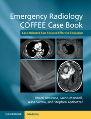
Emergency Radiology COFFEE Case Book
Case-Oriented Fast Focused Effective Education
, pp. 1 - 416
Publisher: Cambridge University Press
Print publication year: 2016

Access options

Get access to the full version of this content by using one of the access options below. (Log in options will check for institutional or personal access. Content may require purchase if you do not have access.)

References

Further reading

Bennett, GL et al. CT Findings in Acute Gangrenous Cholecystitis. AJR 2002; 178:275281.CrossRefGoogle ScholarPubMed
Bortoff, GA et al. Gallbladder Stones: Imaging and Intervention. Radiographics 2000; 20:751766.CrossRefGoogle ScholarPubMed
Catalano, OA et al. MR Imaging of the Gallbladder: A Pictorial Essay. Radiographics 2008; 28:135155.CrossRefGoogle ScholarPubMed
Grayson, DE, Abbott, RM, Levy, AD et al. Emphysematous Infections of the Abdomen and Pelvis: A Pictorial Review. Radiographics 2002; 22:543561.CrossRefGoogle ScholarPubMed
Lassandro, F et al. Clinical Observations: Role of Helical CT in Diagnosis of Gallstone Ileus and Related Conditions. AJR 2005; 185:11591165.CrossRefGoogle Scholar
O’Connor, OJ, Maher, MM. Imaging of Cholecystitis. AJR 2011; 196:W367–374.Google ScholarPubMed
Shakespear, JS et al. Pictorial Essay: CT Findings of Acute Cholecystitis and Its Complications. AJR 2010; 194:15231529.CrossRefGoogle Scholar

Further reading

Blood in the Belly: CT Findings of Hemoperitoneum. Lubner, M, Menias, C, Rucker, C, et al. Radiographics 2007; 27:109125.CrossRefGoogle ScholarPubMed
Genetics and Imaging of Hepatocellular Adenomas: 2011 Update. Katabathina, VS, Menias, CO, Shanbhogue, AK, et al. Radiographics 2011; 31:15291543.CrossRefGoogle ScholarPubMed
Imaging of Nontraumatic Hemorrhagic Hepatic Lesions. Casillas, VJ, Amendola, MA, Gascue, A, et al. Radiographics 2000; 20:367378.CrossRefGoogle ScholarPubMed
Hepatic Adenomas: Imaging and Pathologic Findings. Grazioli, L, Federle, M, Brancatelli, G, et al. Radiographics 2001; 21:877894.CrossRefGoogle ScholarPubMed

Further reading

Balthazar, EJ, Robinson, DL, Megibow, AJ, et al. Acute Pancreatitis: Value of CT in Establishing Prognosis. Radiology 1990; 174:331336.Google Scholar
Balthazar, EJ. Acute Pancreatitis: Assessment of Severity with Clinical and CT Evaluation. Radiology 2002; 223:603613.CrossRefGoogle ScholarPubMed
Bollen, TL, van Santvoort, HC, Besselink, MGH, et al. Update on Acute Pancreatitis: Ultrasound, Computed Tomography, and Magnetic Resonance Imaging Features. Semin Ultrasound CT MRI 2007; 28:371383.CrossRefGoogle ScholarPubMed
Bollen, TL. Imaging of Acute Pancreatitis: Update of the Revised Atlanta Classification. Radiol Clin N Am 2012; 50:429445.CrossRefGoogle ScholarPubMed
Casas, JD, Diaz, R, Valderas, G, et al. Prognostic Value of CT in the Early Assessment of Patients with Acute Pancreatitis. AJR 2004; 182:569574.CrossRefGoogle ScholarPubMed
Lecesne, R, Taourel, P, Bret, P, Atri, M, et al. Acute Pancreatitis: Interobserver Agreement and Correlation of CT and MR Cholangiography with Outcome. Radiology 1999; 211:727735.CrossRefGoogle Scholar
Lenhart, DK, Balthazar, EJ. MDCT of Acute Mild (Nonnecrotizing) Pancreatitis: Abdominal Complications and Fate of Fluid Collections. AJR 2008; 190:643649.CrossRefGoogle ScholarPubMed
O’Connor, OJ, McWilliams, S, Maher, MM. Imaging of Acute Pancreatitis. AJR 2011; 197:W221W225.CrossRefGoogle ScholarPubMed
Sahani, DV, Kalva, SP, Farrell, J, et al. Autoimmune Pancreatitis: Imaging Features. Radiology 2004; 233:345352.CrossRefGoogle ScholarPubMed
Whitcomb, DC. Acute Pancreatitis. N Engl J Med 2006; 354:21422150.CrossRefGoogle ScholarPubMed

Further reading

Hoeffel, C, Azizi, L, Lewin, M, et al. Normal and Pathologic Features of the Postoperative Biliary Tract at 3D MR Cholangiopancreatography and MR Imaging. Radiographics 2006; 26:16031620.CrossRefGoogle ScholarPubMed
Kapoor, V, Baron, RL, Peterson, MS. Pictorial Essay: Bile Leaks After Surgery. AJR 2004; 182:451458.CrossRefGoogle Scholar
O’Connor, OJ, O’Neill, S, Maher, MM. Imaging of Biliary Tract Disease. AJR 2011; 197:551558.CrossRefGoogle ScholarPubMed
Seale, MK, Catalano, OA, Saini, S, et al. Hepatobiliary-specific MR Contrast Agents: Role in Imaging the Liver and Biliary Tree. Radiographics 2009; 29:17251748.CrossRefGoogle ScholarPubMed
Yeh, BM, Liu, PS, Soto, JA, et al. MR Imaging and CT of the Biliary Tract. Radiographics 2009; 29:6691688.CrossRefGoogle ScholarPubMed

Further reading

Antopolsky, M., et al. Splenic infarction: 10 years of experience. American Journal of Emergency Medicine, 27, 262265 (2009).CrossRefGoogle ScholarPubMed
Chaing, I., et al. Splenic abscesses: review of 29 cases. J Med Sci., 19(10), 510515 (2003).Google Scholar
Chew, F., et al. Candidal splenic abscesses. AJR, 156, 474 (1991).CrossRefGoogle ScholarPubMed
Geraghty, M., et al. Large primary splenic cyst: a laparoscopic technique. Journal of Minimal Access Surgery, 5(1), 1416 (2009).Google ScholarPubMed
Giovinale, M., et al. Atypical sarcoidosis: case reports and review of the literature. European Review for Medical and Pharmacological Sciences, 13(1), 3744 (2009).Google ScholarPubMed
Gupta, P., et al. Delayed splenic rupture. J Surg Radiol., 1(2), 110113 (2010).Google Scholar
Lee, W., et al. Abdominal manifestations of extranodal lymphoma: spectrum of imaging findings. AJR, 191(1), 198206 (2008).CrossRefGoogle ScholarPubMed
Moore, N., et al. Systemic candidiasis. Radiographics, 23(5), 12871290 (2003).CrossRefGoogle ScholarPubMed
Nelken, N., et al. Changing clinical spectrum of splenic abscess: a multicenter study and review of the literature. American Journal of Surgery, 154(1), 2734 (1987).CrossRefGoogle ScholarPubMed
Nores, M., et al. The clinical spectrum of splenic infarction. American Surgeon, 64(2), 182188 (1998).Google Scholar
Rabushka, L, et al. Imaging of the spleen: CT with supplemental MR examination. Radiographics, 14(2), 307332 (1994).CrossRefGoogle ScholarPubMed

Further reading

Ba-Salamah, A, Prokop, M, Uffmann, M, et al. Dedicated Multidetector CT of the Stomach: Spectrum of Diseases. Radiographics 2003; 23:625644.CrossRefGoogle Scholar
Burkill, GJ, Badran, M, Al-Muderis, O, et al. Malignant Gastrointestinal Stromal Tumor: Distribution, Imaging Features, and Pattern of Metastatic Spread. Radiology 2003; 226:527532.CrossRefGoogle ScholarPubMed
Hong, X, Choi, H, Loyer, EM, et al. Gastrointestinal Stromal Tumor: Role of CT in Diagnosis and in Response Evaluation and Surveillance after Treatment with Imatinib. Radiographics 2006; 26:481495.CrossRefGoogle ScholarPubMed
Jayaraman, MV, Mayo-Smith, WW, Movson, JS, et al. CT of the Duodenum: An Overlooked Segment Gets Its Due. Radiographics 2001; 21:S147S160.CrossRefGoogle Scholar
Pearl, MS, Hill, MC, Zeman, RK. CT Findings in Duodenal Diverticulitis. AJR 2006; 187:W392W395.CrossRefGoogle ScholarPubMed

Further reading

Eisner, B, et al. Nephrolithiasis: What Surgeons Need to Know. AJR 196: 12741278 (2011).CrossRefGoogle ScholarPubMed
Fowler, K, et al. US for Detecting Renal Calculi with Nonenhanced CT as a Reference Standard. Radiology 222: 109113 (2002).CrossRefGoogle ScholarPubMed
Gershman, B, et al. Causes of Renal Forniceal Rupture. BJU International 108: 19091912 (2011).CrossRefGoogle ScholarPubMed
Glodny, B, et al. Kidney Fusion Anomalies Revisited: Clinical and Radiological Analysis of 209 Cases of Crossed Fused Ectopia and Horseshoe Kidney. BJU International 103: 224235 (2008).CrossRefGoogle ScholarPubMed
Hidas, G, et al. Determination of Renal Stone Composition with Dual-Energy CT: In Vivo Analysis and Comparison with X-ray Diffraction. Radiology 257(2): 394401 (2010).CrossRefGoogle ScholarPubMed
Kawashima, A, et al. CT of Renal Inflammatory Disease. Radiographics 17: 851866 (1997).CrossRefGoogle ScholarPubMed
King, W, et al. Renal Stone Shadowing: An Investigation of Contributing Factors. Radiology 154: 191196 (1985).CrossRefGoogle ScholarPubMed
Messer, J, et al. Multi-institutional Validation of the Ability of Preoperative Hydronephrosis to Predict Advanced Pathologic Tumor Stage in Upper-tract Urothelial Carcinoma. Urologic Oncology: Seminars and Original Investigations 31(6): 904-908 (2013).CrossRefGoogle ScholarPubMed
Moe, O. Kidney Stones: Pathophysiology and Medical Management. Lancet 367: 333344 (2006).CrossRefGoogle ScholarPubMed
Pickhardt, P, et al. Infiltrative Renal Lesions: Radiologic–Pathologic Correlation. Radiographics 20: 215243 (2000).CrossRefGoogle ScholarPubMed
Prando, A, et al. Urothelial Cancer of the Renal Pelvicaliceal System: Unusual Imaging Manifestations. Radiographics 30: 15531566 (2010).CrossRefGoogle ScholarPubMed
Primak, A, et al. Noninvasive Differentiation of Uric Acid versus Non-Uric Acid Kidney Stones Using Dual-Energy CT. Acad Radiol 14: 14411447 (2007).CrossRefGoogle ScholarPubMed
Rao, P. Imaging for Kidney Stones. World J Urol 22: 323327 (2004).CrossRefGoogle ScholarPubMed
Rucker, C, et al. Mimics of Renal Colic: Alternative Diagnoses at Unenhanced Helical CT. Radiographics 24: S11S33 (2004).CrossRefGoogle ScholarPubMed
Yoshimura, K, et al. Emergency Drainage for Urosepsis Associated with Urinary Tract Calculi. J Urology 173: 458462 (2005).CrossRefGoogle ScholarPubMed

Further reading

Cherry, WB, et al. Rectus Sheath Hematoma: Review of 126 Cases at a Single Institution. Medicine 85(2):105110 (2006).CrossRefGoogle Scholar
Chopra, S. Radcases: Genitourinary Imaging. Thieme Medical Publishers, New York: 2012.Google Scholar
Chung, K. Cullen and Grey Turner Signs in Idiopathic Perirenal Hemorrhage. CMAJ 183(16):E1221 (2011).CrossRefGoogle Scholar
Cronin, C, et al. Retroperitoneal Fibrosis: A Review of Clinical Features and Imaging Findings. AJR 191:423431 (2008).CrossRefGoogle ScholarPubMed
Ernst, C. Abdominal Aortic Aneurysm. NEJM 328(16):1167-1172 (1993).Google ScholarPubMed
Federle, M, et al. CT Criteria for Differentiating Abdominal Hemorrhage: Anticoagulation or Aortic Aneurysm Rupture? AJR 188:13241330 (2007).CrossRefGoogle ScholarPubMed
Levine, C, et al. Low Attenuation of Acute Traumatic Hemoperitoneum on CT Scans. AJR 166:10891093 (1996).CrossRefGoogle ScholarPubMed
Ohno, T, et al. Idiopathic Omental Bleeding: Report of a Case. Surg Today 35:493495 (2005).CrossRefGoogle ScholarPubMed
Siegel, C, et al. Abdominal Aortic Aneurysm Morphology: CT Features in Patients with Ruptured and Nonruptured Aneurysms. AJR 163:11231129 (1994).CrossRefGoogle ScholarPubMed
Sunga, K, et al. Spontaneous Retroperitoneal Hematoma: Etiology, Characteristics, Management, and Outcome. J Emerg Med 43(2):e157–161 (August 2012).CrossRefGoogle ScholarPubMed
Van Dyke, J, et al. Review of Iliopsoas Anatomy and Pathology. Radiographics 7(1):5384 (1987).CrossRefGoogle ScholarPubMed
Zhang, J, et al. Etiology of Spontaneous Perirenal Hemorrhage: A Meta-analysis. The Journal of Urology 167(4):15931596 (2002).CrossRefGoogle ScholarPubMed

Further reading

Beer, , et al. Comparison of 16-MDCT and MRI for Characterization of Kidney Lesions. AJR 186:16391650 (2006)CrossRefGoogle ScholarPubMed
Bosniak, , et al. CT Diagnosis of Renal Angiomyolipoma: The Importance of Detecting Small Amounts of Fat. AJR 151:497501 (1988)Google Scholar
Hélénon, , et al. Renal Cell Carcinoma Containing Fat: Demonstration with CT. Radiology 188:429430 (1993)CrossRefGoogle ScholarPubMed
Hélénon, , et al. Unusual Fat-containing Tumors of the Kidney: A Diagnostic Dilemma. Radiographics 17:129144 (1997)CrossRefGoogle ScholarPubMed
Israel, , et al. CT Differentiation of Large Exophytic Renal Angiomyolipomas and Perirenal Liposarcomas. AJR 179:769773 (2002)Google Scholar
Kim, , et al. Renal Angiomyolipoma with Minimal Fat: Differentiation from Other Neoplasms at Double-Echo Chemical Shift FLASH MR Imaging. Radiology 239:174180 (2006)CrossRefGoogle ScholarPubMed
Logue, , et al. Angiomyolipomas in Tuberous Sclerosis. Radiographics 23:241246 (2003)CrossRefGoogle ScholarPubMed
Muttarak, , et al. Renal Angiomyolipoma with Bleeding. Biomed Imaging Interv 3(4):14 (2007)Google ScholarPubMed
Prasad, , et al. Benign Renal Neoplasms in Adults: Cross-Sectional Imaging Findings. AJR 190:158164 (2008)CrossRefGoogle ScholarPubMed
Silverman, , et al. Hyperattenuating Renal Masses: Etiologies, Pathogenesis, and Imaging Evaluation. Radiographics 27:11311143 (2007)CrossRefGoogle ScholarPubMed
Yamakado, , et al. Renal Angiomyolipoma: Relationships between Tumor Size, Aneurysm Formation, and Rupture. Radiology 225:7882 (2002)CrossRefGoogle ScholarPubMed
Zagoria, , et al. Genitourinary Imaging: Case Review Series 2nd Edition. Mosby-Elsevier Publishers, 2006.Google Scholar

Further reading

Long, S.S. et al. Imaging strategies for right lower quadrant pain in pregnancy. AJR: American Journal of Roentgenology 196, 412(2011).Google Scholar
Raja, A.S. et al. Negative appendectomy rate in the era of CT: an 18-year perspective. Radiology 256, 460(2010).Google Scholar
Rybkin, A.V. & Thoeni, R.F. Current concepts in imaging of appendicitis. Radiologic Clinics of North America 45, 411442, vii(2007).CrossRefGoogle ScholarPubMed
Silva, A.C. et al. Spectrum of normal and abnormal CT appearances of the ileocecal valve and cecum with endoscopic and surgical correlation. Radiographics: A Review Publication of the Radiological Society of North America, Inc 27, 10391054(2007).CrossRefGoogle ScholarPubMed
Whitley, S. et al. The appendix on CT. Clinical Radiology 64, 190199(2009).Google Scholar

Further reading

Furukawa, A, Saotome, T, Yamasaki, M, et al. Cross-Sectional Imaging in Crohn Disease. Radiographics 2004; 24:689702.CrossRefGoogle ScholarPubMed
He, JD, Liu, YL, Wang, ZF, et al. Colonoscopy in the Diagnosis of Intestinal Graft versus Host Disease and Cytomegalovirus Enteritis following Allogeneic Haematopoietic Stem Cell Transplantation. Chin Med J 2008; 121:12851289.Google Scholar
Horton, KM, Corl, FM, Fishman, EK. CT Evaluation of the Colon: Inflammatory Disease. Radiographics 2000; 20:399418.CrossRefGoogle ScholarPubMed
Purysko, AS, Remer, HM, LeãoFilho, HM, et al. Beyond Appendicitis: Common and Uncommon Gastrointestinal Causes of Right Lower Quadrant Abdominal Pain at Multidetector CT. Radiographics 2011; 31(4);927947.CrossRefGoogle ScholarPubMed
Tolan, DJM, Greenhalgh, R, Zealley, IA, et al. MR Enterographic Manifestations of Small Bowel Crohn Disease. Radiographics 2010; 30:567584.CrossRefGoogle ScholarPubMed
Wittenberg, J, Harisinghami, MG, Jhaveri, K, et al. Algorithmic Approach to CT Diagnosis of the Abnormal Bowel Wall. Radiographics 2002; 22:10931109.CrossRefGoogle Scholar

Further reading

Eskaros, S. et al., 2009. Correlation of incidental colorectal wall thickening at CT compared to colonoscopy. Emergency radiology, 16(6), pp.473476.CrossRefGoogle ScholarPubMed
Macari, M., Balthazar, E.J. & Megibow, A.J., 1999. The accordion sign at CT: a nonspecific finding in patients with colonic edema. Radiology, 211(3), pp.743746.CrossRefGoogle ScholarPubMed
Moraitis, D. et al., 2006. Colonic wall thickening on computed tomography scan and clinical correlation. Does it suggest the presence of an underlying neoplasia? The American surgeon, 72(3), pp.269271.CrossRefGoogle ScholarPubMed
Taourel, P. et al., 2008. Imaging of ischemic colitis. Radiologic clinics of North America, 46(5), pp.909924, vi.Google Scholar
Thoeni, R.F. & Cello, J.P., 2006. CT imaging of colitis. Radiology, 240(3), pp.623638.CrossRefGoogle ScholarPubMed
Wiesner, W. et al., 2003. CT of acute bowel ischemia. Radiology, 226, p.635.CrossRefGoogle ScholarPubMed
Wolff, J.H. et al., 2008. Clinical significance of colonoscopic findings associated with colonic thickening on computed tomography: is colonoscopy warranted when thickening is detected? Journal of clinical gastroenterology, 42(5), pp.472475.CrossRefGoogle ScholarPubMed

Further reading

Ambrosetti, P., 2008. Acute diverticulitis of the left colon: value of the initial CT and timing of elective colectomy. Journal of gastrointestinal surgery: official journal of the Society for Surgery of the Alimentary Tract, 12(8), pp.13181320.CrossRefGoogle ScholarPubMed
Buckley, O. et al., 2004. Computed tomography in the imaging of colonic diverticulitis. Clinical radiology, 59(11), pp.977983.CrossRefGoogle ScholarPubMed
DeStigter, K.K. & Keating, D.P., 2009. Imaging update: acute colonic diverticulitis. Clinics in colon and rectal surgery, 22(3), p.147.CrossRefGoogle ScholarPubMed
Hjern, F. et al., 2007. CT colonography versus colonoscopy in the follow-up of patients after diverticulitis – a prospective, comparative study. Clinical radiology, 62(7), pp.645650.CrossRefGoogle ScholarPubMed
Praveen, B.V. et al., 2007. Giant colonic diverticulum: an unusual abdominal lump. Journal of surgical education, 64, pp.97100.CrossRefGoogle ScholarPubMed
Thoeni, R.F. & Cello, J.P., 2006. CT imaging of colitis. Radiology, 240(3), pp.623638.CrossRefGoogle ScholarPubMed
Touzios, J.G. & Dozois, E.J., 2009. Diverticulosis and acute diverticulitis. Gastroenterology clinics of North America, 38(3), pp.513525.CrossRefGoogle ScholarPubMed

Further reading

Bennett, G. L., Birnbaum, B. A., & Balthazar, E. J. (2004). CT of Meckel’s diverticulitis in 11 patients. AJR: American Journal of Roentgenology, 182(3), 625.CrossRefGoogle ScholarPubMed
Elsayes, K. M., Menias, C. O., Harvin, H. J., & Francis, I. R. (2007). Imaging manifestations of Meckel’s diverticulum. AJR: American Journal of Roentgenology, 189(1), 8188.CrossRefGoogle ScholarPubMed
Levy, A. D. & Hobbs, C. M. (2004). From the archives of the AFIP. Meckel diverticulum: radiologic features with pathologic correlation. Radiographics: A Review Publication of the Radiological Society of North America, Inc., 24(2), 565587.CrossRefGoogle ScholarPubMed

Further reading

Kamaya, A, Federle, MP, Desser, TS. Imaging Manifestations of Abdominal Fat Necrosis and Its Mimics. Radiographics 2011; 31: 20212034.Google Scholar
Purysko, AS, Remer, HM, LeãoFilho, HM, et al. Beyond Appendicitis: Common and Uncommon Gastrointestinal Causes of Right Lower Quadrant Abdominal Pain at Multidetector CT. Radiographics 2011; 31(4): 927947.CrossRefGoogle ScholarPubMed
Singh, AK, Gervais, DA, Hahn, PF, et al. Acute Epiploic Appendagitis and Its Mimics. Radiographics 2005; 25: 15211534.CrossRefGoogle ScholarPubMed

Further reading

Balthazar, E. et al. Closed-Loop and Strangulating Intestinal Obstruction: CT signs. Radiology. 185:769775 (1992).CrossRefGoogle ScholarPubMed
Boudiaf, M. et al. CT Evaluation of Small Bowel Obstruction. Radiographics. 21:613624 (2001).CrossRefGoogle ScholarPubMed
Ha, H. et al. Differentiation of Simple and Strangulated Small-Bowel Obstruction: Usefulness of Known CT Criteria. Radiology. 204:507512 (1997).Google Scholar
Kim, Y. et al. Adult Intestinal Intussusception: CT Appearances and Identification of a Causative Lead Point. Radiographics. 26: 733-734 (2006).CrossRefGoogle ScholarPubMed
Maglinte, D. et al. Radiology of Small Bowel Obstruction: Contemporary Approach and Controversies. Abdominal Imaging. 30:160178 (2005).Google Scholar
Martin, L. et al. Review of Internal Hernias: Radiographic and Clinical Findings. American Journal of Roentgenology. 186:707717 (2006).Google Scholar
Nicolaou, S. et al. Imaging of Acute Small-Bowel Obstruction. American Journal of Roentgenology. 185:10361044 (2005).CrossRefGoogle ScholarPubMed
Taourel, P. et al. Value of CT in the Diagnosis and Management of Patients with Suspected Acute Small-Bowel Obstruction. American Journal of Roentgenology. 165:11871192 (1995).Google Scholar

Further reading

Blachar, A, et al. Internal Hernia: Clinical and Imaging Findings in 17 Patients with Emphasis on CT Criteria. Radiology 218: 6874 (2001).CrossRefGoogle ScholarPubMed
Bocker, J, et al. Intussusception: An Uncommon Cause of Postoperative Small Bowel Obstruction after Gastric Bypass. Obesity Surgery 14: 116119 (2004).CrossRefGoogle ScholarPubMed
Boudiaf, M, et al. CT Evaluation of Small Bowel Obstruction. Radiographics 21: 613624 (2001).Google Scholar
Carucci, L, et al. Internal Hernia Following Roux-en-Y Gastric Bypass Surgery for Morbid Obesity: Evaluation of Radiographic Findings at Small-Bowel Examination. Radiology 251 (3): 762770 (2009).CrossRefGoogle ScholarPubMed
Higa, K, et al. Internal Hernias after Laparoscopic Roux-en-Y Gastric Bypass: Incidence, Treatment and Prevention. Obesity Surgery 13: 350354 (2003).CrossRefGoogle ScholarPubMed
Lockhart, M, et al. Internal Hernia after Gastric Bypass: Sensitivity and Specificity of Seven CT Signs with Surgical Correlation and Controls. AJR 188: 745750 (2007).CrossRefGoogle ScholarPubMed
Martin, L, et al. Review of Internal Hernias: Radiographic and Clinical Findings. AJR 186: 703717 (2006).CrossRefGoogle ScholarPubMed
Scheirey, C, et al. Radiology of the Laparoscopic Roux-en-Y Gastric Bypass Procedure: Conceptualization and Precise Interpretation of Results. Radiographics 36: 13551371 (2006).CrossRefGoogle Scholar
Takeyama, N, et al. CT of Internal Hernias. Radiographics 25:9971015 (2005).CrossRefGoogle ScholarPubMed
Ximenes, M, et al. Petersen’s Hernia as a Complication of Bariatric Surgery: CT Findings. Abdominal Imaging 36: 126129.CrossRefGoogle Scholar

Further reading

Azar, A. et al. Ileocecal Herniation through the Foramen of Winslow: MDCT Diagnosis. Abdominal Imaging. 35:574577 (2010).Google Scholar
Grisham, A. et al. Image of the Month: Foramen of Winslow Hernia. Archives of Surgery. 146(11):13291330 (2011).CrossRefGoogle ScholarPubMed
Martin, L. et al. Review of Internal Hernias: Radiographic and Clinical Findings. American Journal of Roentgenology. 186:703717 (2006).CrossRefGoogle ScholarPubMed
Takeyama, N. et al. CT of Internal Hernias. Radiographics. 25:9971015 (2005).CrossRefGoogle ScholarPubMed
Wojtasek, D. et al. CT Diagnosis of Cecal Herniation through the Foramen of Winslow. Gastrointestinal Radiology. 16:7779 (1991).Google Scholar

Further reading

Aguirre, D.A. et al. Abdominal Wall Hernias: Imaging Features, Complications, and Diagnostic Pitfalls at Multi-Detector Row CT. Radiographics. 25:1501-1520 (2005).Google Scholar
Lee, G. et al. CT Imaging of Abdominal Hernias. American Journal of Roentgenology. 161:1209-1213 (1993).CrossRefGoogle ScholarPubMed
Rutkow, I.M. Demographic and Socioeconomic Aspects of Hernia Repair in the United States in 2003. Surge Clin North Am. 83(5):1045-1051 (2003).CrossRefGoogle ScholarPubMed
Suzuki, S., Furui, S., Okinaga, K. et al. Differentiation of Femoral versus Inguinal Hernia: CT Findings. AJR: American Journal of Roentgenology. 189(2) (2007), W78–83.CrossRefGoogle ScholarPubMed
Zarvan, N. et al. Abdominal Wall Hernias: CT Findings. American Journal of Roentgenology. 164:1391-1395 (1995).CrossRefGoogle ScholarPubMed

Further reading

Adler, Yolanda et al. Pseudo-obstruction in the Geriatric Population. Radiographics. 6:9951005 (1986).CrossRefGoogle Scholar
Chintapalli, Kedar et al. Diverticulitis versus Colon Cancer: Differentiation with Helical CT Findings. Radiology. 210:429435 (1999).Google Scholar
Choi, JS et al. Colonic Pseudo-obstruction: CT findings. American Journal of Roentgenology. 190:15211526 (2008).Google Scholar
Johnson, CD, Schmit, GD. Mayo Clinic Gastrointestinal Imaging Review. Mayo Clinic Scientific Press (2005)Google Scholar
Levsky, Jeffrey et al. CT Findings of Sigmoid Volvulus. American Journal of Roentgenology. 194:139143 (2010).CrossRefGoogle ScholarPubMed
Moore, Carolyn J. et al. CT of Cecal Volvulus: Unraveling the Image. American Journal of Roentgenology. 177:9598 (2001).CrossRefGoogle ScholarPubMed
Padidar, A. et al. Differentiating Sigmoid Diverticulitis from Carcinoma on CT Scans: Mesenteric Inflammation suggests Diverticulitis. American Journal of Roentgenology. 163:8181 (1994).CrossRefGoogle Scholar
Peterson, Christine et al. Volvulus of the Gastrointestinal Tract: Appearances at Multi-modality Imaging. Radiographics. 29:12811293 (2009).CrossRefGoogle Scholar
Rosenblat, Juliana M. et al. Findings of Cecal Volvulus at CT. Radiology. 256:169175 (2010).CrossRefGoogle ScholarPubMed
Taourel, P. et al. Helical CT of Large Bowel Obstruction. Abdominal Imaging. 28:267275 (2003).Google Scholar

Further reading

Chikarmane, S. A., Khurana, B., Krajewski, K. M., Shinagare, A. B., Howard, S., Sodickson, A., Jagannathan, J., et al. 2012. What the emergency radiologist needs to know about treatment-related complications from conventional chemotherapy and newer molecular targeted agents. Emergency Radiology 19(6):535546.Google Scholar
Hapani, S., Chu, D., and Wu, S.. 2009. Risk of gastrointestinal perforation in patients with cancer treated with bevacizumab: a meta-analysis. Lancet Oncol 10:559568.Google Scholar
Hurwitz, H. I., Saltz, L. B., Van Cutsem, E., Cassidy, J., Wiedemann, J., Sirzen, F., Lyman, G. H., and Rohr, U. P.. 2011. Venous thromboembolic events with chemotherapy plus bevacizumab: a pooled analysis of patients in randomized phase II and III studies. J Clin Oncol 29:17571764.Google Scholar
Merchea, A., Cullinane, D. C., Sawyer, M. D., Iqbal, C. W., Baron, T. H., Wigle, D., Sarr, M. G., Zielinski, M. D.. 2010. Esophagogastroduodenoscopy-associated gastrointestinal perforations: a single-center experience. Surgery 148(4):876880.CrossRefGoogle ScholarPubMed
Saif, M. W., Elfiky, A., and Salem, R. R.. 2007. Gastrointestinal perforation due to bevacizumab in colorectal cancer. Ann Surg Oncol 14:18601869.CrossRefGoogle ScholarPubMed
Torrisi, J. M., Schwartz, L. H., Gollub, M. J., Ginsberg, M. S., Bosl, G. J., and Hricak, H.. 2011. CT findings of chemotherapy-induced toxicity: what radiologists need to know about the clinical and radiologic manifestations of chemotherapy toxicity. Radiology 258:4156.CrossRefGoogle ScholarPubMed

Further reading

Abbas, M. et al. Spontaneous Intramural Small-Bowel Hematoma: Imaging Findings and Outcome. American Journal of Roentgenology. 179:13891394 (2002).CrossRefGoogle ScholarPubMed
Balthazar, E. et al. Closed-Loop and Strangulating Instestinal Obstruction: CT Signs. Radiology. 185:769–775 (1992).CrossRefGoogle Scholar
Furukawa, A. et al. CT Diagnosis of Acute Mesenteric Ischemia from Various Causes. American Journal of Roentgenology. 192:408416 (2009).CrossRefGoogle ScholarPubMed
Ha, H. et al. Differentiation of Simple and Strangulated Small-Bowel Obstruction: Usefulness of Known CT Criteria. Radiology. 204:507512 (1997).CrossRefGoogle ScholarPubMed
Ho, L.M. et al. Pneumatosis Intestinalis in the Adult: Benign to Life-Threatening Causes. American Journal of Roentgenology. 188:16041613 (2007).CrossRefGoogle ScholarPubMed
Kim, J.K. et al. CT Differentiation of Mesenteric Ischemia Due to Vasculitis and Thromboembolic Disease. Journal of Computer Assisted Tomography. 25(4):604611 (2001).CrossRefGoogle ScholarPubMed
Macari, M. et al. CT of Bowel Wall Thickening: Significance and Pitfalls of Interpretation. American Journal of Roentgenology. 176:11051116 (2001).CrossRefGoogle ScholarPubMed
Macari, M. et al. Intestinal Ischemia Versus Intramural Hemorrhage: CT Evaluation. American Journal of Roentgenology. 180:177184 (2003).CrossRefGoogle ScholarPubMed
Rha, S.E. et al. CT and MR Imaging: Findings of Bowel Ischemia from Various Primary Causes. Radiographics. 20:2942 (2000).Google Scholar
Taourel, P. et al. Helical CT of Large Bowel Obstruction. Abdominal Imaging. 2:267275 (2003).Google Scholar
Taourel, P. et al. Imaging of Ischemic Colitis. Radiologic Clinics of North America. 46: 909924 (2008).CrossRefGoogle ScholarPubMed
Wiesner, W. et al. CT of Acute Bowel Ischemia. Radiology. 226:635650 (2003).Google Scholar
Wiesner, W. et al. Pneumatosis Intestinalis and Portomesenteric Venous Gas in Intestinal Ischemia: Correlation of CT findings with Severity of Ischemia and Clinical Outcome. American Journal of Roentgenology. 177:13191323 (2001).CrossRefGoogle ScholarPubMed
Zalcman, M. et al. Helical CT signs in the Diagnosis of Intestinal Ischemia in Small-Bowel Obstruction. American Journal of Roentgenology. 175:16011607(2000).CrossRefGoogle ScholarPubMed

Further reading

Bennet, et al., “Gynecologic Causes of Acute Pelvic Pain: Spectrum of CT Findings,” Radiographics 2002, 22:785.Google Scholar
Jung, et al., “CT and MR Imaging of Ovarian Tumors with Emphasis on Differential Diagnosis,” Radiographics 2002, 22:13051325.Google Scholar
Kaakaji, et al., “Sonography of Obstetric and Gynecological Emergencies,” AJR 2000, 174:651.CrossRefGoogle Scholar
Shulman, et al., “Percutaneous Catheter Drainage of Tuboovarian Abscess,” Obstet Gynecol 1992, 80:55.Google Scholar

Further reading

Atri, M, Leduc, C, Gillett, P, et al. Role of endovaginal sonography in the diagnosis and management of ectopic pregnancy. Radiographics 1996; 16: 755774.CrossRefGoogle ScholarPubMed
Bhatt, S, Ghazale, H, Dogra, VS. Sonographic evaluation of ectopic pregnancy. Radiol Clin North Am 2007; 45: 549560.Google Scholar
Blaivas, M, Lyon, M. Reliability of adnexal mass mobility in distinguishing possible ectopic pregnancy from corpus luteum cysts. J Ultrasound Med 2005; 24: 599603.CrossRefGoogle ScholarPubMed
Dogra, V, Paspulati, RM, Bhatt, S. First trimester bleeding evaluation. Ultrasound 2005; 21: 6985.Google ScholarPubMed
Eyvazzadeh, AD, Levine, D. Imaging of pelvic pain in the first trimester of pregnancy. Radiol Clin North Am 2006; 44: 863877.CrossRefGoogle ScholarPubMed
Levine, D. Ectopic pregnancy. Radiology 2007; 245: 385397.CrossRefGoogle ScholarPubMed
Lin, EP, Bhatt, S, Dogra, V. Diagnostic clues to ectopic pregnancy. Radiographics 2008; 28: 16611671.CrossRefGoogle ScholarPubMed
Lipscomb, GH, Stovall, TG, Ling, FW. Nonsurgical treatment of ectopic pregnancy. N Engl J Med 2000; 343: 13251329.CrossRefGoogle ScholarPubMed
Paspulati, RM, Bhatt, S, Nour, S. Sonographic evaluation of first-trimester bleeding. Radiol Clin North Am 2004; 42: 297314.Google Scholar
Webb, EM, Green, GE, Scoutt, LM. Adnexal mass with pelvic pain. Radiol Clin North Am 2004; 42: 329348.CrossRefGoogle ScholarPubMed

Further reading

Bennet, et al., “Gynecologic Causes of Acute Pelvic Pain: Spectrum of CT Findings,” Radiographics 2002, 22: 785.Google Scholar
Karaosmanoglu, et al., “MDCT of the Ovarian Vein: Normal Anatomy and PathologyAJR 2009, 192: 295299.CrossRefGoogle ScholarPubMed
Sharma, P., Abdi, S., “Ovarian Vein ThrombosisClinical Radiology 2012, 67: 893898.Google Scholar

Further reading

Chang, HC, Bhatt, S, Dogra, VS. Pearls and pitfalls in diagnosis of ovarian torsion. Radiographics. 2008;28(5):13551368.CrossRefGoogle ScholarPubMed
Bennett, GL, Slywotzky, CM, Giovanniello, G. Gynecologic causes of acute pelvic pain: spectrum of CT findings. Radiographics. 2002; 22(4): 785801.CrossRefGoogle ScholarPubMed
Rha, SE, Byun, JY, Jung, SE, et al. CT and MR imaging features of adnexal torsion. Radiographics. 2002; 22(2): 283294.CrossRefGoogle Scholar
Chiou, SY, Lev-Toaff, AS, Masuda, E, Feld, RI, Bergin, D. Adnexal torsion: new clinical and imaging observations by sonography, computed tomography, and magnetic resonance imaging. J Ultrasound Med. 2007;26(10): 12891301.Google Scholar
Singh, A, Danrad, R, Hahn, PF, Blake, MA, Mueller, PR, Novelline, RA. MR imaging of the acute abdomen and pelvis: acute appendicitis and beyond. Radiographics. 2007;27(5):14191431.Google Scholar
Hiller, N, Appelbaum, L, Simanovsky, N, Lev-Sagi, A, Aharoni, D, Sella, T. CT features of adnexal torsion. AJR Am J Roentgenol. 2007;189(1):124129.Google Scholar
Durfee, SM, Frates, MC. Sonographic spectrum of the corpus luteum in early pregnancy: gray-scale, color, and pulsed Doppler appearance. J Clin Ultrasound 1999; 27(2): 5559.3.0.CO;2-T>CrossRefGoogle ScholarPubMed
Albayram, F, Hamper, UM. Ovarian and adnexal torsion: spectrum of sonographic findings with pathologic correlation. J Ultrasound Med. 2001; 20(10): 10831089.CrossRefGoogle ScholarPubMed
Madrazo, BL, Cordes, JF, Cacciarelli, AA. Ultrasound case of the day: right adnexal torsion. Radiographics. 1992; 12(1): 201202.CrossRefGoogle ScholarPubMed
Pena, JE, Ufberg, D, Cooney, N, Denis, AL. Usefulness of Doppler sonography in the diagnosis of ovarian torsion. Fertil Steril. 2000; 73(5): 10471050.Google Scholar
Vijayaraghavan, SB. Sonographic whirlpool sign in ovarian torsion. J Ultrasound Med. 2004; 23(12): 16431649.CrossRefGoogle ScholarPubMed

Further reading

Levenson, RB, Singh, AK, Novelline, RA. Fournier Gangrene: Role of Imaging. Radiographics 2008; 28:519528.Google Scholar
Gore, RM, et al. CT Features of Ulcerative Colitis. AJR Am J Roentgenol 1996; 167:315.Google Scholar
Prasad, SR, Menias, CO, Narra, VR, et al. Cross-sectional Imaging of the Female Urethra: Technique and Results. Radiographics 2005; 25:749761.Google Scholar

Further reading

Adham, WK, Raval, BK, Uzquiano, MC, Lemos, LB. Best cases from the AFIP: bilateral testicular tumors: seminoma and mixed germ cell tumor. Radiographics. 2005 May–-June;25(3):835839.Google Scholar
Benson, CB, Doubilet, PM, Richie, JP. Sonography of the male genital tract. AJR Am J Roentgenol. 1989 Oct.;153(4):705713.Google Scholar
Dogra, VS, Gottlieb, RH, Oka, M, Rubens, DJ. Sonography of the scrotum. Radiology. 2003 Apr.;227(1):1836. Epub 2003 Feb, 28, 2003.Google Scholar
Fernández-Pérez, GC, Tardáguila, FM, Velasco, M, Rivas, C, Dos Santos, J, Cambronero, J, Trinidad, C, San Miguel, P. Radiologic findings of segmental testicular infarction. AJR Am J Roentgenol. 2005 May;184(5):15871593.Google Scholar
Kim, W, Rosen, MA, Langer, JE, Banner, MP, Siegelman, ES, Ramchandani, P. US MR imaging correlation in pathologic conditions of the scrotum. Radiographics. 2007 Sept.-Oct.;27(5):12391253.CrossRefGoogle Scholar
Tan, IB, Ang, KK, Ching, BC, Mohan, C, Toh, CK, Tan, MH. Testicular microlithiasis predicts concurrent testicular germ cell tumors and intratubular germ cell neoplasia of unclassified type in adults: a meta-analysis and systematic review. Cancer. 2010 Oct. 1;116(19):45204532.Google Scholar
Woodward, PJ, Sohaey, R, O’Donoghue, MJ, Green, DE. From the archives of the AFIP: tumors and tumorlike lesions of the testis: radiologic–-pathologic correlation. Radiographics. 2002 Jan.-–Feb.;22(1):189216.Google Scholar

Further reading

Arce, JD, et al. Sonographic diagnosis of acute spermatic cord torsion. Pediatric Radiology. 2002;32: 485491.Google Scholar
Baud, C, et al. Spiral twist of the spermatic cord: a reliable sign of testicular torsion. Pediatric Radiology. 1998;28: 950954.Google Scholar
Dogra, VS, Gottlieb, RH, Oka, M et al Sonography of the scrotum. Radiology. 2003;227 (1): 1836.CrossRefGoogle ScholarPubMed
Karamazyn, B, et al. Clinical and sonographic criteria of acute scrotum in children: A retrospective study of 172 boys. Pediatric Radiology. 2005;35: 302310.CrossRefGoogle Scholar
Paushter, D. Testicular Torsion. Emedicine. July 20, 2004.Google Scholar
Vijayaraghavan, SB. Sonographic differential diagnosis of acute scrotum: Real-time whirlpool sign, a key sign of torsion. J Ultrasound Med. 2006;25: 563574.CrossRefGoogle ScholarPubMed

Further reading

Friedewald, S, Molmenti, E, Friedewald, J, et al. Vascular and nonvascular complications of renal transplants: sonographic evaluation and correlation with other imaging modalities, surgery, and pathology. J Clin Ultrasound. 2005;33:127139.CrossRefGoogle ScholarPubMed
Jimenez, C, Lopez, M, Gonzalez, E, Selgas, R. Ultrasonography in kidney transplantation: values and new developments. Transplant Rev (Orlando). 2009;23:209213.Google Scholar
Singh, AK, Sahani, DV. Imaging of the renal donor and transplant recipient. Radiol Clin North Am. 2008;46:7993.Google Scholar

Further reading

Akpinar, MG, Goodman, LR. Imaging of pulmonary thromboembolism. Clinics in Chest Medicine 2008; 29(1): 107116.Google Scholar
Beckman, MG, Hooper, WC, Critchley, SE, Ortel, TL. Venous thromboembolism: a public health concern. Am J Prev Med. Apr. 2010; 38(4 Suppl): S495–501.Google Scholar
Bettman, MA, Baginski, SG, White, RD, et al. ACR Appropriateness Criteria®. Acute Chest Pain – Suspected Pulmonary Embolism. Available at www.acr.org/~/media/186992e39e754270bb3abbf44a5a5801.pdf. Accessed January 25, 2014.Google Scholar
Castañer, E, Gallardo, X, Ballesteros, E, Andreu, M, Pallardó, Y, Mata, JM, Riera, L. CT diagnosis of chronic pulmonary thromboembolism. Radiographics. 2009 Jan.–Feb. 2009; 29(1): 3150.Google Scholar
Chong, S, Kim, TS, Kim, B-T, Cho, EY, Kim, J. Pulmonary artery sarcoma mimicking pulmonary thromboembolism: integrated FDG PET/CT. AJR June 2007; 188: 16911693.CrossRefGoogle ScholarPubMed
Goldhaber, SZ, Visani, L, De Rosa, M. Acute pulmonary embolism: clinical outcomes in the International Cooperative Pulmonary Embolism Registry (ICOPER). Lancet 1999; 353: 13861389.Google Scholar
Huisman, MV, Klok, FA. How I diagnose acute pulmonary embolism. Blood. 2013 May 30; 121(22): 44434448.CrossRefGoogle Scholar
Wilbur, J, Shian, B. Diagnosis of deep venous thrombosis and pulmonary embolism. Am Fam Physician. Nov. 15, 2012; 86(10): 913919.Google Scholar

Further reading

Young, CA, Menias, CO, Bhalla, S, et al. CT Features of Esophageal Emergencies. Radiographics 2008; 28:15411553.Google Scholar
Zylak, CM, Standen, JR, Barnes, GR, et al. Pneumomediastinum Revisited. Radiographics 2000; 20:10431057.CrossRefGoogle ScholarPubMed

Further reading

Castaner, E, Andreu, M, Gallardo, X, Mata, JM, Cabezuelo, MA, Pallardo, Y. CT in nontraumatic acute thoracic aortic disease: typical and atypical features and complications. Radiographics. Oct. 2003;23 Spec. No.:S93–110.Google Scholar
Hiratzka, LF, Bakris, GL, Beckman, JA, et al. ACCF/AHA/AATS/ACR/ASA/SCA/SCAI/SIR/STS/SVM Guidelines for the diagnosis and management of patients with thoracic aortic disease. J Am Coll Cardiol. Apr. 6, 2010;6;55(14):e27–-e129.CrossRefGoogle ScholarPubMed
Macura, KJ, Corl, FM, Fishman, EK, Bluemke, DA. Pathogenesis in acute aortic syndromes: aortic dissection, intramural hematoma, and penetrating atherosclerotic aortic ulcer. AJR Am J Roentgenol. Aug. 2003;181(2):309-316.CrossRefGoogle ScholarPubMed
Sakamoto, S, Taniguchi, N, Nakajima, S, Takahashi, A. Diagnostic value of nonenhanced multidetector computed tomography for ruling out acute aortic dissection in patients presenting with chest or back pain. Int J Cardiol. Sept. 2013;168(2):734-738.CrossRefGoogle ScholarPubMed
Souza, D, Ledbetter, S. Diagnostic errors in the evaluation of nontraumatic aortic emergencies. Semin Ultrasound CT MR. Aug. 2012;33(4):318-336.CrossRefGoogle ScholarPubMed

Further reading

Mandell, LA, Wunderink, RG, Anzueto, A, et al. Infectious Diseases Society of America/American Thoracic Society consensus guidelines on the management of community-acquired pneumonia. Infect Dis. 2007; 44 (Suppl. 2):S27S72.Google Scholar
Niederman, M. In the clinic: community-acquired pneumonia. Ann Intern Med. 2009; 151(7):ITC4-1.CrossRefGoogle ScholarPubMed
Sharma, S, Maycher, B, Eschun, G. Radiological imaging in pneumonia: recent innovations. Current Opinion in Pulmonary Medicine. 2007; 13 (3):159-169.Google Scholar
Torres, A, et al. Pyogenic bacterial pneumonia and lung abscess. In: Mason, RJ, Broaddus, VC, Martin, TR, et al., eds. Murray & Nadel’s Textbook of Respiratory Medicine. 5th edn. Philadelphia, PA: Saunders Elsevier; 2010: ch. 32.Google Scholar
Van der Poll, T, Opal, SM. Pathogenesis, treatment, and prevention of pneumococcal pneumonia. Lancet. 2009; 374:15431556.CrossRefGoogle ScholarPubMed

Further reading

Ferrer, J, Roldán, J, Teixidor, J, Pallisa, E, Gich, I, Morell, F. Predictors of pleural malignancy in patients with pleural effusion undergoing thoracoscopy. Chest. Mar. 2005; 127 (3): 10171022.Google Scholar
Gaur, P, Dunne, R, Colson, YL, Gill, RR. Bronchopleural fistula and the role of contemporary imaging. J Thorac Cardiovasc Surg. 2014; 148 (1): 341-3473 Dec 16. 5.CrossRefGoogle ScholarPubMed
Gill, RR. Imaging of mesothelioma. Recent Results Cancer Res. 2011; 189: 2743.CrossRefGoogle ScholarPubMed
Light, RW. Parapneumonic effusions and empyema. Proceedings of the American Thoracic Society. 2006; 3 (1): 7580.Google Scholar
Pothula, V, Krellenstein, DJ. Early aggressive surgical management of parapneumonic empyemas. Chest. Mar. 1994; 105 (3): 832836.CrossRefGoogle ScholarPubMed
Qureshi, NR, Rahman, NM, Gleeson, FV. Thoracic ultrasound in the diagnosis of malignant pleural effusion. Thorax. Feb. 2009; 64 (2): 139143.Google Scholar
Tanaka, S, Yajima, T, Mogi, A, Kuwano, H. Successful management of a large bronchopleural fistula after lobectomy: Report of a case. Surg Today. Dec. 2011;41 (12): 16611664. Epub 2011 Oct. 4, 2011.Google Scholar

Further reading

Bartlett, JG. The role of anaerobic bacteria in lung abscess. Clin. Infect. Dis. 2005;40 (7): 923925.Google Scholar
Bartlett, JG, Finegold, SM. Anaerobic pleuropulmonary infections. Medicine (Baltimore). 1972;51 (6): 413450.Google Scholar
Gaur, P, Dunne, R, Colson, YL, Gill, RR. Bronchopleural fistula and the role of contemporary imaging. J Thorac Cardiovasc Surg. 2014; 148(1).CrossRefGoogle ScholarPubMed
Gill, RR, Matsusoka, S, Hatabu, H. Cavities in the lung in oncology patients: imaging overview and differential diagnoses. Applied Radiology. June 2010; 39 (6).Google Scholar
Woodring, JH, Fried, AM. Significance of wall thickness in solitary cavities of the lung: a follow-up study. AJR Am J Roentgenol. 1983; 140: 473474.CrossRefGoogle ScholarPubMed
Woodring, JH, Fried, AM, Chuang, VP. Solitary cavities of the lung: diagnostic implications of cavity wall thickness. AJR Am J Roentgenol. 1980; 135: 12691271.Google Scholar

Further reading

Aberle, DR, et al. Reduced lung-cancer mortality with low-dose computed tomographic screening. The New England Journal of Medicine. 2011;365:395.Google Scholar
Aquino, SL, Chiles, C, Halford, P. Distinction of consolidative bronchioloalveolar carcinoma from pneumonia: do CT criteria work? Am J Roentgenol. 1998; 171:359363.Google Scholar
Byrd, RB, Miller, WE, Carr, DT, Payne, WS, Woolner, LB. The roentgenographic appearance of squamous cell carcinoma of the bronchus. Mayo Clinic Proceedings. 1968;43:327332.Google Scholar
Dahnert, W. Chest Disorders. In: Dahnert, W, ed., Radiology Review Manual. 3rd edn. Baltimore: Williams and Wilkins, 1996:346.Google Scholar
Forster, BB, Muller, NL, Miller, RR, Nelems, B, Evans, KG. Neuroendocrine carcinomas of the lung: clinical, radiologic, and pathologic correlation. Radiology. 1989;170:441445.CrossRefGoogle ScholarPubMed
Fraser, RG, Parre, JAP. Diagnosis of diseases of the chest. 4th edn. Philadelphia, Pa.: W. B. Saunders, 1999:11421143.Google Scholar
Greaves, SM, et al. The new staging system for lung cancer: imaging and clinical implications. Journal of Thoracic Imaging. 2011;26:119.Google Scholar
Johnson, DH, et al. Cancer of the lung: non-small cell lung cancer and small cell lung cancer. In: Abeloff, MD, et al., eds. Abeloff’s Clinical Oncology. 4th edn. Philadelphia, Pa.: Churchill Livingstone, 2008:1307.Google Scholar
Kazerooni, EA, Bhalla, M, Shepard, JA, McLoud, TC. Adenosquamous carcinoma of the lung: radiologic appearance. Am J Roentgenol. 1994;163:301306.CrossRefGoogle ScholarPubMed
Pearlberg, JL, Sandler, MA, Lewis, JW Jr., Beute, GH, Alpern, MB. Small-cell bronchogenic carcinoma: CT evaluation. Am J Roentgenol. 1988;150:265268.Google Scholar
Travis, WD, Brambilla, E, Riely, GJ. New pathologic classification of lung cancer: relevance for clinical practice and clinical trials. J Clin Oncol. 2013:331(8):9921001.Google Scholar

Further reading

Baughman, RP. Current methods of diagnosis. In: Walzer, PD, ed., Pneumocystis carinii pneumonia. 2nd edn. New York: Dekker, 1994:381401.Google Scholar
Kovacs, JA, Hiemenz, JW, Macher, AM, Stover, D, Murray, HW, Shelhamer, J, et al. Pneumocystis carinii pneumonia: a comparison between patients with acquired immunodeficiency syndrome and patients with other immunodeficiencies. Ann Intern Med. 1984;100:663671.CrossRefGoogle ScholarPubMed
Metersky, ML, Colt, HG, Olson, LK, Shanks, TG. AIDS-related spontaneous pneumothorax: risk factors and treatment. Chest. 1995;108:946951.Google Scholar
Opravail, M, Marincek, B, Fuchs, WA, Weber, R, Speich, R, Battegay, M, et al. Shortcomings of chest radiography in detecting Pneumocystis carinii pneumonia. J Acquir Immune Defic Syndr. 1994;7:3945.Google Scholar

Further reading

Bloom, D, et al. Plastic laryngeal foreign bodies in children: a diagnostic challenge. Int J Ped Otorhinolaryngology 69:657662 (2005).Google Scholar
Chawla, A, et al. Clinics in diagnostic imaging case presentation. Singapore Med J 45(8):397403 (2004).Google Scholar
Hajiioannou, J, et al. Iatrogenic migration of an impacted pharyngeal foreign body of the hypopharynx to the prevertebral space. Int J Otolaryngology 1–4:30-34 (2011).Google Scholar
Haliloglu, M, et al. CT virtual bronchoscopy in the evaluation of children with suspected foreign body aspiration. Eur J Rad 48:188192 (2003).Google Scholar
Jhaveri, K, et al. CT and MR imaging findings associated with subacute thyroiditis. AJNR 24:143146 (2003).Google Scholar
Leong, H, et al. Foreign bodies in the upper digestive tract. Singapore Medical Journal 28(2):162165 (1987).Google Scholar
Murtagh, R, et al. CT findings associated with Eagle syndrome. AJNR 22:14011402 (2001).Google Scholar
Savage, J, et al. How I do it: fish bones in the vallecula and tongue base: removal with the rigid nasal endoscope. J Laryngology & Otology 116:842843 (2002).Google Scholar
Schamp, S, et al. Radiological findings in acute adult epiglottitis. Eur Radiology 9: 16291631 (1999).Google Scholar
Thomas, G, et al. Ectopic lingual thyroid: a case report. Int J Oral Maxillofac Surg 32: 219221 (2003).Google Scholar
Toso, A, et al. Lingual thyroid causing dysphagia and dyspnoea: case reports and review of the literature. Acta Otorhinolaryngologica Italica 29:213217 (2009).Google Scholar

Further reading

Baldassari, C. M., Howell, R., Amorn, M., Budacki, R., Choi, S., & Pena, M. (2011). Complications in pediatric deep neck space abscesses. Otolaryngology – head and neck surgery: official journal of American Academy of Otolaryngology – Head and Neck Surgery, 144(4), 592595.Google Scholar
Capps, E., Kinsella, J., Gupta, M., Bhatki, A., & Opatowsky, M. (2010). Emergency imaging asessment of acute, nontraumatic conditions of the head and neck. Radiographics, 30(5), 13351353.Google Scholar
Vieira, F., Allen, S. M., Stocks, R. M. S., & Thompson, J. W. (2008). Deep neck infection. Otolaryngologic clinics of North America, 41(3), 459483.Google Scholar
Wald, E. R. (2011). Peritonsillar cellulitis and abscess. On UpToDate, Post TW (ed.), UpToDate, Waltham, MA (accessed June 29, 2015).Google Scholar

Further reading

Bialek, EJ, Jakubowski, W, Zajkowski, P, et al. US of the major salivary glands: anatomy and spatial relationships, pathologic conditions, and pitfalls. Radiographics. 2006; 26 (3): 745-763.Google Scholar
Gritzmann, N. Sonography of the salivary glands. AJR Am J Roentgenol. 1989; 153 (1): 161-166.CrossRefGoogle ScholarPubMed
Som, PM, Curtin, HD. Head and Neck Imaging. 4th edn. St. Louis, MO: Mosby, 2003.Google Scholar
Sumi, M, Izumi, M, Yonetsu, K, et al. The MR imaging assessment of submandibular gland sialoadenitis secondary to sialolithiasis: correlation with CT and histopathologic findings. AJNR Am J Neuroradiol. 1999; 20 (9): 1737-1743.Google Scholar
Witt, RL. Salivary Gland Diseases, Surgical and Medical Management. New York; Thieme Medical Pub., 2006.Google Scholar

Further reading

Brock, I. Microbiology and antimicrobial treatment of orbital and intracranial complications of sinusitis in children and their management. Int J Pediatr Otorhinolaryngol Sept. 2009; 73(9):11831186.Google Scholar
Brock, I. Microbiology of intracranial abscesses and their associated sinusitis. Arch Otolaryngol Head Neck Surg Nov. 2005; 131(11):10171019.Google Scholar
Luthra, G et al. Comparative evaluation of fungal, tubercular and pyogenic brain abscesses with conventional and diffusional MR imaging and proton MR spectroscopy. AJNR Aug. 2007; 28:13321338.Google Scholar
Muzumdar, D et al. Brain abscess: an overview. Int J Surgery 2011; 9(2):136144.Google Scholar

Further reading

Eastwood, JD, Hudgins, PA, Malone, D. Retropharyngeal effusion in acute calcific prevertebral tendinitis: diagnosis with CT and MR imaging. AJNR 1998; 19:1789-1792.Google Scholar
Hoang, JK, Branstetter IV, BF, Eastwood, JD, Glastonbury, CM. Multiplanar CT and MRI of collections in the retropharyngeal space: is it an abscess? AJR 2011; 196:W426W432.CrossRefGoogle ScholarPubMed

Further reading

Blackham, KA et al. Endovascular therapy of acute ischemic stroke: report of the Standards of Practice Committee of the Society of Neurointerventional Surgery. J Neurointerventional Surgery 2012; 4: 8793Google Scholar
Del Zoppo, GJ et al. Expansion of the time window for treatment of acute ischemic stroke with intravenous tissue plasminogen activator: a science advisory from the American Heart Association/American Stroke Association. Stroke 2009; 40: 14Google Scholar
Nagar, VA, McKinney, AM, Karagulle, AT, Truwit, CL. Reperfusion phenomenon masking acute and subacute infarcts at dynamic perfusion CT: confirmation by fusion of CT and diffusion-weighted MR images. AJNR 2009; 193(6): 16291638Google Scholar
Srinivasan, A et al. State-of-the-art imaging of acute stroke. Radiographics 2006; 26: 575595Google Scholar

Further reading

Lantos, PL et al. Tumors of the nervous system. In Graham, DI, Lantos, PL, eds. Greenfield’s neuropathology. 7th edn. Boca Raton, Fla.: CRC Press, 2002, 789-798Google Scholar
Wen, PY, Kesari, S. Malignant gliomas in adults. NEJM July 2008; 359(5): 492-507Google Scholar
Lacroix, M et al. A multivariate analysis of 416 patients with glioblastoma multiforme: prognosis, extent of resection, and survival. J Neurosurgery Aug. 2001; 95(2): 190-198Google Scholar
Ryken, TC et al. Surgical management of newly diagnosed glioblastoma in adults: role of cytoreductive surgery. J Neurooncology Sept. 2008; 89(3): 271-286Google Scholar

Further reading

Hegde, A N, Mohan, S, Lath, N, Lim, C. Differential diagnosis for bilateral abnormalities of the basal ganglia and thalamus. Radiographics 2011; 31(1): 5.Google Scholar
Schaefer, PW, Grant, PE, Gonzalez, RG. Diffusion-weighted MR imaging of the brain. Radiology 2000; 217: 331345.Google Scholar
Ukisu, R, Kushihashi, T, Kitanosono, T, et al. Serial diffusion-weighted MRI of Creutzfeldt–Jakob disease. AJR 2005; 184: 560566.Google Scholar
Ukisu, R, Kushihashi, T, Tanaka, E, et al. Diffusion-weighted imaging of early-stage Creutzfeldt–Jakob disease: typical and atypical manifestations. Radiographics 2006; 26 (suppl 1): S191S204.Google Scholar
Vitali, P, Maccagnano, E, Caverzasi, E, et al. Diffusion-weighted MRI hyperintensity patterns differentiate CJD from other rapid dementias. Neurology 2011; 76: 17111719.Google Scholar

Further reading

Schievink, WI. Spontaneous spinal cerebrospinal fluid leaks and intracranial hypotension. JAMA 2006; 295: 2286-2296.Google Scholar
Schievink, WI, Maya, MM, Louy, C, et al. Diagnostic criteria for spontaneous spinal CSF leaks and intracranial hypotension. AJNR 2008; 29: 853-856.Google Scholar
Smirniotopoulos, JG, Murphy, FM, Rushing, EJ, Rees, JH, Schroeder, JW. From the Archives of the AFIP: patterns of contrast enhancement in the brain and meninges. Radiographics 2007; 27: 525-551.Google Scholar

Further reading

Chaudhuri, A, Kennedy, PGE. Diagnosis and treatment of viral encephalitis. Postgrad Med J 2002; 78:575583.Google Scholar
Deresiewicz, RL, Thaler, SJ, Hsu, L, Zamani, AA. Clinical and neuroradiological manifestations of eastern equine encephalitis. NEJM 1997; 26:18671874.Google Scholar
Kennedy, PGE. Viral encephalitis: causes, differential diagnosis, and management. J Neurol Neurosurg Psychiatry 2004; 75:i10–15.Google Scholar

Further reading

Dentali, F et al. Natural history of cerebral vein thrombosis: a systemic review. Blood 2006; 108(4):1129-1134Google Scholar
Ferro, JM et al. Prognosis of cerebral vein and dural sinus thrombosis: results of the International Study on Cerebral Vein and Dural Sinus Thrombosis. Stroke 2004; 35(3):664-670Google Scholar
Martinelli, I et al. How I treat rare venous thromboses. Blood 2008; 112(13):4818-4823Google Scholar
Stam, J. Thrombosis of the cerebral veins and sinuses. NEJM 2005; 352(17):1791-1798Google Scholar

Further reading

Fisher, C, Kistler, J, Davis, J. Relation of cerebral vasospasm to subarachnoid hemorrhage visualized by computerized tomographic scanning. Neurosurgery 1980; 6(1): 19Google Scholar
Flaherty, ML et al. Perimesencephalic subarachnoid hemorrhage: incidence, risk factors and outcome. J Stroke Cerebrovasc Dis 2005; 14(6): 267271Google Scholar
Hunt, WE, Hess, RM. Surgical risk as related to time of intervention in the repair of intracranial aneurysms. Journal of Neurosurgery 1968; 28(1): 1420Google Scholar
Keyrouz, SG, Diringer, MN. Clinical review: prevention and therapy of vasospasm in subarachnoid hemorrhage. Critical Care 2007; 11(4): 220Google Scholar
Le Roux, P, Winn, R, Newell, D. Management of cerebral aneurysms. Philadelphia, PA: Saunders 1st edn. 2003Google Scholar
Molyneux, A, et al. International subarachnoid aneurysm trial (ISAT) of neurosurgical clipping vs endovascular clipping of 2143 patients with ruptured intracranial aneurysms: a randomized trial. Lancet 2002; 360: 12671274CrossRefGoogle Scholar
Molyneux, A, et al. ISAT trial: coiling or clipping of intracranial aneurysm. Lancet 2005; 366: 783785Google Scholar
Osborn, AG. Diagnostic cerebral angiography. Philadelphia, PA: Lippincott Williams and Wilkins, 2nd edn. 1998Google Scholar
van Gijn, J, Kerr, RS, Rinkel, GJ. Subarachnoid haemorrhage. Lancet 2007; 369(9558): 306318Google Scholar

Further reading

Cordonnier, C., Klijn, C. J. M., van Beijnum, J., & Al-Shahi Salman, R. (2010). Radiological investigation of spontaneous intracerebral hemorrhage: systematic review and trinational survey. Stroke: a journal of cerebral circulation, 41(4), 685690.Google Scholar
Delgado Almandoz, J. E., Schaefer, P. W., Forero, N. P., Falla, J. R., Gonzalez, R. G., & Romero, J. M. (2009). Diagnostic accuracy and yield of multidetector CT angiography in the evaluation of spontaneous intraparenchymal cerebral hemorrhage. AJNR: American journal of neuroradiology, 30(6), 12131221.Google Scholar
Fischbein, N. J., & Wijman, C. A. C. (2010). Nontraumatic intracranial hemorrhage. Neuroimaging clinics of North America, 20(4), 469492.Google Scholar
Yousem, D. Y. (2011). Imaging of hemorrhage. Neuroradiology – The Requisites, ed. Youssem, D. Y., Zimmerman, R. D., Grossman, R. I.. Maryland Heights, MO: Mosby. 3rd edn. (pp. 111).Google Scholar

Further reading

Bartynski, WS. Posterior Reversible Encephalopathy Syndrome, part 1: fundamental imaging and clinical features. AJNR 2008; 29: 10361042.Google Scholar
Geibpraset, S, Gallucci, M, Krings, T. Addictive illegal drugs: structural neuroimaging. AJNR 2001; 31: 803808.Google Scholar
Hagan, IG, Burney, K. Radiology of recreational drug abuse. Radiographics 2007; 27: 919940.CrossRefGoogle ScholarPubMed
Maloof, NM, Harik, SI. Clinical reasoning: a 33-year-old woman with severe post-partum occipital headache. Neurology 2012; 78: 366369.Google Scholar
Miller, TR, Shivashankar, R, Mosse-Basha, M, Gandhi, D. Reversible cerebral vasoconstriction syndrome, part 1: Epidemiology, pathogenesis, and clinical course. AJNR 2015; 36: 13921399.Google Scholar

Further reading

Dziurzynska-Bialek, E, Kruk-Bachonko, J, Guz, W, et al. Diagnostic Difficulties Resulting from Morphological Image Variation in Spondylodiscitis MR Imaging. Pol J Radiol 2012; 77(3):2534.Google Scholar
Fardon, DF, Milette, PC. Nomenclature and Classification of Lumbar Disc Pathology: Recommendations of the Combined Task Forces of the North American Spine Society, American Society of Spine Radiology, and American Society of Neuroradiology. Spine 2001; 26:E93E113.Google Scholar
Hong, SH, Choi, J, Lee, JW, et al. Diagnostic MR Imaging Assessment of the Spine: Infection or an Imitation? Radiographics 2009; 29:599612.Google Scholar
Meyer, CA, Vagal, AS, Seaman, D. Put Your Back into It: Pathologic Conditions of the Spine at Chest CT. Radiographics 2011; 31:14251441.Google Scholar
Numaguchi, Y, Rigamonti, D, Rothman, MI, et al. Spinal Epidural Abscess: Evaluation with Gadolinium-enhanced MR Imaging. Radiographics 1993; 13:545559.CrossRefGoogle ScholarPubMed

Further reading

Fugitt, JB, Puckett, ML, Quigley, MM, Kerr, SM. Necrotizing Fasciitis. Radiographics 2004; 24:14721476Google Scholar
Hasham, S, Matteucci, P, Stanley, PR, Hart, NB. Necrotising Fasciitis. BMJ 2005; 330:830833 [Erratum in BMJ 2005; 330:1143]Google Scholar
Mulcahy, H, Richardson, ML. Imaging of Necrotizing Fasciitis: Self-Assessment Module. AJR Integrative Imaging 2010; 195:S66–69Google Scholar
Schulze, M, Kotter, I, Ernemann, U, et al. MRI Findings in Inflammatory Muscle Diseases and their Noninflammatory Mimics. AJR 2009; 192(6):1708-1716Google Scholar

Save book to Kindle

To save this book to your Kindle, first ensure coreplatform@cambridge.org is added to your Approved Personal Document E-mail List under your Personal Document Settings on the Manage Your Content and Devices page of your Amazon account. Then enter the ‘name’ part of your Kindle email address below. Find out more about saving to your Kindle.

Note you can select to save to either the @free.kindle.com or @kindle.com variations. ‘@free.kindle.com’ emails are free but can only be saved to your device when it is connected to wi-fi. ‘@kindle.com’ emails can be delivered even when you are not connected to wi-fi, but note that service fees apply.

Find out more about the Kindle Personal Document Service.

Available formats
×

Save book to Dropbox

To save content items to your account, please confirm that you agree to abide by our usage policies. If this is the first time you use this feature, you will be asked to authorise Cambridge Core to connect with your account. Find out more about saving content to Dropbox.

Available formats
×

Save book to Google Drive

To save content items to your account, please confirm that you agree to abide by our usage policies. If this is the first time you use this feature, you will be asked to authorise Cambridge Core to connect with your account. Find out more about saving content to Google Drive.

Available formats
×