
Book contents
- The Brain and Behavior
- The Brain and Behavior
- Copyright page
- Dedication
- Contents
- Preface to the Fourth Edition
- Abbreviations
- Chapter 1 Introduction
- Chapter 2 Anatomy of the Gross Brain
- Chapter 3 Histology
- Chapter 4 Occipital and Parietal Lobes
- Chapter 5 Temporal Lobe: Neocortical Structures
- Chapter 6 Frontal Lobe
- Chapter 7 Basal Ganglia
- Chapter 8 Diencephalon: Hypothalamus and Epithalamus
- Chapter 9 Diencephalon: Thalamus
- Chapter 10 Brainstem
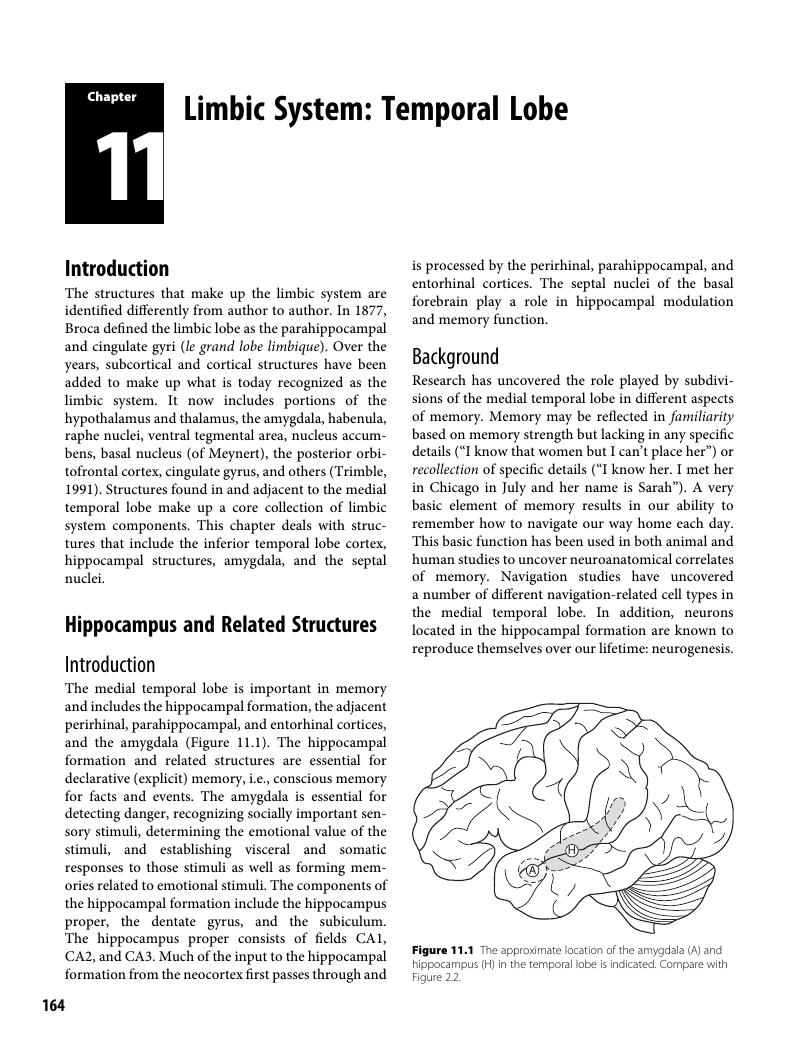
- Chapter 11 Limbic System: Temporal Lobe
- Chapter 12 Limbic System: Cingulate Cortex
- Chapter 13 Asymmetry and Interhemispheric Connections
- Index
- References
Chapter 11 - Limbic System: Temporal Lobe
Published online by Cambridge University Press: 22 February 2018

Book contents
- The Brain and Behavior
- The Brain and Behavior
- Copyright page
- Dedication
- Contents
- Preface to the Fourth Edition
- Abbreviations
- Chapter 1 Introduction
- Chapter 2 Anatomy of the Gross Brain
- Chapter 3 Histology
- Chapter 4 Occipital and Parietal Lobes
- Chapter 5 Temporal Lobe: Neocortical Structures
- Chapter 6 Frontal Lobe
- Chapter 7 Basal Ganglia
- Chapter 8 Diencephalon: Hypothalamus and Epithalamus
- Chapter 9 Diencephalon: Thalamus
- Chapter 10 Brainstem
- Chapter 11 Limbic System: Temporal Lobe
- Chapter 12 Limbic System: Cingulate Cortex
- Chapter 13 Asymmetry and Interhemispheric Connections
- Index
- References
Summary
A summary is not available for this content so a preview has been provided. Please use the Get access link above for information on how to access this content.
- Type
- Chapter
- Information
- The Brain and BehaviorAn Introduction to Behavioral Neuroanatomy, pp. 164 - 196Publisher: Cambridge University PressPrint publication year: 2018
References
Select Bibliograpy
Aggleton, J. P. (Ed.) (2000). The Amygdala: A Functional Analysis, 2nd edn. Oxford, UK: Oxford University Press.CrossRefGoogle Scholar
Andersen, P., Morris, R., Amaral, D., Bliss, T., and O’Keefe, J. (Eds.) (2007). The Hippocampus Book. New York, NY: Oxford University Press.Google Scholar
Bartsch, T. (Ed.) (2012). The Clinical Neurobiology of the Hippocampus. Oxford, UK, Oxford University Press.Google Scholar
Numan, R. (Ed.) (2000). The Behavioral Neuroscience of the Septal Region. New York, NY: Springer-Verlag.Google Scholar
Rolls, E. T. (2008). Memory, Attention, and Decision-making. A Unifying Computational Neuroscience Approach. New York, NY: Oxford University Press.Google Scholar
Shinnick-Gallagher, P., Pitkänen, A., Shekhar, A., and Cahill, L. (Eds.) (2003). The Amygdala in Brain Function: Basic and Clinical Approaches. New York, NY: New York Academy of Sciences, Vol. 985.Google Scholar
Whalen, P. J., and Phelps, E. A. (Eds.) (2009). The Human Amygdala. New York, NY: Guilford Press.Google Scholar
Yilmazer-Hanke, D. M. (2012). Amygdala. In Mai, J. K., and Paxinos, G. (Eds.), The Human Nervous System (3rd edn., pp. 759–835), New York, NY: Elsevier.Google Scholar
References
Adolphs, R. (2003). Cognitive neuroscience of human social behaviour. Nat. Rev. Neurosci., 4(3), 165–178. doi:10.1038/nrn1056Google Scholar
Adolphs, R., Tranel, D., Damasio, H., and Damasio, A. (1994). Impaired recognition of emotion in facial expressions following bilateral damage to the human amygdala. Nature, 372, 669–672. doi:10.1038/372669a0CrossRefGoogle Scholar
Adolphs, R., Tranel, D., Damasio, H., and Damasio, A. R. (1995). Fear and the human amygdala. J. Neurosci., 15(9), 5879–5891. Retrieved from: www.ncbi.nlm.nih.gov/pubmed/7666173Google Scholar
Aimone, J. B., Wiles, J., and Gage, F. H. (2009). Computational influence of adult neurogenesis on memory encoding. Neuron, 61, 187–202. doi:10.1016/j.neuron.2008.11.026Google Scholar
Almeida, J. R., Versace, A., Mechelli, A., Hassel, S., Quevedo, K., Kupfer, D. J., and Phillips, M. L. (2009). Abnormal amygdala-prefrontal effective connectivity to happy faces differentiates bipolar from major depression. Biol. Psychiatry, 66, 451–459. doi:10.1016/j.biopsych.2009.03.024CrossRefGoogle ScholarPubMed
Alvarez, R. P., Chen, G., Bodurka, J., Kaplan, R., and Grillon, C. (2011). Phasic and sustained fear in humans elicits distinct patterns of brain activity. Neuroimage, 55, 389–400. doi:10.1016/j.neuroimage.2010.11.057CrossRefGoogle ScholarPubMed
Amaral, D., and Lavenex, P. (2007). Hippocampal neuroanatomy In Andersen, P., Morris, R., Amaral, D., Bliss, T., and O’Keefe, J.. (Eds.) The Hippocampus Book. (pp. 37–114) New York, NY: Oxford University Press.Google Scholar
Andy, O. J., and Stephan, H. (1968). The septum in the human brain. J. Comp. Neurol. 133(3), 383–410. doi:10.1002/cne.901330308Google Scholar
Arnsten, A. F. (2009). Stress signalling pathways that impair prefrontal cortex structure and function. Nat. Rev. Neurosci., 10, 410–422. doi:10.1038/nrn2648CrossRefGoogle ScholarPubMed
Astur, R. S., St. Germain, S. A., Tolin, D., Ford, J., Russell, D., and Stevens, M. 2006. Hippocampus function predicts severity of post-traumatic stress disorder. Cyberpsychol. Behav., 9, 234–240. doi:10.1089/cpb.2006.9.234CrossRefGoogle ScholarPubMed
Ballmaier, M., Narr, K. L., Toga, A. W., Elderkin-Thompson, V., Thompson, P. M., Hamilton, L.,… Kuman, A. (2008). Hippocampal morphology and distinguishing late-onset from early-onset elderly depression. Am. J. Psychiatry, 165, 229–237. doi:10.1176/appi.ajp.2007.07030506CrossRefGoogle ScholarPubMed
Baldi, E., and Bucherelli, C. (2015). Brain sites involved in fear memory reconsolidation and extinction of rodents. Neurosci. Biobehav. Rev., 53, 160–190. doi:10.1016/j.neubiorev.2015.04.003CrossRefGoogle ScholarPubMed
Bar, M., and Aminoff, E. (2003). Cortical analysis of visual context. Neuron, 38, 347–358. doi:10.1016/S0896-6273(03)00167–3CrossRefGoogle ScholarPubMed
Baron-Cohen, S., Ring, H. A., Bullmore, E. T., Wheelwright, S., Ashwin, C., and Williams, S. C. R. (2000). The amygdala theory of autism. Neurosci. Biobehav. Rev., 24, 355–364. doi:10.1016/S0149-7634(00)00011–7Google Scholar
Barrash, J., Damasio, H., Adolphs, R., and Tranel, D. 2000. The neuroanatomical correlates of route learning impairment. Neuropsychologia, 38, 820–836. doi:10.1016/S0028-3932(99)00131–1CrossRefGoogle ScholarPubMed
Bartolomei, F., Barbeau, E., Gavaret, M., Guye, M., McGonigal, A., Régis, J., and Chauvel, P. (2004). Cortical stimulation study of the role of rhinal cortex in déjà vu and reminiscence of memories. Neurol., 636, 858–864. doi:10.1212/01CrossRefGoogle Scholar
Bartsch, T., Döhring, J., Rohr, A., Jansen, O., and Deuschl, G. (2011). CA1 neurons in the human hippocampus are critical for autobiographical memory, mental time travel, and autonoetic consciousness. Proc. Natl. Acad. Sci. U.S.A., 108, 17562–17567. doi:10.1073/pnas.1110266108CrossRefGoogle ScholarPubMed
Bauman, M. L., and Kemper, T. L. (1985). Histoanatomic observations of the brain in early infantile autism. Neurology, 35, 866–874. doi:10.1212/WNL.35.6.866Google Scholar
Bauman, M. L., LeMay, M., Bauman, R. A., and Rosenberger, P. (1985). Computerized tomographic (CT) observations of the posterior fossa in early infantile autism. Neurology, 35 (Suppl. 1), 247. Retrieved from http://insights.ovid.com/neurology/neur/1985/04/001/computerized-tomographic-ct-observations-posterior/572/00006114Google Scholar
Baur, V., Hänggi, J., and Jäncke, L. (2012). Volumetric associations between uncinate fasciculus, amygdala, and trait anxiety. BMC Neuroscience 13, 4. doi:10.1186/1471–2202-13–4.Google Scholar
Bear, D. (1986). Behavioural changes in temporal lobe epilepsy: Conflict, confusion, challenge. In: Trimble, M. R. and Bolwig, T. G. (Eds.), Aspects of Epilepsy and Psychiatry (pp. 19–30). Chichester, England: Wiley.Google Scholar
Bennett, M. R., Gibson, W. G., and Robinson, J. (1994). Dynamics of the CA3 pyramidal neuron autoassociative memory network in the hippocampus. Philos. Trans. R. Soc. Lond. B Biol. Sci., 343, 167–187. doi:10.1098/rstb.1994.0019Google ScholarPubMed
Biagini, G., D’Antuono, M., Venini, R., de Guzman, P., Longo, D., and Avoli, M. (2013). Perirhinal cortex and temporal lobe epilepsy. Front. Cell. NeuroSci., 7, doi:10.3389/fncel.2013.00130.Google Scholar
Biedermann, S. V., Fuss, J., Steinle, J., Auer, M. K., Dormann, C., Falfán-Melgoza, C.,… Weber-Fahr, W. (2014). The hippocampus and exercise: histological correlates of MR-detected volume changes. Brain Struct., 221(3), 1353–1363, doi:10.1007/s00429-014–0976-5.Google Scholar
Blumberg, H. P., Donegan, N. H., Sanislow, C. A., Collins, S., Lacadie, C., Skudlarski, P.,… Gore, J. C., and Krystal, J. H. (2005). Preliminary evidence for medication effects on functional abnormalities in the amygdala and anterior cingulate in bipolar disorder. Psychopharmacology (Berl), 183, 308–313. doi:10.1007/s00213-005–0156-7CrossRefGoogle ScholarPubMed
Blümcke, I., Suter, B., Behle, K., Kuhn, R., Schramm, I., Elger, C. E, and Wiestler, O. D. (2000). Loss of hilar mossy cells in Ammon’s horn sclerosis. Epilepsia, 41, S174–180. doi:10.1111/j.1528–1157.2000.tb01577.xGoogle Scholar
Bouton, M. E., and Bolles, R. C. (1979). Role of conditioned contextual stimuli in reinstatement of extinguished fear. J. Exp. Psychol. Anim. Behav. Process., 5, 368–378. doi:10.1037/0097–7403.5.4.368Google Scholar
Bremner, J. D. (2003). Functional neuroanatomical correlates of traumatic stress revisited 7 years later, this time with data. Psychopharmacol. Bull., 37, 6–25. doi:10.1016/S0079-6123(07)67012–5Google Scholar
Bremner, J. D., Randall, P., Vermetten, E., Staib, L., Bronen, R. B., Mazure, C.,… Charney, D. S. (1977). MRI-based measurement of hippocampal volume in posttraumatic stress disorder related to childhood physical and sexual abuse – a preliminary report. Biol. Psychiatry, 41, 23–32. doi:10.1176/appi.ajp.158.8.1248Google Scholar
Brown, J. E., Yates, B. J., and Taube, J. S. (2002). Does the vestibular system contribute to head direction cell activity in the rat? Physiol. Behav., 77, 743–748. doi:10.1016/S0031-9384(02)00928–9CrossRefGoogle ScholarPubMed
Buckner, R. L. (2000). Neural origins of “I remember.” Nat. Neurosci., 3, 1068–1069. doi:10.1038/80569Google Scholar
Burwell, R. D. (2002). The parahippocampal region: Corticocortical connectivity. Ann. N.Y. Acad. Sci., 911, 23–42. doi:10.1111/j.1749–6632.2000.tb06717.xGoogle Scholar
Bussey, T. J., Saksida, L. M., and Murray, E. A. (2005). The perceptual-mnemonic/feature conjunction model of perirhinal cortex function. Q.J. Exp. Psychol. B, 58(3–4), 269–282. doi:10.1080/02724990544000004CrossRefGoogle ScholarPubMed
Cameron, H. A., and McKay, R. D. (2001). Adult neurogenesis produces a large pool of new granule cells in the dentate gyrus. J. Comp. Neurol., 435, 406–417. doi:10.1002/cne.1040Google Scholar
Canteras, N. S., and Graeff, F. G. (2014). Executive and modulatory neural circuits of defensive reactions: Implications for panic disorder. Neurosci. Biobehav. Rev., 46, 352–364. doi:10.1016/j.neubiorev.2014.03.020Google Scholar
Cirillo, M. A., and Seidman, L. J. (2003). Verbal declarative memory dysfunction in schizophrenia: From clinical assessment to genetics and brain mechanisms. Neuropsychol. Rev., 13, 43. Retrieved from www.ncbi.nlm.nih.gov/pubmed/12887039Google Scholar
Curlik, D. M. 2nd, and Shors, T. J. (2013). Training your brain: Do mental and physical (MAP) training enhance cognition through the process of neurogenesis in the hippocampus? Neuropharm., 64, 506–514. doi:10.1016/j.neuropharm.2012.07.027Google Scholar
Dabrowska, J., Hazra, R., Guo, J-D., Dewitt, S., and Rainnie, D. G. (2013). Central CRF neurons are not created equal: Phenotypic differences in CRF-containing neurons of the rat paraventricular hypothalamus and the bed nucleus of the stria terminalis. Front. Neurosci., 7, 156–170. doi:10.3389/fnins.2013.00156Google Scholar
Davis, M., Walker, D. L., Miles, L., and Grillon, C. (2010). Phasic vs sustained fear in rats and humans: Role of the extended amygdala in fear vs anxiety. Neuropsychopharmacology, 35, 105–135. doi:10.1038/npp.2009.109Google Scholar
de Lange, F. P., Koers, A., Kalkman, J. S., Bleijenberg, G., Hagoort, P., van der Meer, J. W., and Toni, I. (2008). Increase in prefrontal cortical volume following cognitive behavioural therapy in patients with chronic fatigue syndrome. Brain 131, 2172–2180. doi:10.1093/brain/awn140Google Scholar
Debiec, J., and LeDoux, J. E. (2006). Noradrenergic signaling in the amygdala contributes to the reconsolidation of fear memory: Treatment implications for PTSD. Ann. N. Y. Acad. Sci., 1071, 521–524. doi:10.1196/annals.1364.056CrossRefGoogle Scholar
Delacourte, A, David, J. P., Sergeant, N., Buée, L., Wattez, A., Vermersch, P.,… Di Menza, C. (1999). The biochemical pathway of neurofibrillary degeneration in aging and Alzheimer’s disease. Neurology, 52, 672–673. doi:10.1212/WNL.54.2.538Google Scholar
Delvecchio, G., Fossati, P., Boyer, P., Brambilla, P., Falkai, P., Gruber, O.,… Frangou, S. (2012). Common and distinct neural correlates of emotional processing in bipolar disorder and major depressive disorder: A voxel-based meta-analysis of functional magnetic resonance imaging studies. Eur. Neuropsychopharmacol., 22, 100–113. doi:10.1016/j.euroneuro.2011.07.003CrossRefGoogle ScholarPubMed
Derdikman, D., and Moser, E. I. (2010). A manifold of spatial maps in the brain. Trends Cogn. Sci., 14, 561–569. doi:10.1016/j.tics.2010.09.004Google Scholar
deToledo-Morrell, L., Stoub, T. R., Bulgakova, M., Wilson, R. S., Bennett, D. A., Leurgans, S.,… Turner, D. A. (2004). MRI-derived entorhinal volume is a good predictor of conversion from MCI to AD. Neurobiol. Aging, 25, 1197–1203. doi:10.1016/j.neurobiolaging.2003.12.007CrossRefGoogle ScholarPubMed
Diana, R. A., Yonelinas, A. P., and Ranganath, C. (2007). Imaging recollection and familiarity in the medial temporal lobe: a three-component model. Trends Cog. Sci. 11(9):379–386. doi:10.1016/j.tics.2007.08.001Google Scholar
Diana, R. A., Yonelinas, A. P., and Ranganath, C. (2013). Parahippocampal cortex activation during context reinstatement predicts item recollection. J. Exp. Psychol. Gen., 142(4), 1287–1297. doi:10.1037/a0034029Google Scholar
Dichter, G. S. (2012). Functional magnetic resonance imaging of autism spectrum disorders. Dialogues Clin. Neurosci., 14, 319–351. Retrieved from www.ncbi.nlm.nih.gov/pmc/articles/PMC3513685Google Scholar
Didic, M., Barbeau, E. J., Felician, O., Tramoni, E., Guedj, E., Poncet, M., and Ceccaldi, M. (2011). Which memory system is impaired first in Alzheimer’s disease? J. Alzheimer’s Disease, 27, 11–22. doi:10.3233/JAD-2011–110557Google Scholar
Ding, S-L. (2013). Comparative anatomy of the prosubiculum, subiculum, presubiculum, postsubiculum, and parasubiculum in human, monkey, and rodent. J. Comp. Neurol., 52, 4145–4162. doi:10.1002/cne.23416CrossRefGoogle Scholar
Ding, S-L., and Van Hoesen, G. W. (2010). Borders, extent, and topography of human perirhinal cortex as revealed using multiple modern neuroanatomical and pathological markers. Hum. Brain Mapp., 31, 1359–1379. doi:10.1002/hbm.20940Google Scholar
Drevets, W. C. (2003). Neuroimaging abnormalities in the amygdala in mood disorders. Ann. N.Y. Acad. Sci., 985, 420–444. doi:10.1111/j.1749–6632.2003.tb07098.xViewCrossRefGoogle ScholarPubMed
Driessen, M., Herrmann, J., Stahl, K., Zwaan, M., Meier, S., Hill, A.,… Petersen, D. (2000). Magnetic resonance imaging volumes of the hippocampus and the amygdala in women with borderline personality disorder and early traumatization. Arch. Gen. Psychiatry, 57, 1115–1122. doi:10.1001/archpsyc.57.12.1115Google Scholar
Duncan, K., Ketz, N., Inati, S., and Davachi, L. (2012). Evidence for area CA1 as a match/mismatch detector: A high-resolution fMRI study of the human hippocampus. Hippocampus, 22, 389–398. doi:10.1002/hipo.20933Google Scholar
Dwyer, T. A., Servatius, R. J., and Pang, K. C. (2007). Noncholinergic lesions of the medial septum impair sequential learning of different spatial locations. J. Neurosci., 27, 299–303. doi:10.1523/JNEUROSCI.4189–06.2007Google Scholar
Eack, S. M., Hogarty, G. E., Cho, R. Y., Prasad, K. M., Greenwald, D. P., Hogarty, S. S., and Keshavan, M. S. (2010). Neuroprotective effects of cognitive enhancement therapy against gray matter loss in early schizophrenia: Results from a 2-year randomized controlled trial. Arch. Gen. Psychiatry, 67, 674–682. doi:10.1001/archgenpsychiatry.2010.63Google Scholar
Eden, A. S., Schreiber, J., Anwander, A., Keuper, K., Laeger, I., Zwanzger, P.,… Dobel, C. (2015). Emotion regulation and trait anxiety are predicted by the microstructure of fibers between amygdala and prefrontal cortex. J. Neurosci., 35, 6020–6027. doi:10.1523/JNEUROSCI.3659–14.2015Google Scholar
Ego-Stengel, V., and Wilson, M. A. (2010). Disruption of ripple-associated hippocampal activity during rest impairs spatial learning in the rat. Hippocampus, 20, 1–10. doi:10.1002/hipo.20707CrossRefGoogle ScholarPubMed
Eichenbaum, H.,Yonelinas, A. R., and Ranganath, C. (2007). The medial temporal lobe and recognition memory. Ann. Rev. Neurosci., 30, 123–152. doi:10.1146/annurev.neuro.30.051606.094328Google Scholar
Ekstrom, A. D., Kahana, M. J., Caplan, J. B., Fields, T. A., Isham, E. A., Newman, E. L., and Freid, I. (2003). Cellular networks underlying human spatial navigation. Nature, 425, 184–188. doi:10.1038/nature01964Google Scholar
Elliot, B., Joyce, E., and Shorvon, S. (2009). Delusions, illusions and hallucinations in epilepsy: 1. Elementary phenomena. Epilepsy Res., 85(2–3), 162–171. doi:10.1016/j.eplepsyres.2009.03.018Google Scholar
Elliot, F. A. (1992). Violence: The neurological contribution: An overview. Arch. Neurol. 49(6), 595–603. doi:10.1001/archneur.1992.00530300027006CrossRefGoogle Scholar
Epstein, R., and Kanwisher, N. (1998). A cortical representation of the local visual environment. Nature, 392, 598–601. doi:10.1038/33402Google Scholar
Erb, S., Salmaso, N., Rodaros, D., and Stewart, J. (2001). A role for the CRF-containing pathway from central nucleus of the amygdala to bed nucleus of the stria terminalis in the stress-induced reinstatement of cocaine seeking in rats. Psychopharmacol., 158, 360–365. doi:10.1007/s002130000642Google Scholar
Eren, I., Tukel, R., Polat, A., Karaman, R., and Unal, S. (2003). Evaluation of regional cerebral blood flow changes in panic disorder with Te99 m-HMPAOSPECT. Psychiatry Res., 123, 135–143. doi:10.1016/S0925-4927(03)00062–3Google Scholar
Fanselow, M. S., and Dong, H. W. (2010). Are the dorsal and ventral hippocampus functionally distinct structures? Neuron, 65, 7–19. doi:10.1016/j.neuron.2009.11.031Google Scholar
Farruggia, S., and Babcock, D. (1981). The cavum septi pellucidi: Its appearance and incidence with cranial ultrasonography in infancy. Radiology, 139, 147–150. doi:10.1148/radiology.139.1.7208915Google Scholar
Fernandez, M., Pissiota, A., Frans, O., von Knorring, L., Fischer, H., and Fredrikson, M. (2001). Brain function in a patient with torture related post-traumatic stress disorder before and after fluoxetine treatment: A positron emission tomography provocation study. Neurosci. Lett., 297, 101–104. doi:10.1016/S0304-3940(00)01674–8CrossRefGoogle Scholar
Fitzgerald, D. A., Angstadt, M., Jeisone, L. M., Nathan, P. J., and Phan, K. L. (2006). Beyond threat: Amygdala reactivity across multiple expressions of facial affect. Neuroimage, 30, 1441–1448. doi:10.1016/j.neuroimage.2005.11.003Google Scholar
Flood, D. G., Buell, S. J., Horwitz, G. J., and Coleman, P. D. (1987). Dendritic extent in human dentate gyrus granule cells in normal aging and senile dementia. Brain Res., 402, 205–216. doi:10.1016/0006–8993(87)90027–8Google Scholar
Fransén, E., Alonso, A. A., and Hasselmo, M. E. (2002). Simulations of the role of the muscarinic-activated calcium-sensitive nonspecific cation current INCM in entorhinal neuronal activity during delayed matching tasks. J. Neurosci., 22, 1081–1097. doi:10.1152/jn.00911.2007Google Scholar
Freese, J. L., and Amaral, D. G. (2009). Neuroanatomy of the Primate Amygdala. In Whalen, P. J., and Phelps, E. A. (Eds.) The Human Amygdala (pp. 1–42). New York, NY: Gilford.Google Scholar
Fu, C. H., Williams, S. C., Cleare, A. J., Brammer, M. J., Walsh, N. D., Kim, J.,… Bullmore, E. T. (2004). Attenuation of the neural response to sad faces in major depression by antidepressant treatment: A prospective, event-related functional magnetic resonance imaging study. Arch. Gen. Psychiatry, 61, 877–889. doi:10.1001/archpsyc.61.9.877Google Scholar
Fudge, J. L., and Haber, S. N. (2001). Bed nucleus of the stria terminalis and extended amygdala inputs to dopamine subpopulations in primates. Neurosci., 104, 807–827. doi:10.1002/cne.23340Google Scholar
Fudge, J. L., deCampo, D. M., and Becoats, K. T. (2012). Revisiting the hippocampal-amygdala pathway in primates: Association with immature-appearing neurons. Neurosci., 14, 212–219. doi:10.1016/j.neuroscience.2012.03.040Google Scholar
Fuentemilla, L., Barnes, G. R., Düzel, E., and Levine, B. (2014). Theta oscillations orchestrate medial temporal lobe and neocortex in remembering autobiographical memories. NeuroImage, 85, 730–737. doi:10.1016/j.neuroimage.2013.08.029Google Scholar
Gage, F. H. (2000). Mammalian neural stem cells. Science, 287, 1433–1438. doi:10.1016/j.stem.2015.09.Google Scholar
Ge, S., Pradhan, D. A., Ming, G. L., and Song, H. (2007). GABA sets the tempo for activity-dependent adult neurogenesis. Trends Neurosci., 30, 1–8. doi:10.1016/j.tins.2006.11.001CrossRefGoogle ScholarPubMed
Ge, S., Yang, C. H., Hsu, K. S., Ming, G. L., and Song, H. (2007). A critical period for enhanced synaptic plasticity in newly generated neurons of the adult brain. Neuron, 54, 559–566. doi:10.1016/j.neuron.2007.05.002Google Scholar
Gilboa, A., Winocur, G., Grady, C. L., Hevenor, S. J., and Moscovitch, M. (2004). Remembering our past: Functional neuroanatomy of recollection of recent and very remote personal events. Cereb. Cortex, 14, 1214–1225. doi:10.1093/cercor/bhh082Google Scholar
Gogolla, N., Caroni, P., Lüthi, A., and Herry, C. (2009). Perineuronal nets protect fear memories from erasure. Science, 325, 1258–1261. doi:10.1126/science.1174146CrossRefGoogle ScholarPubMed
Gómez-Isla, T., Price, J. L., McKeel, D. W. Jr., Morris, J. C., Growdon, J. H., and Hyman, B. T. (1996). Profound loss of layer II entorhinal cortex neurons occurs in very mild Alzheimer’s disease. J. Neurosci., 16, 4491–4500. Retrieved from www.ncbi.nlm.nih.gov/pubmed/8699259Google Scholar
Goossens, L., Kukolja, J., Onur, O. A., Fink, G. R., Maier, W., Griez, E.,… Hurlemann, R. (2009). Selective processing of social stimuli in the superficial amygdala. Human Brain Mapping, 30, 3332–3338. doi:10.1002/hbm.20755Google Scholar
Greer, S. M., Goldstein, A. N., and Walker, M. P. (2013). The impact of sleep deprivation on food desire in the human brain. Nat. Commun., 4, 2259. doi:10.1038/ncomms3259Google Scholar
Groenewegen, H. J., Vermeulen-Van der Zee, E., te Kortschot, A., and Witter, M. P. (1987). Organization of the projections from the subiculum to the ventral striatum in the rat. A study using anterograde transport of Phaseolus vulgaris leucoagglutinin. Neurosci., 23, 103–120. doi:10.1016/0306–4522(87)90275–2Google Scholar
Guedj, E., Aubert, S., McConigal, A., Mundler, O., and Bartolomei, F. (2010). Déjà-vu in temporal lobe epilepsy: Metabolic pattern of cortical involvement in patients with normal brain MRI. Neuropsychologia, 48, 2174–2181. doi:10.1016/j.neuropsychologia.2010.04.009Google Scholar
Guptill, J. T., Booker, A. B., Gibbs, T. T., Kemper, T. L., Bauman, M. L., and Blatt, G. J. (2007). [3H]-flunitrazepam-labeled benzodiazepine binding sites in the hippocampal formation in autism: A multiple concentration autoradiographic study. J. Autism Dev. Disord., 37, 911–920. doi:10.1007/s10803-006–0226-7Google Scholar
Gurvits, T. G., Shenton, M. E., Hokama, H., Ohta, H., Lasko, N. B., Gilbertson, M. W.,… Pitman, R. K. (1996). Magnetic resonance imaging study of hippocampal volume in chronic combat-related posttraumatic stress disorder. Biol. Psychiatry, 40, 192–199. doi:10.1016/S0006-3223(96)00229–6Google Scholar
Haghdoost-Yazdi, H., Pasbakhsh, P., Vatanparast, J., Rajaei, F., and Behzadi, G. (2009). Topographical and quantitative distribution of the projecting neurons to main divisions of the septal area. Neurol. Res., 31(5), 503–513. doi:10.1179/174313208X353712Google Scholar
Hales, J. B., Israel, S. L., Swann, N. C., and Brewer, J. B. (2009). Dissociation of frontal and medial temporal lobe activity in maintenance and binding of sequentially presented paired associates. J. Cogn. Neurosci., 21, 1244–1254. doi:10.1162/jocn.2009.21096Google Scholar
Hasler, G., Drevets., W. C., Manji, H. K., and Charney, D. S. (2004). Discovering endophenotypes for major depression. NeuropsychoPharmacol., 29, 1765–1781. doi:10.1038/mp.2011.23Google Scholar
Hasselmo, M. E., and Schnell, E. (1994). Laminar selectivity of the cholinergic suppression of synapse transmission in rat hippocampal region CA1: Computational modeling and brain slice physiology. J. Neurosci., 14, 3898–3914. Retrieved from http://people.bu.edu/hasselmo/HasselmoSchnell.pdfGoogle Scholar
Heath, R., Dempsy, C., Fontana, C., and Myers, W. (1978). Cerebellar stimulation: Effects on septal region, hippocampus, and amygdala of cats and rats. Biol. Psychiatry, 13(5), 501–529. Retrieved from www.ncbi.nlm.nih.gov/pubmed/728506Google Scholar
Henseler, I., Galkai, P., and Gruber, O. (2009). A systematic fMRI investigation of the brain systems subserving different working memory components in schizophrenia. Eur. J. Neurosci., 30, 693–702. doi:10.1111/j.1460–9568.2009.06850.xGoogle Scholar
Hitier, M., Besnard, S., and Smith, P. F. (2014). Vestibular pathways involved in cognition. Front. Integr. Neurosci. doi: 10.3389/fnint.2014.00059Google Scholar
Hoang, L. T., Lister, J. P., and Barnes, C. A. (2012). The ageing hippocampus. In Bartsch, T. (Ed.), The Clinical Neurobiology of the Hippocampus. (pp. 153–171). Oxford, UK, Oxford University Press.Google Scholar
Insausti, R., and Amaral, D. G. (2012). Hippocampal Formation. In Mai, J. K. and Paxinos, G. (Eds.) The Human Nervous System (pp. 896–942). Boston, MA: Elsevier Academic Press.Google Scholar
Jackson, G. D., Briellmann, R. S., and Kuzniecky, R. I. (2005). Temporal lobe epilepsy. In R. I. Kuzniecky and G. D. Jackson (Eds.), Magnetic resonance in epilepsy (2nd edn.) (pp. 99–1760). London: Elsevier.CrossRefGoogle Scholar
Jacobs, J. (2013). Hippocampal theta oscillations are slower in humans than in rodents: Implications for models of spatial navigation and memory. Phil. Trans. R. Soc. B, 369, 20130304. doi:10.1098/rstb.2013.0304Google Scholar
Jacobs, J., and Kahana, M. J. (2010). Direct brain recordings fuel advances in cognitive electrophysiology. Trends Cognitive Sci., 14, 162–171. doi:10.1016/j.tics.2010.01.005Google Scholar
Johnson, P. L., Federici, L. M., and Shekhar, A. (2014). Etiology, triggers and neurochemical circuits associated with unexpected, expected, and laboratory-induced panic attacks. Neurosci. Biobehav. Rev., 46, 420–454. doi:10.1016/j.neubiorev.2014.07.027Google Scholar
Jones, B. F., and Witter, M. P. (2007). Cingulate cortex projections to the parahippocampal region and hippocampal formation in the rat. Hippocampus, 17(10), 957–976. doi:10.1002/hipo.20330Google Scholar
Jones, J. E., Jackson, D. C., Chambers, K. L., Dabbs, K., Hsu, D. A., Stafstrom, C. E.,… Hermann, B. P. (2015). Children with epilepsy and anxiety: Subcortical and cortical differences. Epilepsia, 56, 283–290. doi:10.1111/epi.12832Google Scholar
Karl, A., Schaefer, M., Malta, L. S., Dörfel, D., Rohleder, N., and Werner, A. (2006). A meta-analysis of structural brain abnormalities in PTSD. Neurosci. Biobehav. Rev., 30, 1004–1031. doi:10.1016/j.neubiorev.2006.03.004Google Scholar
Kempermann, G. (2002). Why new neurons? Possible functions for adult hippocampal neurogenesis. J. Neurosci., 22, 635–638. Retrieved from www.ncbi.nlm.nih.gov/pubmed/11826092Google Scholar
Keshavarzi, S., Sullivan, R. K. P., Ianno, D. J., and Sah, P. (2014). Functional properties and projections of neurons in the medial amygdala. J. Neurosci., 34, 8699–8715. doi:10.1523/JNEUROSCI.1176–14.2014Google Scholar
Khan, Z. U., Martin-Montañez, E., and Baxter, M. G. (2011). Visual perception and memory systems: From cortex to medial temporal lobe. Cell. Mol. Life Sci., 68, 1737–1754. doi:10.1007/s00018-011–0641-6Google Scholar
Kile, S. J. Ellis, W. G., Olichney, J. M., Farias, S., and DeCarli, C. (2009). Alzheimer abnormalities of the amygdala with Klüver-Bucy syndrome symptoms: An amygdaloid variant of Alzheimer disease. Arch. Neurol., 66(1), 125–129. doi:10.1001/archneurol.2008.517Google Scholar
Kishi, T., Tsumori, T., Yokota, S. and Yasui, Y. (2006). Topographical projection from the hippocampal formation to the amygdala: A combined anterograde and retrograde tracing study in the rat. J. Comp. Neurol., 496(3), 349–368. doi:10.1002/cne.20919Google Scholar
Knierim, J. J., Neunuebel, J. P., and Deshmukh, S. S. (2014). Functional correlates of the lateral and medial entorhinal cortrex: Objects, path integration and local-global reference frames. Philos. Trans. R. Soc. Lond. B Biol. Sci., 369, 20130369. doi:10.1098/rstb.2013.0369.Google Scholar
Knoth, R., Singec, I., Ditter, M., Pantazis, G., Capetian, P., Meyer, R. P.,… Kempermann, G. (2010). Murine features of neurogenesis in the human hippocampus across the lifespan from 0 to 100 years. PLoS One. doi:10.1371/journal.pone.0008809.Google Scholar
Lahmann, C., Henningsen, P., Brandt, T., Strupp, M., Jahn, K., Dietench, M.,… Schmid, G. (2015). Psychiatric comorbidity and psychosocial impairment among patients with vertigo and dizziness. J. Neurol. Neurosurg. Psychiatry, 86(3), 302–308. doi:10.1136/jnnp-2014–307601Google Scholar
Lai, C. H., Hsu, Y. Y., and Wu, Y. T. (2010). First episode drug-naïve major depressive disorder with panic disorder: Gray matter deficits in limbic and default network structures. Eur. Neuropsychopharmacol., 20, 676–682. doi:10.1016/j.euroneuro.2010.06.002Google Scholar
Landin-Romero, R., Sarró, S., Fernández-Corcuera, P., Moro, N., Manuel Goikolea, J., Isabel Carrión, M.… Radua, J. (2015). Prevalence of cavum vergae in psychosis and mood spectrum disorders. J. Affect. Disord., 186, 53–57. doi:10.1016/j.jad.2015.07.020Google Scholar
Lanfumey, L, Mongeau, R, Cohen-Salmon, C., and Hamon, M. (2008). Corticosteroid-serotonin interactions in the neurobiological mechanisms of stress-related disorders. Neurosci. Biobehav. Rev., 32, 1174–1184. doi:10.1016/j.neubiorev.2008.04.006Google Scholar
Lavenex, P. (2012). Functional anatomy, development, and pathology of the hippocampus. In Bartsch, T. (Ed.), The Clinical Neurobiology of the Hippocampus: An Integrative View. (pp. 10–38). Oxford, UK: Oxford University Press.Google Scholar
Lavenex, P., Sugden, S. G., Davis, R. R., Gregg, J. P., and Lavenex, P. B. (2011). Developmental regulation of gene expression and astrocytic processes may explain selective hippocampal vulnerability. Hippocampus, 21, 142–149. doi:10.1002/hipo.20730Google Scholar
Leão, R., Targino, Z. H., Colom, L. V., and Fisahn, A. (2015). Interconnection and synchronization of neuronal populations in the mouse medial septum/diagonal band of Broca. J. Neurophysiol., 113, 971–980. doi:10.1152/jn.00367.2014Google Scholar
Lebow, M. A., and Chen, A. (2016). Overshadowed by the amygdala: The bed nucleus of the stria terminalis emerges as key to psychiatric disorders. Mol. Psychiatry, 21, 450–463. doi:10.1038/mp.2016.1Google Scholar
Lee, I., and Knierim, J. J. (2007). The relationship between the field-shifting phenomenon and representational coherence of place cells in CA1 and CA3 in a cue-altered environment. Learn. Mem., 14, 807–815. doi:10.1101/lm.706207Google Scholar
Lesse, H., and Harper, R. K. (1985). Frequency-related, bidirectional limbic response to cocaine: Comparisons with amphetamine and lidocaine. Brain Res., 335, 21–31. Retrieved from www.ncbi.nlm.nih.gov/pubmed/4005544Google Scholar
Leuner, B., Glasper, E. R., and Gould, E. (2010). Sexual experience promotes adult neurogenesis in the hippocampus despite an initial elevation in stress hormones. PLoS ONE 5(7), e11597 doi:10.1371/journal.pone.0011597Google Scholar
Liddell, B. J., Brown, K. J., Kemp, A. H., Barton, M. J., Das, P., Peduto, A.,… Williams, L. M. (2005). A direct brainstem-amygdala-cortical “alarm” system for subliminal signals of fear. Neuroimage, 24, 235–243. doi:10.1016/j.neuroimage.2004.08.016Google Scholar
Lisman, J. E. (2007). Role of the dual entorhinal inputs to hippocampus: A hypothesis based on cue/action (non-self/self) couplets. Prog. Brain Res., 163, 615–625. doi:10.1016/S0079-6123(07)63033–7. 615Google Scholar
Liu, Y. C., Cheng, J. K., and Lien, C. C. (2014). Rapid dynamic changes of dendritic inhibition in the dentate gyrus by presynaptic activity patterns. J. Neurosci., 34, 1344–1357. doi:10.1523/JNEUROSCI.2566–13.2014Google Scholar
Lott, I. T., and Dierssen, M. (2010). Cognitive deficits and associated neurological complications in individuals with Down’s syndrome. Lancet Neurol., 9, 623–633. doi:10.1016/S1474-4422(10)70112–5Google Scholar
Lowenstein, D. H., Thomas, M. J., Smith, D. H., and McIntosh, T. K. (1992). Selective vulnerability of dentate hilar neurons following traumatic brain injury: A potential mechanistic link between head trauma and disorders of the hippocampus. J. Neurosci., 12, 4846–4853. doi:10.1016/j.expneurol.2009.08.019Google Scholar
Lucassen, P. J., Fuchs, E., and Czéh, B. (2004). Antidepressant treatment with tianeptine reduces apoptosis in the hippocampal dentate gyrus and temporal cortex. Eur. J. Neurosci., 14, 161–166. doi:10.1016/j.biopsych.2003.12.014Google Scholar
Luo, A. H., Tahsili-Fahadan, P., Wise, R. A., Lupica, C. R., and Aston-Jones, G. (2011). Linking context with reward: A functional circuit from hippocampal CA3 to ventral tegmental area. Science, 333, 353–357. doi:10.1126/science.1204622Google Scholar
MacFall, J. R., Payne, M. E., Provenzale, J. E., and Krishnan, K. R. (2001). Medial orbital frontal lesions in late-onset depression. Biol. Psychiatry, 49, 803–806. doi:10.1016/S0006-3223(00)01113–6Google Scholar
MacMillan, S., Szeszko, P. R., Moore, G. J., Madden, R., Lorch, E., Ivey, J.,… Rosenberg, D. R. (2003). Increased amygdala: Hippocampal volume ratios associated with severity of anxiety in pediatric major depression. J. Child Adolesc. Psychopharmacol., 13, 65–73. doi:10.1089/104454603321666207Google Scholar
MacQueen, G. M., and Frodl, T. (2012). The hippocampus in major depression. In Bartsch, T. (Ed.), The Clinical Neurobiology of the Hippocampus. (pp. 273–287). Oxford, UK: Oxford University Press.Google Scholar
Maguire, E. A., Woollett, K., and Spiers, H. J. (2006). London taxi drivers and bus driver: A structural MRI and neuropsychological analysis. Hippocampus, 16, 1091–1101. doi:10.1002/hipo.20233Google Scholar
Makino, S., Gold, P. W., and Schulkin, J. (1994). Effects of corticosterone on CRH mRNA and content in the bed nucleus of the stria terminalis; comparison with the effects in the central nucleus of the amygdala and the paraventricular nucleus of the hypothalamus. Brain Res., 42, 25–28. Retrieved from www.ncbi.nlm.nih.gov/pubmed/7820612Google Scholar
Malberg, J. E., and Duman, R. S. (2003). Cell proliferation in adult hippocampus is decreased by inescapable stress: Reversal by fluoxetine treatment. Neuropsychopharmacol., 28, 1562–1571. doi:10.1038/sj.npp.1300234CrossRefGoogle ScholarPubMed
Marazziti, D., and Conti, L. (1991). Aggression, hyperactivity, and platelet IMI-binding. Acta Psychiatr. Scand., 84, 209–211. doi:10.1111/j.1600–0447.1991.tb03130Google Scholar
Markwald, R. R., Melanson, E. L., Smith, M. R., Higgins, J., Perreault, L., Eckel, R. H., and Wright, K. P. Jr. (2013). Impact of insufficient sleep on total daily energy expenditure, food intake, and weight gain. Proc. Natl. Acad. Sci. U. S. A., 110(14), 5695–5700. doi:10.1073/pnas.1216951110Google Scholar
Maunder, L., Schoemaker, D., and Pruessner, J. C. (2016). Frequency of penile-vaginal intercourse is associated with verbal recognition performance in adult women. Arch. Sex. Behav., Epub ahead of print. doi:10.1007/s10508-016–0890-4Google Scholar
McIntyre, C. K., Power, A. E., Roozendaal., B., and McGaugh, J. L. (2003). Role of the basolateral amygdala in memory consolidation. Ann. N.Y. Acad. Sci., 985, 273–293. doi:10.1016/S0301-0082(03)00104–7Google Scholar
McKinnon, M. C., Yucel, K., Nazarov, A., and MacQueen, G. M. (2009). A meta-analysis examining clinical predictors of hippocampal volume in patients with major depressive disorder. J. Psychiatry Neurosci., 34, 41–54. doi:10.1007/s00234-008–0383-9Google Scholar
McNaughton, N. M., Bernat, K., and Hajós, M. (2007). Elicited hippocampal theta rhythm: A screen for anxiolytic and procognitive drugs through changes in hippocampal function? Behav. Pharmacol., 18, 329–346. doi:10.1097/FBP.0b013e3282ee82e3Google Scholar
Mégevand, P., Groppe, D. M., Goldfinger, M. S., Hwang, S. T., Kingsley, P. B., Davidesco, I., and Mehta, A. D. (2014). Seeing scenes: Topographic visual hallucinations evoked by direct electrical stimulation of the parahippocampal place area. J. Neurosci., 34, 5399–5406. doi:10.1523/JNEUROSCI.5202–13.2014Google Scholar
Mendez, M. F., and Cummings, J. L. (2003). Dementia: A Clinical Approach (3rd edn.). Philadelphia, PA: Butterworth-Heinemann.Google Scholar
Mendez, M. F., and Foti, D. J. (1997). Lethal hyperoral behaviour from Klüver-Bucy syndrome. J. Neural. Neurosurg. Psychiatry, 63(3), 293–294. doi: 0.1136/jnnp.62.3.293-aGoogle Scholar
Mesulam, M-M., Mufson, E. J., Wainer, D. B. H., and Levey, A. I. (1983). Central cholinergic pathways in the rat: An overview based on an alternative nomenclature (Ch1-Ch6). Neurosci., 10 (4), 1185–1201. Retrieved from www.ncbi.nlm.nih.gov/pubmed/6320048Google Scholar
Milad, M. R., Orr, S. P., Lasko, N. B., Chang, Y., Rauch, S. L., and Pitman, R. K. (2007). Presence and acquired origin of reduced recall for fear extinction in PTSD: Results of a twin study. J. Psychiatry Res., 42, 515–520. doi:10.1016/j.jpsychires.2008.01.017Google Scholar
Milad, M. R., Rauch, S. L., Pitman, R. K., and Quirk, G. J. (2006). Fear extinction in rats: Implications for human brain imaging and anxiety disorders. Biol. Psychol., 73, 61–71. doi:10.1016/j.biopsycho.2006.01.008Google Scholar
Mitsueda-Ono, T., Ikeda, A., Inouchi, M., Takaya, S., Matsumoto, R., Hanakawa, T.,… Takahashi, R. (2011). Amygdalar enlargement in patients with temporal lobe epilepsy. J. Neurol. Neurosurg. Psychiatry, 82, 652–657. doi:10.1136/jnnp.2010.206342.Google Scholar
Moreau, D., Morrison, A. B., and Conway, A. R. (2015). An ecological approach to cognitive enhancement: Complex motor training. Acta Psychol. (Amst), 157C, 44–55. doi:10.1016/j.actpsy.2015.02.007Google Scholar
Motzkin, J. C., Phillippi, C. L., Wolf, R. C., Baskaya, M. K., and Koenigs, M. (2015). Ventromedial prefrontal cortex is critical for the regulation of amygdala activity in humans. Biol. Psychiatry, 77, 276–284. doi:10.1016/j.biopsych.2014.02.014Google Scholar
Moutsiana, C., Johnstone, T., Murray, L., Fearon, P., Cooper, P. J., Pliatsikas, C.,… Halligan, S. L. (2015). Insecure attachment during infancy predicts greater amygdala volumes in early adulthood. J. Child Psychol. Psychiatry, 56(5), 540–548. doi:10.1111/jcpp.12317Google Scholar
Murray, E. A., and Wise, S. P. (2012). Why is there a special issue on perirhinal cortex in a journal called Hippocampus? The perirhinal cortex in historical perspective. Hippocampus, 22, 1941–1951. doi:10.1002/hipo.22055Google Scholar
Murray, E. A., Bussey, T. J., and Saksida, L. M. (2007). Visual perception and memory: A new view of medial temporal lobe function in primates and rodents. Annu. Rev. Neurosci., 30, 99–122. doi:10.1146/annurev.neuro.29.051605.113046Google Scholar
Murray, E. A., Wise, S. P., and Drevets, W. C. (2011). Localization of dysfunction in major depressive disorder: Prefrontal cortex and amygdala. Biol. Psychiatry, 69, e43–e54.Google Scholar
Nadel, L. (2003). Down’s syndrome: a genetic disorder in biobehavioral perspective. Genes, Brain, Behav., 2, 156–166. doi:10.1016/j.biopsych.2010.09.041Google Scholar
Nakashiba, T., Cushman, J. D., Pelkey, K. A., Renaudineau, S., Derek, L. Buhl, D. L.,… Tonegawal, S. (2012). Young dentate granule cells mediate pattern separation, whereas old granule cells facilitate pattern completion. Cell, 149, 188–201. doi:10.1016/j.cell.2012.01.046Google Scholar
Neunuebel, J. P., and Knierim, J. J. (2014). CA3 retrieves coherent representations from degraded input: Direct evidence for CA3 pattern completion and dentate gyrus pattern separation. Neuron, 81, 416–427. doi:10.1016/j.neuron.2013.11.017Google Scholar
Nitschke, J. B., Sarinopoulos, I., Oathes, D. J., Johnstone, T., Whalen, P. J., Davidson, R. J., and Kalin, N. H. (2009). Anticipatory activation in the amygdala and anterior cingulate in generalized anxiety disorder and prediction of treatment response. Am. J. Psychiatry, 166, 302–310. doi:10.1176/appi.ajp.2008.07101682Google Scholar
Nopoulos, P., Swayze, V., Flaum, M., Ehrhardt, J. C., Yuh, W. T., and Andreasen, N. C. (1997). Cavum septi pellucidi in normal and patients with schizophrenia as detected by magnetic resonance imaging, Biol. Psychiatry, 41(11), 1102–1108. doi:10.1016/S0006-3223(96)00209–0Google Scholar
Norris, C. M., and Scheff, S. W. (2009). Recovery of afferent function and synaptic strength in hippocampal CA1 following traumatic brain injury. J. Neurotrauma, 26, 2269–2278. doi:10.1089/neu.2009.1029Google Scholar
O’Daly, O. G., Trick, L., Scaife, J., Marshall, J., Ball, D., Phillips, M. L.,… Duka, T. (2012). Withdrawal-associated increases and decreases in functional neural connectivity associated with altered emotional regulation in alcoholism. Neuropsychopharmacology, 37, 2267–2276. doi:10.1038/npp.2012.77Google Scholar
Oblak, A. L., Gibbs, T. T., and Blatt, G. J. (2010). Decreased GABA(B) receptors in the cingulate cortex and fusiform gyrus in autism. J. Neurochem., 114, 1414–1423. doi:10.1111/j.1471–4159.2010.06858.xGoogle Scholar
Pang, P. T., and Lu, B. (2004). Regulation of late-phase LTP, long-term memory in normal and aging hippocampus: Role of secreted proteins tPA and BDNF. Ageing Res. Rev., 3, 407–430. doi:10.1016/j.arr.2004.07.002Google Scholar
Paz, R., and Pare, D. (2013). Physiological basis for emotional modulation of memory circuits by the amygdala. Curr. Opin. Neurobiol., 23, 381–386. doi:10.1016/j.conb.2013.01.008Google Scholar
Phelps, E. A., and LeDoux, J. E. (2005). Contributions of the amygdala to emotion processing: From animal models to human behavior. Neuron, 48, 175–187. doi:10.1016/j.neuron.2005.09.025Google Scholar
Phillips, M. L., and Swartz, H. A. (2014). A critical appraisal of neuroimaging studies of bipolar disorder: Toward a new conceptualization of underlying neural circuitry and roadmap for future research. Am. J. Psychiatry, 171, 829–843. doi:10.1176/appi.ajp.2014.13081008Google Scholar
Phillips, M. L., Drevets, W. C., Rauch, S. L., and Lane, R. (2003). Neurobiology of emotion perception II: Implications for major psychiatric disorders. Biol. Psychiatry, 54, 515–528. doi:10.1016/S0006-3223(03)00171–9Google Scholar
Pinter, J. D., Brown, W. E., Eliez, S., Schmitt, J. E., Capone, G. T., and Reiss, A. L. (2001). Amygdala and hippocampal volumes in children with Down syndrome: A high-resolution MRI study. Neurology, 10, 972–974. doi:10.1212/WNL.56.7.972Google Scholar
Ploner, C. J., Gaymard, B. M., Rivaud-Péchoux, S., Baulac, M., Clémenceau, S., Samson, S., and Pierrot-Deseilligny, C. (2000). Lesions affecting the parahippocampal cortex yield spatial memory deficits in humans. Cereb. Cortex, 10, 1211–1216. doi:10.1093/cercor/10.12.1211Google Scholar
Pritchard, P. B. III, Holmstrom, V. L., and Roitzsch, J. C. (1985). Epileptic amnestic attacks: Benefits from antiepileptic drugs. Neurology, 35(8), 1188–1189. doi:10.1212/WNL.35.8Google Scholar
Quirk, G. J., and Gehlert, D. R. (2003). Inhibition of the amygdala: Key to pathological states? Ann. N.Y. Acad. Sci., 985, 263–272. doi:10.1111/j.1749–6632.2003.tb07087.xGoogle Scholar
Radwa, A. B., and Jackson, G. D., (2012). Temporal lobe epilepsy and the hippocampus. In Bartsch, T. (Ed.), The Clinical Neurobiology of the Hippocampus. (pp. 225–261). Oxford, UK: Oxford University Press.Google Scholar
Raghavachari, S., Kahana, M. J., Rizzuto, D. S., Caplan, J. B., Kirschen, M. P., Bourgeois, B.,… Lisman, J. E. (2001). Gating of human theta oscillations by a working memory task. J. Neurosci., 21, 3175–3183. Retrieved from www.ncbi.nlm.nih.gov/pubmed/11312302Google Scholar
Rao, U., Chen, L. A., Bidesi, A. S., Shad, M. U., Thomas, M. A., and Hammen, C. L. (2010). Hippocampal changes associated with early-life adversity and vulnerability to depression. Biol. Psychiatry, 67, 357–364. doi:10.1016/j.biopsych.2009.10.017Google Scholar
Rauch, S. L., Shin, L. M., and Phelps, E. A. (2006). Neurocircuitry models of posttraumatic stress disorder and extinction: Human neuroimaging research – past, present, and future. Biol. Psychiatry, 60, 376–382. doi:10.1016/j.biopsych.2006.06.004Google Scholar
Rauch, S. L., van der Kilk, B. A., Fisler, R. E., Alpert, N. M., Orr, S. P., Savage, C. R.,… Pitman, R. K. (1996). A symptom provocation study of posttraumatic stress disorder using positron emission tomography and script-driven imagery. Arch. Gen. Psychiatry, 53(5), 380–386. doi:10.1001/archpsyc.1996.01830050014003Google Scholar
Risch, N., Herrell, R., Lehner, T., Liang, K. Y., Eaves, L, Hoh, J.,… Merikangas, K. R. (2009). Interaction between the serotonin transporter gene (5-HTTLPR), stressful life events, and risk of depression: A meta-analysis. J.A.M.A. 301, 2462–2471. doi:10.1001/jama.2009.878.Google Scholar
Rolls, E. T. (2000). Memory systems in the brain. Ann. Rev. Psychol., 51, 599–630. doi:10.1146/annurev.psych.51.1.599 doi:10.1016/S0140-6736(03)12987-XGoogle Scholar
Rolls, E. T. (2010). A computational theory of episodic memory formation in the hippocampus. Behav. Brain Res., 205, 180–196. doi:10.1016/j.bbr.2010.03.027Google Scholar
Rolls, E. T. (2013). The mechanisms for pattern completion and pattern separation in the hippocampus. Front. Systems Neurosci., 7. doi:10.3389/fnsys.2013.00074Google Scholar
Rudebeck, P. H., and Murray, E. A. (2014). The orbitofrontal oracle: Cortical mechanisms for the prediction and evaluation of specific behavioral outcomes. Neuron, 84, 1143–1156. doi:10.1016/j.neuron.2014.10.049.Google Scholar
Sakai, Y., Kumano., H., Nishikawa, M., Sakano, Y., Kaiya, H., Imabayashi, E.,… Kuboki, T. (2005). Cerebral glucose metabolism associated with a fear network in panic disorder. Neuroreport, 16, 927–931. doi:10.1097/00001756–200506210-00010Google Scholar
Santarelli, L., Saxe, M., Gross, C., Surget, A., Battaglia, F., Dulawa, S., Weisstaub, N. Lee, J., Duman, R., Arancio, O., Belzung, C., and Hen, R. (2003). Requirement of hippocampal neurogenesis for the behavioral effects of antidepressants. Science, 301, 805–809. doi:10.1126/science.1083328Google Scholar
Santoro, A. (2013). Reassessing pattern separation in the dentate gyrus. Front. Behav. Neurosci,. 7, 96. doi:10.3389/fnbeh.2013.00096Google Scholar
Sasaki, T., Leutgeb, S., and Leutgeb, J. K. (2015). Spatial and memory circuits in the medial entorhinal cortex. Curr. Opinion Neurobiol., 32, 16–23. doi:10.1016/j.conb.2015.03.002Google Scholar
Scharfman, H. E., and Myers, C. E. (2012). Hilar mossy cells of the dentate gyrus: A historical perspective. Front.Neural Circuits. doi:10.3389/fncir.2012.00106Google Scholar
Scheff, S. W., Price, D. A., Schmitt, F. A., DeKosky, S. T., and Mufson, E. J. (2007). Synaptic alterations in CA1 in mild Alzheimer disease and mild cognitive impairment. Neurology 68, 1501–1508. doi:10.1212/01.wnl.0000260698.46517.8fGoogle Scholar
Schienle, A., Ebner, F., and Schäfer, A. (2011). Localized gray matter volume abnormalities in generalized anxiety disorder. Eur. Arch. Psychiatry Clin. Neurosci., 26, 303–307. doi:10.1007/s00406-010–0147-5Google Scholar
Schmidt, B., Marrone, D. F., and Markus, E. J. (2012). Disambiguating the similar: The dentate gyrus and pattern separation. Behav. Brain Res., 226, 56–65. doi:10.1016/j.bbr.2011.08.039Google Scholar
Schuff, N., Marmar, C. R., Weiss, D. S., Neylan, T. C., Schoenfeld, F., Fein, G., and Weiner, M. W. (1997). Reduced hippocampal volume and n-acetyl aspartate in posttraumatic stress disorder. Ann. N.Y. Acad. Sci., 821, 516–520. doi:10.1111/j.1749–6632.1997.tb48319.xGoogle Scholar
Seidenberg, M., Kelly, K. G., Parrish, J., Geary, E., Dow, C., Rutecki, P., and Hermann, B. (2005). Ipsilateral and contralateral MRI volumetric abnormalities in chronic unilateral temporal lobe epilepsy and their clinical correlates. Epilepsia, 46, 420–430. doi:10.1111/j.0013–9580.2005.27004.xGoogle Scholar
Seminowicz, D. A., Shpaner, M., Keaser, M. L., Krauthamer, G. M., Mantegna, J., Dumas, J. A.,… Naylor, M. R. (2013). Cognitive-behavioral therapy increases prefrontal cortex gray matter in patients with chronic pain. J. Pain, 14, 1573–1584. doi:10.1016/j.jpain.2013.07.020Google Scholar
Sewards, T. V. (2011). Neural structures and mechanisms involved in scene recognition: A review and interpretation. Neuropsychologia, 49, 277–298. doi:10.1016/j.neuropsychologia.2010.11.018Google Scholar
Shaabani, M., Lotfi, Y., Karimian, S. M., Rahgozar, M., and Hooshmandi, M (2016). Short-term galvanic vestibular stimulation promotes functional recovery and neurogenesis in unilaterally labyrinthectomized rats. Brain Res., 1648(Pt A), 152–162. doi: 10.1016/j.brainres.2016.07.029Google Scholar
Sheehan, T. P., Chambers, R. A., and Russell, D. S. (2004). Regulation of affect by the lateral septum: Implications for neuropsychiatry. Brain Resh. Rev., 46, 71–117. doi:10.1016/j.brainresrev.2004.04.009Google Scholar
Sheline, Y. I., Barch, D. M., Donnelly, J. M., Ollinger, J. M., Snyder, A. Z., and Mintun, M. A. (2001). Increased amygdala response to masked emotional faces in depressed subjects resolves with antidepressant treatment: An fMRI study. Biol. Psychiatry, 50, 651–658. doi:10.1016/S0006-3223(01)01263-XGoogle Scholar
Shi, F, Liu, B., Zhou, Y., Yu, C., and Jiang, T. (2009). Hippocampal volume and asymmetry in mild cognitive impairment and Alzheimer’s disease: Meta-analyses of MRI studies. Hippocampus, 19, 1055–1064. doi:10.1002/hipo.20573Google Scholar
Shiflett, M. W., and Balleine, B. W. (2010). At the limbic-motor interface: Disconnection of basolateral amygdala from nucleus accumbens core and shell reveals dissociable components of incentive motivation. Eur. J. Neurosci., 32, 1735–1743. doi:10.1111/j.1460–9568.2010.07439.xGoogle Scholar
Shin, L. M., and Liberzon, I. (2010). The neurocircuitry of fear, stress, and anxiety disorders. Neuropsychopharmacol., 35, 169–191. doi:10.1038/npp.2009.83Google Scholar
Shin, L. M., Orr, S. P., Carson, M. A., Rauch, S. L., Macklin, M. L., Lasko, N. B.,… Pitman, R. K. (2004). Regional cerebral blood flow in the amygdala and medial prefrontal cortex during traumatic imagery in male and female Vietnam veterans with PTSD. Arch. Gen. Psychiatry, 61, 168–176. doi:10.1001/archpsyc.61.2.168Google Scholar
Shin, L. M., Whalen, P. J., Pitman, R. K., Bush, G., Macklin, M. L., Lasko, N. B.,… Rauch, S. L. (2001). An fMRI study of anterior cingulate function in posttraumatic stress disorder. Biol. Psychiatry, 50, 932–942. doi:10.1016/S0006-3223(01)01215-XGoogle Scholar
Shors, T. J., Anderson, M. L., Curlik, D. M. 2nd, and Nokia, M. S. (2012). Use it or lose it: How neurogenesis keeps the brain fit for learning. Behav. Brain Res., 227, 450–458. doi:10.1016/j.bbr.2011.04.023Google Scholar
Siegle, G. J., Steinhauer, S. R., Thase, M. E., Stenger, V. A., and Carter, C. S. (2002). Can’t shake that feeling: fMRI assessment of sustained amygdala activity in response to emotional information in depressed individuals. Biol. Psychiatry, 51, 693–707. doi:10.1016/S0006-3223(02)01314–8Google Scholar
Sim, K., DeWitt, I., Ditman, T., Zalesak, M., Greenhouse, I., Goff, D.,… Heckers, S. (2006). Hippocampal and parahippocampal volumes in schizophrenia: A structural MRI study. Schizophrenia Bull., 32, 332–340. doi:10.1093/schbul/sbj030Google Scholar
Simpson, J. R. Jr., Drevets, W. C., Snyder, A. Z., Gusnard, D. A., and Raichle, M. E. (2001). Emotion-induced changes in human medial prefrontal cortex: II. During anticipatory anxiety. Proc. Natl. Acad. Sci. U. S. A., 98, 688–693. doi:10.1073/pnas.98.2.683Google Scholar
Sinha, S., McGovern, R. A., and Sheth, S. A. (2015). Deep brain stimulation for severe autism: From pathophysiology to procedure. Neurosurg. Focus, 38(6), E3. doi:10.3171/2015.3.FOCUS1548Google Scholar
Sismanlar, S. G., Anik, Y., Coskun, A., Agaoglu, B., Karakaya, I., and Yavuz, C. I. (2009). The volumetric differences of the frontotemporal region in young offspring of schizophrenic patients. Eur. Child Adolesc. Psychiatry, 5, 151–157. doi:10.1007/s00787-009–0052-5Google Scholar
Small, S. A., Chawla, M. K., Buonocore, M., Rapp, P. R., and Barnes, C. A. (2004). Imaging correlates of brain function in monkeys and rats isolates a hippocampal subregion differentially vulnerable to aging. Proc. Natl. Acad. Sci. U. S. A., 101, 7181–7186. doi:10.1073/pnas.0400285101Google Scholar
Small, S. A., Wu., E. X., Bartsch, D., Perera, B. M., Lacefield, C. O., DeLaPaz, R.,… Kandel, E. R. (2000). Imaging physiologic dysfunction of individual hippocampal subregions in humans and genetically modified mice. Neuron, 28, 653–664. doi:10.1016/S0896-6273(00)00144–6Google Scholar
Śmigielska-Kuzia, J., Boćkowski, L., Sobaniec, W., Sendrowski, K., Olchowik, B.,… łebkowska, U. (2011). A volumetric magnetic resonance imaging study of brain structures in children with Down syndrome. Neurol. Neurochirurgia Polska, 4, 363–369. doi:10.1016/S0028-3843(14)60107–9Google Scholar
Smith, P. F. (2017). The vestibular system and cognition. Curr. Opin. Neurol., 30(1), 84–89. doi:10.1097/WCO.0000000000000403Google Scholar
Solstad, T., Boccara, C. N., Kropff, E., Moser, M. B., and Moser, E. I. (2008). Representation of geometric borders in the entorhinal cortex. Science, 322, 1865–1868. doi:10.1126/science.1166466Google Scholar
Sotres-Bayon, F., Bush, D. E. A., and LeDoux, J. E. (2004). Emotional perseveration: An update on prefrontal-amygdala interactions in fear extinction. Learn. Mem., 11, 525–535. doi:10.1101/lm.79504Google Scholar
Spaulding, K. L., Bergmann, O., Alkass, K., Bernard, S., Salehpour, M., Huttner, H. B.,… Frisén, J. (2013). Dynamics of hippocampal neurogenesis in adult humans. Cell, 153, 1219–1227. doi:10.1016/j.cell.2013.05.002Google Scholar
Steen, R. G., Mull, C., McClure, R., Hamer, R. M., and Lieberman, J. A. (2006). Brain volume in first-episode schizophrenia: Systematic review and meta-analysis of magnetic resonance imaging studies. Brit. J. Psychiatry, 188, 510–518. doi:10.1192/bjp.188.6.510Google Scholar
Stevens, W. D., Kahn, I., Wig, G. S., and Schacter, D. L. (2012). Hemispheric asymmetry of visual scene processing in the human brain: Evidence from repetition priming and intrinsic activity. Cereb. Cortex, 22, 1935–1949. doi:10.1093/cercor/bhr273CrossRefGoogle ScholarPubMed
Stuber, G. D., Britt, J. P., and Bonci, A. (2012). Optogenetic modulation of neural circuits that underlie reward seeking. Biol. Psychiatry, 71, 1061–1067. doi:10.1093/cercor/bhr273Google Scholar
Stuhrmann, A., Dohm, K., Kugel, H., Zwanzger, P., Redlich, R., Grotegerd, D.,… Dannlowski, U. (2013). Mood-congruent amygdala responses to subliminally presented facial expressions in major depression: Associations with anhedonia. J. Psychiatry Neurosci., 38, 249–258. doi:10.1503/jpn.120060Google Scholar
Suzuki, W. A., and Naya, Y. (2014). The perirhinal cortex. Ann. Rev. Neurosci., 37, 39–53. doi:10.1146/annurev-neuro-071013–014207Google Scholar
Sweatt, J. (1999). Toward a molecular explanation for long-term potentiation. Learn. Mem., 6, 399–416. doi:10.1101/lm.6.5.399Google Scholar
Takei, K., Yamasue, H., Abe, O., Yamada, H., Inoue, H., Suga, M.,… Kasi, K. (2008). Disrupted integrity of the fornix is associated with impaired memory organization in schizophrenia. Schizophrenia Resh., 103, 52–61. doi:10.1016/j.schres.2008.03.008Google Scholar
Tamminga, C. A., Stan, A. D., and Wagner, A. D. (2010). The hippocampal formation in schizophrenia. Am. J. Psychiatry, 167, 1178–1193. doi:10.1016/j.nicl.2014.08.015Google Scholar
Tate, D. F., and Bigler, E. D. (2000). Fornix and hippocampal atrophy in traumatic brain injury. Learn Mem., 7, 442–446. doi:10.1101/lm.33000Google Scholar
Taylor, K. I., and Probst, A. (2008). Anatomic localization of the tranentorhinal region of the perirhinal cortex. Neurobiol. Aging, 29, 1591–1596. doi:10.1016/j.neurobiolaging.2007.03.024Google Scholar
Thom, M., Eriksson, S., Martinian, L., Caboclo, L. O., McEvoy, A. W., Duncas, J. S., and Sisodiya, S. M. (2009). Temporal lobe sclerosis associated with hippocampal sclerosis in temporal lobe epilepsy: Neuropathological features. J. Neuropathol. Exp. Neurol., 68, 928–938. doi:10.1097/NEN.0b013e3181b05d67Google Scholar
Thomaes, K., Dorrepaal, E., Draijer, N., de Ruiter, M. B., Elzinga, B. M., Sjoerds, Z.,… Veltman, D. J. (2011). Increased anterior cingulate cortex and hippocampus activation in Complex PTSD during encoding of negative words. Soc. Cogn. Affect. Neurosci., 8, 190–200. doi:10.1093/scan/nsr084Google Scholar
Thompson, C. L., Pathak, S. D., Jeromin, A., Ng, L. L., MacPherson, C. R., Mortrud, M. T.,… Lein, E. S. (2008). Genomic anatomy of the hippocampus. Neuron, 60(6), 1010–1021. doi:10.1016/j.neuron.2008.12.008Google Scholar
Tovote, P., Fadok, J. P., and Lüthi, A. (2015). Neuronal circuits for fear and anxiety. Nat. Rev. Neurosci., 16, 317–331. doi:10.1038/nrn4028Google Scholar
Treves, A., and Rolls., E. T. (1992). Computational constraints suggest the need for two distinct input systems to the hippocampal CA3 network. Hippocampus, 2, 189–199. Retrieved from www.ncbi.nlm.nih.gov/pubmed/1308182Google Scholar
Trimble, M. R., Mendez, M. F., and Cummings, J. L. (1997). Neuropsychiatric symptoms from the temporolimbic lobes. J. Neuropsychiatry Clin. Neurosci., 9(3), 429–438. doi:10.1176/jnp.9.3.429Google Scholar
van der Laan, L. N., de Ridder, D. T. D., Viergever, M. A., and Smeets, P. A. M. (2011). The first taste is always with the eyes: A meta-analysis on the neural correlates of processing visual food cues. Neuroimage, 55(1), 296–303. doi:10.1016/j.neuroimage.2010.11.055Google Scholar
van Praag, H. (2008). Neurogenesis and exercise: past and future directions. Neurol. Med., 10(2), 128–130. doi:10.1007/sl2017-008–8028-zGoogle Scholar
Vermetten, E., and Bremner., J. D. (2002). Circuits and systems in stress. II. Applications to neurobiology and treatment of PTSD. Depress. Anxiety, 16, 14–38. doi:10.1002/da.10017Google Scholar
Vertes, R. P., Hoover, W. B., and di Prisco, G. V. (2004). Theta rhythm of the hippocampus: Subcortical control and functional significance. Behav. Cogn. Neurosci. Rev. 3, 173–200. doi:10.1177/1534582304273594Google Scholar
Viard, A., Piolino, P., Desgranges, B., Chetelat, G., Lebreton, K., Landeau, B.,… Eustache, F. (2007). Hippocampal activation for autobiographical memories over the entire lifetime in healthy aged subjects: An fMRI study. Cereb. Cortex, 17, 2453–2467. doi:10.1093/cercor/bhl153Google Scholar
Villeda, S. A., Luo, J., Mosher, K. I., Zou, B., Britschgi, M., Bieri, G.,… Wyss-Coray, T. (2011). The ageing systemic milieu negatively regulates neurogenesis and cognitive function. Nature, 477, 90–94. doi:10.1038/nature10357Google Scholar
Von Der Heide, R. J., Skipper, L. M., and Olson, I. R. (2013). Anterior temporal face patches: A meta-analysis and empirical study. Front. Human Neurosci., 7, 17. doi:10.3389/FNHUM.2013.00017.Google Scholar
Wang, Z, Neylan, T. C., Mueller, S. G., Lenoci, M., Truran, D., Marmar, C. R.,… Schuff, N. (2010). Magnetic resonance imaging of hippocampal subfields in posttraumatic stress disorder. Arch. Gen. Psychiatry, 67, 266–303. doi:10.1001/archgenpsychiatry.2009.205Google Scholar
Weniger, G., Siemerkus, J., Schmidt-Samoa, C., Mehitz, M., Baudewig, J., Dechent, P., and Irle, E. (2010). The human parahippocampal cortex subserves egocentric spatial learning during navigation in a virtual maze. Neurobiol. Learn. Mem., 93, 46–55. doi:10.1016/j.nlm.2009.08.003Google Scholar
West, M. J., Kawas, C. H., Stewart, W. F., Rudow, G. L., and Troncoso, J. C. (2004). Hippocampal neurons in pre-clinical Alzheimer’s disease. Neurobiol. Aging, 25, 1205–1212. doi:10.1186/1741–7015-10–127Google Scholar
Wieser, H. G. (2000). Mesial temporal lobe epilepsy versus amygdalar epilepsy: Late seizure recurrence after initially successful amygdalotomy and regained seizure control following hippocampectomy. Epileptic Disord., 2, 141–152. Retrieved from http://europepmc.org/abstract/med/11022139Google Scholar
Witter, M. P., Wouterlood, F. G., Naber, P. A., and van Haeften, T. (2000). Anatomical organization of the parahippocampal-hippocampal network. Ann. N. Y. Acad. Sci., 911, 1–24. doi:10.1111/j.1749–6632.2000.tb06716.xGoogle Scholar
Wolf, S. S., Hyde, T. M., and Weinberger, D. R. (1994). Malformations of the septum pellucidum: Two distinctive cases in association with schizophrenia. J. Psychiatry Neurosci., 19(2), 140–144. Retrieved from www.ncbi.nlm.nih.gov/pmc/articles/PMC1188578/pdf/jpn00054-0062.pdfGoogle ScholarPubMed
Woollett, K., and Maguire, E A. (2009). Navigational expertise may compromise anterograde associative memory. Neuropsychologia, 47, 1088–1095. doi:10.1016/j.neuropsychologia.2008.12.036Google Scholar
Woollett, K., and Maguire, E. A. (2011). Acquiring “The Knowledge” of London’s layout drives structural brain changes. Curr. Biol., 21, 2109–2114. doi:10.1016/j.cub.2011.11.018Google Scholar
Wrase, J., Makris, N., Braus, D. F., Mann, K., Smolka, M. N., Kennedy, D. N.,… Heinz, A. (2008). Amygdala volume associated with alcohol abuse relapse and craving. Am. J. Psychiatry, 165, 1179–1184. doi:10.1176/appi.ajp.2008.07121877Google Scholar
Xue, Y. X., Xue, L. F., Liu, J. F., He, J., Deng, J. H., Sun, S. C.,… Lu., L. (2014). Depletion of perineuronal nets in the amygdala to enhance the erasure of drug memories. J. Neurosci., 34, 6647–6658. doi:10.1523/JNEUROSCI.5390–13.2Google Scholar
Yadin, E., Thomas, E., Grishkat, H. L., and Strickland, C. E. (1993). The role of the lateral septum in anxiolysis. Physiol. Behav., 53, 1077–1083. doi:10.1016/0031–9384(93)90362-JGoogle Scholar
Yassa, M. A., and Stark, C. E. (2011). Pattern separation in the hippocampus. Trends Neurosci., 34, 515–525. doi:10.1016/j.tins.2011.06.006Google Scholar
Yeung, M., Treit, D., and Dickson, C. T. (2012). A critical test of the hippocampal theta model of anxiolytic drug action. Neuropharmacol., 62, 155–160. doi:10.1016/j.neuropharm.2011.06.011Google Scholar
Yilmazer-Hanke, D. M., O’Loughlin, E., and McDermott, K. (2016). Contribution of amygdala pathology to comorbid emotional disturbances in temporal lobe epilepsy. J. Neurosci. Res., 94, 486–503/ doi:10.1002/jnr.23689Google Scholar
Yilmazer-Hanke, D. M., Wolf, H. K., Schramm, J., Elger, C. E., Wiestler, O. D., and Blümcke, I. (2000). Subregional pathology of the amygdala complex and entorhinal region in surgical specimens from patients with pharmacoresistant temporal lobe epilepsy. J. Neuropathol. Exp. Neurol., 59, 907–920. doi:10.1093/jnen/59.10.907Google Scholar
Yoo, S-S., Gujar, N., Hu, P., Jolesz, F. A., and Walker, M. P. (2007). The human emotional brain without sleep – A prefrontal amygdala disconnect. Curr. Biol., 17(20), R877–R878. doi:10.1016/j.cub.2007.08.007Google Scholar
Yu, T. S., Zhang, G., Liebl, D. J., and Kernie, S. G. (2008). Traumatic brain injury-induced hippocampal neurogenesis requires activation of early nestin-expressing progenitors. J. Neurosci., 28, 12901–12912. doi:10.1523/JNEUROSCI.4629–08.2008Google Scholar
Zaborszky, L., Hoemke, L., Mohlberg, H., Schleicher, A., Amunts, K., and Zilles, K. (2008). Stereotaxic probabilistic maps of the of the magnocellular cell groups in human basal forebrain. NeuroImage, 42, 1127–1141. doi:10.1016/j.neuroimage.2008.05.055Google Scholar
Zhang, L., Kerich, M., Schwandt, M. L., Rawlings, R. R., McKellar, J. D.,… George, D. T. (2013). Smaller right amygdala in Caucasian alcohol-dependent male patients with a history of intimate partner violence: A volumetric imaging study. Addict. Biol., 18, 537–547. doi:10.1111/j.1369–1600.2011.00381.xGoogle Scholar