Skip to main content Accessibility help
×
Hostname: page-component-8448b6f56d-cfpbc Total loading time: 0 Render date: 2024-04-23T11:22:45.264Z Has data issue: false hasContentIssue false

Chapter 3 - Neuroimaging in dementia

Published online by Cambridge University Press:  01 December 2016

Bruce L. Miller
Affiliation:
University of California, San Francisco
Bradley F. Boeve
Affiliation:
Mayo Clinic, Minnesota
Get access

Summary

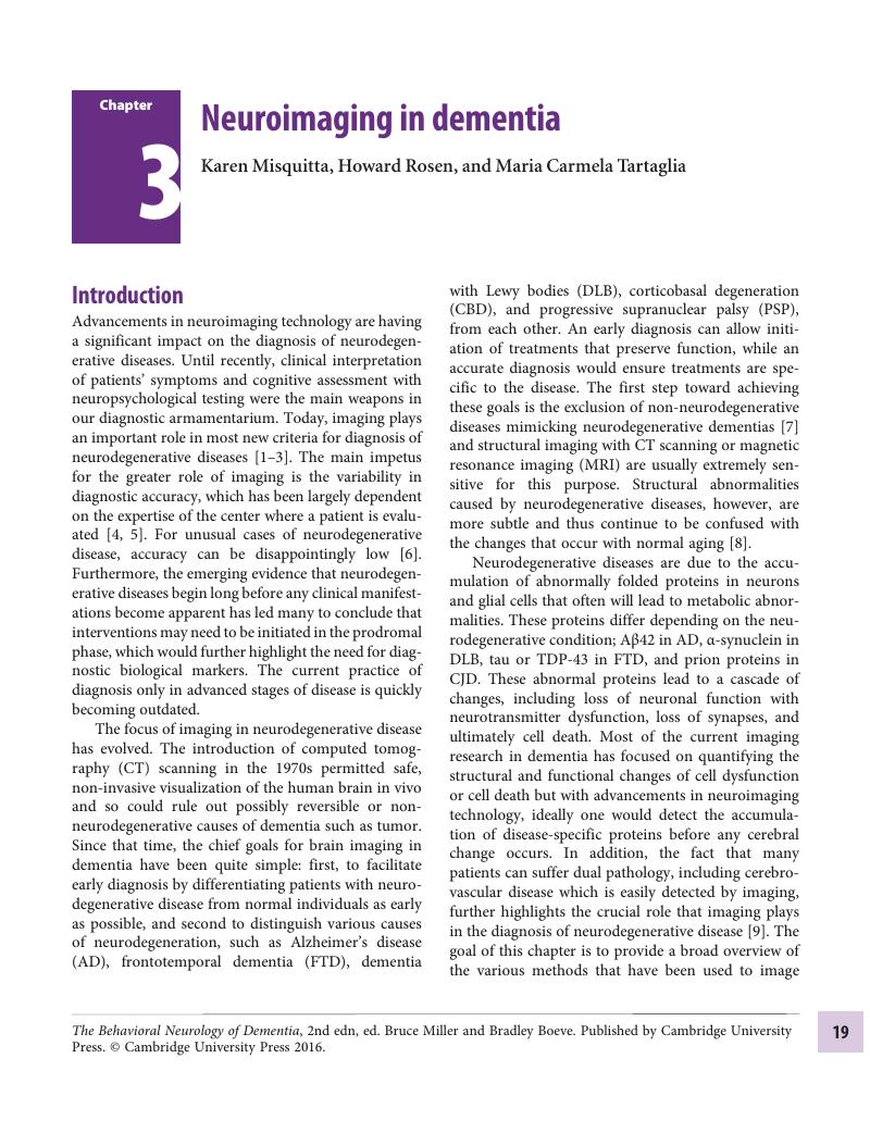
Image of the first page of this content. For PDF version, please use the ‘Save PDF’ preceeding this image.'
Type
Chapter
Information
Publisher: Cambridge University Press
Print publication year: 2016

Access options

Get access to the full version of this content by using one of the access options below. (Log in options will check for institutional or personal access. Content may require purchase if you do not have access.)

References

McKhann, G.M., Changing concepts of Alzheimer disease. JAMA, 2011. 305(23): 2458–9.CrossRefGoogle ScholarPubMed
Rascovsky, K., et al., Sensitivity of revised diagnostic criteria for the behavioural variant of frontotemporal dementia. Brain, 2011. 134(Pt 9): 2456–77.CrossRefGoogle ScholarPubMed
Gorno-Tempini, M.L., et al., Classification of primary progressive aphasia and its variants. Neurology, 2011. 76(11): 1006–14.CrossRefGoogle ScholarPubMed
Knopman, DS and Roberts, R., Estimating the number of persons with frontotemporal lobar degeneration in the US population. J Mol Neurosci, 2011. 45(3): 330–5.CrossRefGoogle ScholarPubMed
Litvan, I., et al., What are the obstacles for an accurate clinical diagnosis of Pick’s disease? A clinicopathologic study. Neurology, 1997. 49(1): 62–9.CrossRefGoogle ScholarPubMed
Mendez, M.F., et al., Frontotemporal dementia versus Alzheimer’s disease: differential cognitive features. Neurology, 1996. 47(5): 1189–94.CrossRefGoogle ScholarPubMed
Clarfield, A.M., The decreasing prevalence of reversible dementias: an updated meta-analysis. Arch Intern Med, 2003. 163(18): 2219–29.CrossRefGoogle ScholarPubMed
Suarez, J., et al., Characterizing radiology reports in patients with frontotemporal dementia. Neurology, 2009. 73(13): 1073–4.CrossRefGoogle ScholarPubMed
Attems, J. and Jellinger, K.A., The overlap between vascular disease and Alzheimer’s disease – lessons from pathology. BMC Med, 2014. 12: 206.CrossRefGoogle ScholarPubMed
Faulstich, M.E., Brain imaging in dementia of the Alzheimer type. Int J Neurosci, 1991. 57(1–2): 3949.CrossRefGoogle ScholarPubMed
Jack, C.R. Jr., et al., 11C PiB and structural MRI provide complementary information in imaging of Alzheimer’s disease and amnestic mild cognitive impairment. Brain, 2008. 131(Pt 3): 665–80.CrossRefGoogle ScholarPubMed
Rapoport, S.I., Positron emission tomography in normal aging and Alzheimer’s disease. Gerontology, 1986. 32 Suppl 1: 613.CrossRefGoogle ScholarPubMed
Silverman, D.H., Small, G.W., and Phelps, M.E., Clinical value of neuroimaging in the diagnosis of dementia. Sensitivity and specificity of regional cerebral metabolic and other parameters for early identification of Alzheimer’s disease. Clin Positron Imaging, 1999. 2(3): 119130.CrossRefGoogle ScholarPubMed
Mielke, R. and Heiss, W.D., Positron emission tomography for diagnosis of Alzheimer’s disease and vascular dementia. J Neural Transm Suppl, 1998. 53: 237–50.CrossRefGoogle ScholarPubMed
Minoshima, S., et al., Metabolic reduction in the posterior cingulate cortex in very early Alzheimer’s disease. Ann Neurol, 1997. 42(1): 8594.CrossRefGoogle ScholarPubMed
Devous, M.D. Sr., Functional brain imaging in the dementias: role in early detection, differential diagnosis, and longitudinal studies. Eur J Nucl Med Mol Imaging, 2002. 29(12): 1685–96.CrossRefGoogle ScholarPubMed
O’Brien, J.T., et al., 18F-FDG PET and perfusion SPECT in the diagnosis of Alzheimer and Lewy body dementias. J Nucl Med, 2014. 55(12): 1959–65.CrossRefGoogle ScholarPubMed
Spehl, T.S., et al., Role of semiquantitative assessment of regional binding potential in 123I-FP-CIT SPECT for the differentiation of frontotemporal dementia, dementia with Lewy bodies, and Alzheimer’s dementia. Clin Nucl Med, 2015. 40(1): e27–33.CrossRefGoogle ScholarPubMed
Hoffman, J.M., et al., Interpretation variability of 18FDG-positron emission tomography studies in dementia. Invest Radiol, 1996. 31(6): 316–22.CrossRefGoogle ScholarPubMed
Van Heertum, R.L. and Tikofsky, R.S., Positron emission tomography and single-photon emission computed tomography brain imaging in the evaluation of dementia. Semin Nucl Med, 2003. 33(1): 7785.CrossRefGoogle ScholarPubMed
Jack, C.R. Jr., et al., Hypothetical model of dynamic biomarkers of the Alzheimer’s pathological cascade. Lancet Neurol, 2010. 9(1): 119–28.CrossRefGoogle ScholarPubMed
Petersen, R.C., et al., Current concepts in mild cognitive impairment. Arch Neurol, 2001. 58(12): 1985–92.CrossRefGoogle ScholarPubMed
Mosconi, L., Brain glucose metabolism in the early and specific diagnosis of Alzheimer’s disease. FDG-PET studies in MCI and AD. Eur J Nucl Med Mol Imaging, 2005. 32(4): 486510.CrossRefGoogle ScholarPubMed
Anchisi, D., et al., Heterogeneity of brain glucose metabolism in mild cognitive impairment and clinical progression to Alzheimer disease. Arch Neurol, 2005. 62(11): 1728–33.CrossRefGoogle ScholarPubMed
Drzezga, A., et al., Prediction of individual clinical outcome in MCI by means of genetic assessment and (18)F-FDG PET. J Nucl Med, 2005. 46(10): 1625–32.Google Scholar
Chetelat, G., et al., FDG-PET measurement is more accurate than neuropsychological assessments to predict global cognitive deterioration in patients with mild cognitive impairment. Neurocase, 2005. 11(1): 1425.CrossRefGoogle ScholarPubMed
Johnson, K.A., et al., Single photon emission computed tomography perfusion differences in mild cognitive impairment. J Neurol Neurosurg Psychiatry, 2007. 78(3): 240–7.Google ScholarPubMed
Cohn-Hokke, E., et al., Genetics of dementia: update and guidelines for the clinician. Am J Med Genet B Neuropsychiatr Genet, 2012. 159B(6): 628–43.CrossRefGoogle ScholarPubMed
Seelaar, H., et al., Brain perfusion patterns in familial frontotemporal lobar degeneration. Neurology, 2011. 77(4): 384–92.CrossRefGoogle ScholarPubMed
Vanitallie, T.B., Preclinical sporadic Alzheimer’s disease: target for personalized diagnosis and preventive intervention. Metabolism, 2013. 62 Suppl 1: S30–3.CrossRefGoogle ScholarPubMed
Kadir, A., et al., Dynamic changes in PET amyloid and FDG imaging at different stages of Alzheimer’s disease. Neurobiol Aging, 2012. 33(1): 198 e1–14.CrossRefGoogle ScholarPubMed
Mosconi, L., et al., Hypometabolism exceeds atrophy in presymptomatic early-onset familial Alzheimer’s disease. J Nucl Med, 2006. 47(11): 1778–86.Google ScholarPubMed
Lista, S., et al., CSF Abeta1-42 combined with neuroimaging biomarkers in the early detection, diagnosis and prediction of Alzheimer’s disease. Alzheimers Dement, 2014. 10(3): 381–92.CrossRefGoogle ScholarPubMed
Landau, S.M., et al., Comparing predictors of conversion and decline in mild cognitive impairment. Neurology, 2010. 75(3): 230–8.CrossRefGoogle ScholarPubMed
Cruchaga, C., et al., Cortical atrophy and language network reorganization associated with a novel progranulin mutation. Cereb Cortex, 2009. 19(8): 1751–60.CrossRefGoogle ScholarPubMed
Jagust, W.J., et al., Clinical-physiologic correlates of Alzheimer’s disease and frontal lobe dementia. Am J Physiol Imaging, 1989. 4(3): 8996.Google ScholarPubMed
Salmon, E., et al., Differential diagnosis of Alzheimer’s disease with PET. J Nucl Med, 1994. 35(3): 391–8.Google ScholarPubMed
Frisoni, G.B., et al., Dementia of the frontal type: neuropsychological and [99Tc]-HM-PAO SPET features. J Geriatr Psychiatry Neurol, 1995. 8(1): 42–8.Google ScholarPubMed
Miller, B.L., et al., Frontal lobe degeneration: clinical, neuropsychological, and SPECT characteristics. Neurology, 1991. 41(9): 1374–82.CrossRefGoogle ScholarPubMed
Edwards-Lee, T., et al., The temporal variant of frontotemporal dementia. Brain, 1997. 120 (Pt 6): 1027–40.CrossRefGoogle ScholarPubMed
Foster, N.L., et al., FDG-PET improves accuracy in distinguishing frontotemporal dementia and Alzheimer’s disease. Brain, 2007. 130(Pt 10): 2616–35.CrossRefGoogle ScholarPubMed
Albin, R.L., et al., Fluoro-deoxyglucose positron emission tomography in diffuse Lewy body disease. Neurology, 1996. 47(2): 462–6.CrossRefGoogle ScholarPubMed
Lobotesis, K., et al., Occipital hypoperfusion on SPECT in dementia with Lewy bodies but not AD. Neurology, 2001. 56(5): 643–9.CrossRefGoogle Scholar
Gilman, S., et al., Differentiation of Alzheimer’s disease from dementia with Lewy bodies utilizing positron emission tomography with [18F]fluorodeoxyglucose and neuropsychological testing. Exp Neurol, 2005. 191 (Suppl 1): S95S103.CrossRefGoogle ScholarPubMed
Talbot, P.R., et al., A clinical role for 99mTc-HMPAO SPECT in the investigation of dementia? J Neurol Neurosurg Psychiatry, 1998. 64(3): 306–13.CrossRefGoogle ScholarPubMed
Read, S.L., et al., SPECT in dementia: clinical and pathological correlation. J Am Geriatr Soc, 1995. 43(11): 1243–7.CrossRefGoogle ScholarPubMed
Teipel, S.J., et al., Reliability of multicenter magnetic resonance imaging: Results of a phantom test and in vivo measurements by the German Dementia Competence Network. Nervenarzt, 2006. 77(9): 1086–95.Google ScholarPubMed
Schroeter, M.L., et al., Dissociating behavioral disorders in early dementia – An FDG-PET study. Psychiatry Res, 2011. 194(3): 235–44.CrossRefGoogle ScholarPubMed
de Leon, M.J., et al., Positron emission tomographic studies of aging and Alzheimer disease. AJNR Am J Neuroradiol, 1983. 4(3): 568–71.Google ScholarPubMed
Haxby, J.V., et al., Relations between neuropsychological and cerebral metabolic asymmetries in early Alzheimer’s disease. J Cereb Blood Flow Metab, 1985. 5(2): 193200.CrossRefGoogle ScholarPubMed
Mosconi, L., et al., Hippocampal hypometabolism predicts cognitive decline from normal aging. Neurobiol Aging, 2008. 29(5): 676–92.CrossRefGoogle ScholarPubMed
Salmon, E., et al., Neural correlates of anosognosia for cognitive impairment in Alzheimer’s disease. Hum Brain Mapp, 2006. 27(7): 588–97.CrossRefGoogle ScholarPubMed
Benoit, M., et al., Brain perfusion in Alzheimer’s disease with and without apathy: a SPECT study with statistical parametric mapping analysis. Psychiatry Res, 2002. 114(2): 103–11.CrossRefGoogle Scholar
Mueller, S.G., et al., Ways toward an early diagnosis in Alzheimer’s disease: the Alzheimer’s Disease Neuroimaging Initiative (ADNI). Alzheimers Dement, 2005. 1(1): 5566.CrossRefGoogle ScholarPubMed
Stefanova, E., et al., Longitudinal PET evaluation of cerebral glucose metabolism in rivastigmine-treated patients with mild Alzheimer’s disease. J Neural Transm, 2006. 113(2): 205–18.CrossRefGoogle ScholarPubMed
Bozzao, A., et al., Diffusion and perfusion MR imaging in cases of Alzheimer’s disease: correlations with cortical atrophy and lesion load. AJNR Am J Neuroradiol, 2001. 22(6): 1030–6.Google ScholarPubMed
Sandson, T.A., et al., Noninvasive perfusion MRI in Alzheimer’s disease: a preliminary report. Neurology, 1996. 47(5): 1339–42.CrossRefGoogle ScholarPubMed
Johnson, N.A., et al., Pattern of cerebral hypoperfusion in Alzheimer disease and mild cognitive impairment measured with arterial spin-labeling MR imaging: initial experience. Radiology, 2005. 234(3): 851–9.CrossRefGoogle ScholarPubMed
Du, A.T., et al., Hypoperfusion in frontotemporal dementia and Alzheimer disease by arterial spin labeling MRI. Neurology, 2006. 67(7): 1215–20.CrossRefGoogle ScholarPubMed
Hu, W.T., et al., Distinct cerebral perfusion patterns in FTLD and AD. Neurology, 2010. 75(10): 881–8.CrossRefGoogle ScholarPubMed
Fox, J.H. and Huckman, M.S., Computerized tomography: A recent advance in evaluating senile dementia. Geriatrics, 1975. 30(11): 97100.Google ScholarPubMed
Farina, E., Pomati, S., and Mariani, C., Observations on dementias with possibly reversible symptoms. Aging (Milano), 1999. 11(5): 323–8.Google ScholarPubMed
Anstey, K.J. and Maller, J.J., The role of volumetric MRI in understanding mild cognitive impairment and similar classifications. Aging Ment Health, 2003. 7(4): 238–50.CrossRefGoogle ScholarPubMed
Atiya, M., et al., Structural magnetic resonance imaging in established and prodromal Alzheimer disease: a review. Alzheimer Dis Assoc Disord, 2003. 17(3): 177–95.CrossRefGoogle ScholarPubMed
Chetelat, G. and Baron, J.C., Early diagnosis of Alzheimer’s disease: contribution of structural neuroimaging. Neuroimage, 2003. 18(2): 525–41.CrossRefGoogle ScholarPubMed
Good, C.D., Dementia and ageing. Br Med Bull, 2003. 65: 159–68.CrossRefGoogle ScholarPubMed
Petrella, J.R., Coleman, R.E., and Doraiswamy, P.M., Neuroimaging and early diagnosis of Alzheimer disease: a look to the future. Radiology, 2003. 226(2): 315–36.CrossRefGoogle ScholarPubMed
Small, S.A., Imaging Alzheimer’s disease. Curr Neurol Neurosci Rep, 2003. 3(5): 385–92.CrossRefGoogle ScholarPubMed
Norfray, J.F. and Provenzale, J.M., Alzheimer’s disease: neuropathologic findings and recent advances in imaging. AJR Am J Roentgenol, 2004. 182(1): 313.CrossRefGoogle ScholarPubMed
Kantarci, K., Magnetic resonance markers for early diagnosis and progression of Alzheimer’s disease. Expert Rev Neurother, 2005. 5(5): 663–70.CrossRefGoogle ScholarPubMed
Ramani, A., Jensen, J.H., and Helpern, J.A., Quantitative MR imaging in Alzheimer disease. Radiology, 2006. 241(1): 2644.CrossRefGoogle ScholarPubMed
Frisoni, G.B., et al., The clinical use of structural MRI in Alzheimer disease. Nat Rev Neurol, 2010. 6(2): 6777.CrossRefGoogle ScholarPubMed
Braskie, M.N. and Thompson, P.M., A focus on structural brain imaging in the Alzheimer’s disease neuroimaging initiative. Biol Psychiatry, 2014. 75(7): 527–33.CrossRefGoogle ScholarPubMed
Braak, H. and Braak, E., Neuropathological stageing of Alzheimer-related changes. Acta Neuropathol (Berl), 1991. 82(4): 239–59.CrossRefGoogle ScholarPubMed
Bobinski, M., et al., Atrophy of hippocampal formation subdivisions correlates with stage and duration of Alzheimer disease. Dementia, 1995. 6(4): 205–10.Google ScholarPubMed
Fox, N.C., Freeborough, P.A., and Rossor, M.N., Visualisation and quantification of rates of atrophy in Alzheimer’s disease. Lancet, 1996. 348(9020): 94–7.CrossRefGoogle ScholarPubMed
Jack, C.R. Jr., et al., Medial temporal atrophy on MRI in normal aging and very mild Alzheimer’s disease. Neurology, 1997. 49(3): 786–94.CrossRefGoogle ScholarPubMed
Juottonen, K., et al., Volumes of the entorhinal and perirhinal cortices in Alzheimer’s disease. Neurobiol Aging, 1998. 19(1): 1522.CrossRefGoogle ScholarPubMed
Krasuski, J.S., et al., Volumes of medial temporal lobe structures in patients with Alzheimer’s disease and mild cognitive impairment (and in healthy controls). Biol Psychiatry, 1998. 43(1): 60–8.CrossRefGoogle ScholarPubMed
Csernansky, J.G., et al., Early DAT is distinguished from aging by high-dimensional mapping of the hippocampus. Dementia of the Alzheimer type. Neurology, 2000. 55(11): 1636–43.Google ScholarPubMed
Du, A.T., et al., Magnetic resonance imaging of the entorhinal cortex and hippocampus in mild cognitive impairment and Alzheimer’s disease. J Neurol Neurosurg Psychiatry, 2001. 71(4): 441–7.CrossRefGoogle ScholarPubMed
Killiany, R.J., et al., MRI measures of entorhinal cortex vs. hippocampus in preclinical AD. Neurology, 2002. 58(8): 1188–96.CrossRefGoogle ScholarPubMed
Bobinski, M., et al., MRI of entorhinal cortex in mild Alzheimer’s disease. Lancet, 1999. 353(9146): 3840.CrossRefGoogle ScholarPubMed
Hampel, H., et al., Corpus callosum atrophy is a possible indicator of region- and cell type-specific neuronal degeneration in Alzheimer disease: a magnetic resonance imaging analysis. Arch Neurol, 1998. 55(2): 193–8.CrossRefGoogle ScholarPubMed
Teipel, S.J., et al., Progression of corpus callosum atrophy in Alzheimer disease. Arch Neurol, 2002. 59(2): 243–8.CrossRefGoogle ScholarPubMed
Rusinek, H., et al., Alzheimer disease: measuring loss of cerebral gray matter with MR imaging. Radiology, 1991. 178(1): 109–14.CrossRefGoogle ScholarPubMed
Jones, B.F., et al., Differential regional atrophy of the cingulate gyrus in Alzheimer disease: a volumetric MRI study. Cereb Cortex, 2006. 16(12): 1701–8.Google ScholarPubMed
Jack, C.R. Jr., et al., Prediction of AD with MRI-based hippocampal volume in mild cognitive impairment. Neurology, 1999. 52(7): 1397–403.CrossRefGoogle ScholarPubMed
Killiany, R.J., et al., Use of structural magnetic resonance imaging to predict who will get Alzheimer’s disease. Ann Neurol, 2000. 47(4): 430–9.3.0.CO;2-I>CrossRefGoogle ScholarPubMed
Rosen, H.J., et al., Patterns of brain atrophy in frontotemporal dementia and semantic dementia. Neurology, 2002. 58(2): 198208.CrossRefGoogle ScholarPubMed
Liu, W., et al., Behavioral disorders in the frontal and temporal variants of frontotemporal dementia. Neurology, 2004. 62(5): 742–8.CrossRefGoogle ScholarPubMed
Krueger, C.E., et al., Longitudinal rates of lobar atrophy in frontotemporal dementia, semantic dementia, and Alzheimer’s disease. Alzheimer Dis Assoc Disord, 2010. 24(1): 43–8.CrossRefGoogle ScholarPubMed
Moller, C., et al., More atrophy of deep gray matter structures in frontotemporal dementia compared to Alzheimer’s disease. J Alzheimers Dis, 2015. 44(2): 635–47.CrossRefGoogle ScholarPubMed
Ashburner, J. and Friston, K.J., Voxel-based morphometry – the methods. Neuroimage, 2000. 11(6 Pt 1): 805–21.CrossRefGoogle ScholarPubMed
Phillips, M.L., et al., Neurobiology of emotion perception I: The neural basis of normal emotion perception. Biol Psychiatry, 2003. 54(5): 504–14.CrossRefGoogle ScholarPubMed
Kril, J.J. and Halliday, G.M., Clinicopathological staging of frontotemporal dementia severity: correlation with regional atrophy. Dement Geriatr Cogn Disord, 2004. 17(4): 311–15.CrossRefGoogle ScholarPubMed
Ibach, B., et al., Contrasting metabolic impairment in frontotemporal degeneration and early onset Alzheimer’s disease. Neuroimage, 2004. 23(2): 739–43.CrossRefGoogle ScholarPubMed
Tranel, D. and Damasio, H., Neuroanatomical correlates of electrodermal skin conductance responses. Psychophysiology, 1994. 31(5): 427–38.CrossRefGoogle ScholarPubMed
Friston, K.J., et al., Detecting activations in PET and fMRI: levels of inference and power. Neuroimage, 1996. 4(3 Pt 1): 223–35.CrossRefGoogle ScholarPubMed
Whitwell, J.L., et al., Magnetic resonance imaging signatures of tissue pathology in frontotemporal dementia. Arch Neurol, 2005. 62(9): 1402–8.CrossRefGoogle ScholarPubMed
Rabinovici, G.D., et al. Voxel-based morphometry in autopsy-proven frontotemporal lobar degeneration and Alzheimer’s disease. In 58th Annual Meeting of the American Academy of Neurology, 2006. San Diego, CA.Google Scholar
Cahn, D.A., et al., Structural MRI correlates of recognition memory in Alzheimer’s disease. J Int Neuropsychol Soc, 1998. 4(2): 106–14.CrossRefGoogle ScholarPubMed
Boxer, A.L., et al., Focal right inferotemporal atrophy in AD with disproportionate visual constructive impairment. Neurology, 2003. 61(11): 1485–91.CrossRefGoogle ScholarPubMed
Brambati, S.M., et al., The anatomy of category-specific object naming in neurodegenerative diseases. J Cogn Neurosci, 2006. 18(10): 1644–53.CrossRefGoogle ScholarPubMed
Rosen, H.J., et al., Neuroanatomical correlates of impaired recognition of emotion in dementia. Neuropsychologia, 2006. 44(3): 365–73.CrossRefGoogle ScholarPubMed
Rankin, K.P., et al., Structural anatomy of empathy in neurodegenerative disease. Brain, 2006. 129(Pt 11): 2945–56.CrossRefGoogle ScholarPubMed
Gorno-Tempini, M.L., et al., Cognitive and behavioral profile in a case of right anterior temporal lobe neurodegeneration. Cortex, 2004. 40(4–5): 631–44.CrossRefGoogle Scholar
Rosen, H.J., et al., Neuroanatomical correlates of behavioural disorders in dementia. Brain, 2005. 128(Pt 11): 2612–25.CrossRefGoogle ScholarPubMed
Woolley, J.D., et al., Binge eating is associated with right orbitofrontal-insular-striatal atrophy in frontotemporal dementia. Neurology, 2007. 69(14): 1424–33.CrossRefGoogle ScholarPubMed
Rolls, E.T., The orbitofrontal cortex and reward. Cereb Cortex, 2000. 10(3): 284–94.CrossRefGoogle ScholarPubMed
Singh, V., et al., Spatial patterns of cortical thinning in mild cognitive impairment and Alzheimer’s disease. Brain, 2006. 129(Pt 11): 2885–93.CrossRefGoogle ScholarPubMed
Henderson, G., Tomlinson, B.E., and Gibson, H., Cell counts in human cerebral cortex in normal adults throughout life using an image analysing computer. J Neurol Sci, 1980. 46(1): 113–36.CrossRefGoogle ScholarPubMed
Davies, C.A., et al., A quantitative morphometric analysis of the neuronal and synaptic content of the frontal and temporal cortex in patients with Alzheimer’s disease. J Neurol Sci, 1987. 78(2): 151–64.CrossRefGoogle ScholarPubMed
Regeur, L., Increasing loss of brain tissue with increasing dementia: a stereological study of post-mortem brains from elderly females. Eur J Neurol, 2000. 7(1): 4754.CrossRefGoogle ScholarPubMed
Du, A.T., et al., Different regional patterns of cortical thinning in Alzheimer’s disease and frontotemporal dementia. Brain, 2007. 130(Pt 4): 1159–66.Google ScholarPubMed
Dickerson, B.C., Wolk, D.A..Alzheimer’s Disease Neuroimaging: MRI cortical thickness biomarker predicts AD-like CSF and cognitive decline in normal adults. Neurology, 2012. 78(2): 8490.CrossRefGoogle ScholarPubMed
Trzepacz, T., et al., Comparison of neuroimaging modalities for the prediction of conversion from mild cognitive impairment to Alzheimer’s dementia. Neurobiol Aging, 2014. 35(1): 143–51.CrossRefGoogle ScholarPubMed
Kawachi, T., et al., Comparison of the diagnostic performance of FDG-PET and VBM-MRI in very mild Alzheimer’s disease. Eur J Nucl Med Mol Imaging, 2006. 33(7): 801–9.CrossRefGoogle ScholarPubMed
Yuan, Y., Gu, Z.X., and Wei, W.S., Fluorodeoxyglucose-positron-emission tomography, single-photon emission tomography, and structural MR imaging for prediction of rapid conversion to Alzheimer disease in patients with mild cognitive impairment: a meta-analysis. AJNR Am J Neuroradiol, 2009. 30(2): 404–10.CrossRefGoogle ScholarPubMed
Young, G.S., et al., Diffusion-weighted and fluid-attenuated inversion recovery imaging in Creutzfeldt-Jakob disease: high sensitivity and specificity for diagnosis. AJNR Am J Neuroradiol, 2005. 26(6): 1551–62.Google ScholarPubMed
Zeidler, M., et al., The pulvinar sign on magnetic resonance imaging in variant Creutzfeldt-Jakob disease. Lancet, 2000. 355(9213): 1412–18.CrossRefGoogle ScholarPubMed
Moseley, M.E., et al., Diffusion-weighted MR imaging of anisotropic water diffusion in cat central nervous system. Radiology, 1990. 176(2): 439–45.CrossRefGoogle ScholarPubMed
Beaulieu, C. and Allen, P.S., Determinants of anisotropic water diffusion in nerves. Magn Reson Med, 1994. 31(4): 394400.CrossRefGoogle ScholarPubMed
Henkelman, R.M., et al., Anisotropy of NMR properties of tissues. Magn Reson Med, 1994. 32(5): 592601.CrossRefGoogle ScholarPubMed
Basser, P.J., Mattiello, J., and LeBihan, D., Estimation of the effective self-diffusion tensor from the NMR spin echo. J Magn Reson B, 1994. 103(3): 247–54.CrossRefGoogle ScholarPubMed
Hanyu, H., et al., Diffusion-weighted MR imaging of the hippocampus and temporal white matter in Alzheimer’s disease. J Neurol Sci, 1998. 156(2): 195200.CrossRefGoogle ScholarPubMed
Sandson, T.A., et al., Diffusion-weighted magnetic resonance imaging in Alzheimer’s disease. Dement Geriatr Cogn Disord, 1999. 10(2): 166–71.CrossRefGoogle ScholarPubMed
Bozzali, M., et al., Quantification of tissue damage in AD using diffusion tensor and magnetization transfer MRI. Neurology, 2001. 57(6): 1135–7.CrossRefGoogle ScholarPubMed
Ray, K.M., et al., Mild cognitive impairment: apparent diffusion coefficient in regional gray matter and white matter structures. Radiology, 2006. 241(1): 197205.CrossRefGoogle ScholarPubMed
Zhang, Y., et al., Diffusion tensor imaging of cingulum fibers in mild cognitive impairment and Alzheimer disease. Neurology, 2007. 68(1): 1319.CrossRefGoogle ScholarPubMed
Huang, J. and Auchus, A.P., Diffusion tensor imaging of normal appearing white matter and its correlation with cognitive functioning in mild cognitive impairment and Alzheimer’s disease. Ann N Y Acad Sci, 2007. 1097: 259–64.CrossRefGoogle ScholarPubMed
Firbank, M.J., et al., Diffusion tensor imaging in dementia with Lewy bodies and Alzheimer’s disease. Psychiatry Res, 2007. 155(2): 135–45.CrossRefGoogle ScholarPubMed
Borroni, B., et al., Evidence of white matter changes on diffusion tensor imaging in frontotemporal dementia. Arch Neurol, 2007. 64(2): 246–51.CrossRefGoogle ScholarPubMed
Zhang, Y., et al., MRI signatures of brain macrostructural atrophy and microstructural degradation in frontotemporal lobar degeneration subtypes. J Alzheimers Dis. 2013, 33(2): 431–44.Google ScholarPubMed
Galantucci, S., et al., White matter damage in primary progressive aphasias: a diffusion tensor tractography study. Brain, 2011. 134(Pt 10): 3011–29.CrossRefGoogle ScholarPubMed
Huang, W., et al., Brain metabolite concentration and dementia severity in Alzheimer’s disease: a (1)H MRS study. Neurology, 2001. 57(4): 626–32.CrossRefGoogle ScholarPubMed
Schuff, N., et al., Selective reduction of N-acetylaspartate in medial temporal and parietal lobes in AD. Neurology, 2002. 58(6): 928–35.CrossRefGoogle ScholarPubMed
Klunk, W.E., et al., N-acetyl-L-aspartate and other amino acid metabolites in Alzheimer’s disease brain: a preliminary proton nuclear magnetic resonance study. Neurology, 1992. 42(8): 1578–85.CrossRefGoogle Scholar
Miller, B.L., et al., Alzheimer disease: depiction of increased cerebral myo-inositol with proton MR spectroscopy. Radiology, 1993. 187(2): 433–7.CrossRefGoogle ScholarPubMed
Schuff, N., et al., Alzheimer disease: quantitative H-1 MR spectroscopic imaging of frontoparietal brain. Radiology, 1998. 207(1): 91102.CrossRefGoogle ScholarPubMed
Kantarci, K., et al., Regional metabolic patterns in mild cognitive impairment and Alzheimer’s disease: A 1H MRS study. Neurology, 2000. 55(2): 210–17.CrossRefGoogle ScholarPubMed
Jessen, F., et al., Proton MR spectroscopy detects a relative decrease of N-acetylaspartate in the medial temporal lobe of patients with AD. Neurology, 2000. 55(5): 684–8.CrossRefGoogle ScholarPubMed
Schuff, N., et al., Different patterns of N-acetylaspartate loss in subcortical ischemic vascular dementia and AD. Neurology, 2003. 61(3): 358–64.CrossRefGoogle ScholarPubMed
Franczak, M., et al., Proton magnetic resonance spectroscopy of the hippocampus in patients with mild cognitive impairment: a pilot study. J Comput Assist Tomogr, 2007. 31(5): 666–70.CrossRefGoogle ScholarPubMed
Coulthard, E., et al., Proton magnetic resonance spectroscopy in frontotemporal dementia. J Neurol, 2006. 253(7): 861–8.CrossRefGoogle ScholarPubMed
Garrard, P., et al., Posterior cingulate neurometabolite profiles and clinical phenotype in frontotemporal dementia. Cogn Behav Neurol, 2006. 19(4): 185–9.CrossRefGoogle ScholarPubMed
Macfarlane, R.G., et al., Neuroimaging findings in human prion disease. J Neurol Neurosurg Psychiatry, 2007. 78(7): 664–70.Google ScholarPubMed
Gomez-Anson, B., et al., Decreased frontal choline and neuropsychological performance in preclinical Huntington disease. Neurology, 2007. 68(12): 906–10.Google ScholarPubMed
van den Bogaard, S.J., et al., Longitudinal metabolite changes in Huntington’s disease during disease onset. J Huntingtons Dis, 2014. 3(4): 377–86.Google ScholarPubMed
Schifitto, G., et al., Memantine and HIV-associated cognitive impairment: a neuropsychological and proton magnetic resonance spectroscopy study. AIDS, 2007. 21(14): 1877–86.CrossRefGoogle ScholarPubMed
Mihara, M., et al., Magnetic resonance spectroscopic study of Alzheimer’s disease and frontotemporal dementia/Pick complex. Neuroreport, 2006. 17(4): 413–16.CrossRefGoogle ScholarPubMed
Ackl, N., et al., Hippocampal metabolic abnormalities in mild cognitive impairment and Alzheimer’s disease. Neurosci Lett, 2005. 384(1–2): 23–8.CrossRefGoogle ScholarPubMed
Petersen, S.E., et al., Positron emission tomographic studies of the processing of singe words. J Cogn Neurosci, 1989. 1(2): 153–70.CrossRefGoogle ScholarPubMed
Logothetis, N.K., MR imaging in the non-human primate: studies of function and of dynamic connectivity. Curr Opin Neurobiol, 2003. 13(5): 630–42.CrossRefGoogle ScholarPubMed
Okonkwo, O.C., et al., Amyloid burden, neuronal function, and cognitive decline in middle-aged adults at risk for Alzheimer’s disease. J Int Neuropsychol Soc, 2014. 20(4): 422–33.CrossRefGoogle ScholarPubMed
Saykin, A.J., et al., Neuroanatomic substrates of semantic memory impairment in Alzheimer’s disease: patterns of functional MRI activation. J Int Neuropsychol Soc, 1999. 5(5): 377–92.CrossRefGoogle ScholarPubMed
Prvulovic, D., et al., Functional imaging of visuospatial processing in Alzheimer’s disease. Neuroimage, 2002. 17(3): 1403–14.CrossRefGoogle ScholarPubMed
Gron, G., et al., Subjective memory complaints: objective neural markers in patients with Alzheimer’s disease and major depressive disorder. Ann Neurol, 2002. 51(4): 491–8.CrossRefGoogle ScholarPubMed
Small, S.A., et al., Differential regional dysfunction of the hippocampal formation among elderly with memory decline and Alzheimer’s disease. Ann Neurol, 1999. 45(4): 466–72.3.0.CO;2-Q>CrossRefGoogle ScholarPubMed
Kato, T., Knopman, D., and Liu, H., Dissociation of regional activation in mild AD during visual encoding: a functional MRI study. Neurology, 2001. 57(5): 812–16.CrossRefGoogle ScholarPubMed
Rombouts, S.A., et al., Loss of frontal fMRI activation in early frontotemporal dementia compared to early AD. Neurology, 2003. 60(12): 1904–8.CrossRefGoogle ScholarPubMed
Dickerson, B.C., et al., Medial temporal lobe function and structure in mild cognitive impairment. Ann Neurol, 2004. 56(1): 2735.CrossRefGoogle ScholarPubMed
Dickerson, B.C., et al., Increased hippocampal activation in mild cognitive impairment compared to normal aging and AD. Neurology, 2005. 65(3): 404–11.CrossRefGoogle ScholarPubMed
Petrella, J.R., et al., Cortical deactivation in mild cognitive impairment: high-field-strength functional MR imaging. Radiology, 2007. 245(1): 224–35.CrossRefGoogle ScholarPubMed
Taylor, J.P., et al., Visual cortex in dementia with Lewy bodies: magnetic resonance imaging study. Br J Psychiatry, 2012. 200(6): 491–8.CrossRefGoogle ScholarPubMed
Virani, K., et al., Functional neural correlates of emotional expression processing deficits in behavioural variant frontotemporal dementia. J Psychiatry Neurosci, 2013. 38(3): 174–82.CrossRefGoogle ScholarPubMed
Bangen, K.J., et al., Assessment of Alzheimer’s disease risk with functional magnetic resonance imaging: an arterial spin labeling study. J Alzheimers Dis, 2012. 31 (Suppl 3): S59–74.CrossRefGoogle ScholarPubMed
Westerberg, C., et al., Distinct medial temporal contributions to different forms of recognition in amnestic mild cognitive impairment and Alzheimer’s disease. Neuropsychologia, 2013. 51(12): 2450–61.CrossRefGoogle ScholarPubMed
Diamond, E.L., et al., Relationship of fMRI activation to clinical trial memory measures in Alzheimer disease. Neurology, 2007. 69(13): 1331–41.CrossRefGoogle ScholarPubMed
Wright, C.I., et al., A functional magnetic resonance imaging study of amygdala responses to human faces in aging and mild Alzheimer’s disease. Biol Psychiatry, 2007. 62(12): 1388–95.CrossRefGoogle ScholarPubMed
Johnson, S.C., et al., Activation of brain regions vulnerable to Alzheimer’s disease: the effect of mild cognitive impairment. Neurobiol Aging, 2006. 27(11): 1604–12.CrossRefGoogle ScholarPubMed
Trivedi, M.A., et al., fMRI activation changes during successful episodic memory encoding and recognition in amnestic mild cognitive impairment relative to cognitively healthy older adults. Dement Geriatr Cogn Disord, 2008. 26(2): 123–37.CrossRefGoogle ScholarPubMed
Sugarman, M.A., et al., Functional magnetic resonance imaging of semantic memory as a presymptomatic biomarker of Alzheimer’s disease risk. Biochim Biophys Acta, 2012. 1822(3): 442–56.Google ScholarPubMed
Woodard, J.L., et al., Lifestyle and genetic contributions to cognitive decline and hippocampal structure and function in healthy aging. Curr Alzheimer Res, 2012. 9(4): 436–46.Google ScholarPubMed
Buckley, R.F., et al., Autobiographical narratives relate to Alzheimer’s disease biomarkers in older adults. Int Psychogeriatr, 2014. 26(10): 1737–46.CrossRefGoogle ScholarPubMed
Greicius, M.D., et al., Functional connectivity in the resting brain: a network analysis of the default mode hypothesis. Proc Natl Acad Sci USA, 2003. 100(1): 253–8.CrossRefGoogle Scholar
Greicius, M.D., et al., Default-mode network activity distinguishes Alzheimer’s disease from healthy aging: evidence from functional MRI. Proc Natl Acad Sci U S A, 2004. 101(13): 4637–42.CrossRefGoogle ScholarPubMed
Rombouts, S.A., et al., Altered resting state networks in mild cognitive impairment and mild Alzheimer’s disease: an fMRI study. Hum Brain Mapp, 2005. 26(4): 231–9.CrossRefGoogle ScholarPubMed
Sorg, C., et al., Selective changes of resting-state networks in individuals at risk for Alzheimer’s disease. Proc Natl Acad Sci U S A, 2007. 104(47): 18760–5.CrossRefGoogle ScholarPubMed
Seeley, W.W., et al., Dissociable intrinsic connectivity networks for salience processing and executive control. J Neurosci, 2007. 27(9): 2349–56.CrossRefGoogle ScholarPubMed
Seeley, W.W., et al., Neurodegenerative diseases target large-scale human brain networks. Neuron, 2009. 62(1): 4252.CrossRefGoogle ScholarPubMed
Zhou, J., et al., Divergent network connectivity changes in behavioural variant frontotemporal dementia and Alzheimer’s disease. Brain, 2010. 133(Pt 5): 1352–67.CrossRefGoogle ScholarPubMed
Seeley, W.W., et al., Early frontotemporal dementia targets neurons unique to apes and humans. Ann Neurol, 2006. 60(6): 660–7.CrossRefGoogle ScholarPubMed
Kikuchi, T., et al., Cerebral acetylcholinesterase imaging: development of the radioprobes. Curr Top Med Chem, 2007. 7(18): 1790–9.CrossRefGoogle ScholarPubMed
Iyo, M., et al., Measurement of acetylcholinesterase by positron emission tomography in the brains of healthy controls and patients with Alzheimer’s disease. Lancet, 1997. 349(9068): 1805–9.CrossRefGoogle ScholarPubMed
Kadir, A., et al., Changes in brain 11C-nicotine binding sites in patients with mild Alzheimer’s disease following rivastigmine treatment as assessed by PET. Psychopharmacology (Berl), 2007. 191(4): 1005–14.CrossRefGoogle ScholarPubMed
Hilker, R., et al., Dementia in Parkinson disease: functional imaging of cholinergic and dopaminergic pathways. Neurology, 2005. 65(11): 1716–22.CrossRefGoogle ScholarPubMed
Siemers, E., et al., Role of biochemical Alzheimer’s disease biomarkers as end points in clinical trials. Biomark Med, 2010. 4(1): 81–9.CrossRefGoogle ScholarPubMed
Verhoeff, N.P., et al., In-vivo imaging of Alzheimer disease beta-amyloid with [11C]SB-13 PET. Am J Geriatr Psychiatry, 2004. 12(6): 584–95.Google ScholarPubMed
Klunk, W.E., et al., Imaging brain amyloid in Alzheimer’s disease with Pittsburgh Compound-B. Ann Neurol, 2004. 55(3): 306–19.CrossRefGoogle ScholarPubMed
Small, G.W., et al., PET of brain amyloid and tau in mild cognitive impairment. N Engl J Med, 2006. 355(25): 2652–63.CrossRefGoogle ScholarPubMed
Ng, S., et al., Visual assessment versus quantitative assessment of 11C-PIB PET and 18F-FDG PET for detection of Alzheimer’s disease. J Nucl Med, 2007. 48(4): 547–52.CrossRefGoogle ScholarPubMed
Kemppainen, N.M., et al., PET amyloid ligand [11C]PIB uptake is increased in mild cognitive impairment. Neurology, 2007. 68(19): 1603–6.CrossRefGoogle ScholarPubMed
Rabinovici, G.D., et al., 11C-PIB PET imaging in Alzheimer disease and frontotemporal lobar degeneration. Neurology, 2007. 68(15): 1205–12.CrossRefGoogle ScholarPubMed
Engler, H., et al., Two-year follow-up of amyloid deposition in patients with Alzheimer’s disease. Brain, 2006. 129(Pt 11): 2856–66.CrossRefGoogle ScholarPubMed
Boxer, A.L., et al., Amyloid imaging in distinguishing atypical prion disease from Alzheimer disease. Neurology, 2007. 69(3): 283–90.CrossRefGoogle ScholarPubMed
Maetzler, W., et al., [11C]PIB binding in Parkinson’s disease dementia. Neuroimage, 2008. 39(3): 1027–33.CrossRefGoogle ScholarPubMed
Wong, D.F., et al., In vivo imaging of amyloid deposition in Alzheimer disease using the radioligand 18F-AV-45 (florbetapir [corrected] F 18). J Nucl Med, 2010. 51(6): 913–20.CrossRefGoogle Scholar
Johnson, K.A., et al., Florbetapir (F18-AV-45) PET to assess amyloid burden in Alzheimer’s disease dementia, mild cognitive impairment, and normal aging. Alzheimers Dement, 2013. 9(5 Suppl): S72–83.CrossRefGoogle ScholarPubMed
Rosenberg, P.B., et al., Cognition and amyloid load in Alzheimer disease imaged with florbetapir F 18(AV-45) positron emission tomography. Am J Geriatr Psychiatry, 2013. 21(3): 272–8.CrossRefGoogle ScholarPubMed
Beach, T.G., et al., Theoretical impact of Florbetapir (18F) amyloid imaging on diagnosis of Alzheimer dementia and detection of preclinical cortical amyloid. J Neuropathol Exp Neurol, 2014. 73(10): 948–53.CrossRefGoogle ScholarPubMed
Mintun, M.A., et al., [11C]PIB in a nondemented population: potential antecedent marker of Alzheimer disease. Neurology, 2006. 67(3): 446–52.CrossRefGoogle Scholar
Pike, K.E., et al., Beta-amyloid imaging and memory in non-demented individuals: evidence for preclinical Alzheimer’s disease. Brain, 2007. 130(Pt 11): 2837–44.CrossRefGoogle ScholarPubMed
Bennett, D.A., et al., Neuropathology of older persons without cognitive impairment from two community-based studies. Neurology, 2006. 66(12): 1837–44.CrossRefGoogle ScholarPubMed
Goedert, M. and Jakes, R., Mutations causing neurodegenerative tauopathies. Biochim Biophys Acta, 2005. 1739(2–3): 240–50.Google ScholarPubMed
Roberson, E.D., et al., Reducing endogenous tau ameliorates amyloid beta-induced deficits in an Alzheimer’s disease mouse model. Science, 2007. 316(5825): 750–4.CrossRefGoogle Scholar
Delaere, P., et al., Tau, paired helical filaments and amyloid in the neocortex: a morphometric study of 15 cases with graded intellectual status in aging and senile dementia of Alzheimer type. Acta Neuropathol, 1989. 77(6): 645–53.CrossRefGoogle ScholarPubMed
Arriagada, P.V., et al., Neurofibrillary tangles but not senile plaques parallel duration and severity of Alzheimer’s disease. Neurology, 1992. 42(3 Pt 1): 631–9.CrossRefGoogle ScholarPubMed
Rowe, C.C., et al., Imaging beta-amyloid burden in aging and dementia. Neurology, 2007. 68(20): 1718–25.CrossRefGoogle ScholarPubMed
Villemagne, V.L., et al., Longitudinal assessment of Abeta and cognition in aging and Alzheimer disease. Ann Neurol, 2011. 69(1): 181–92.CrossRefGoogle ScholarPubMed
Price, J.L., et al., Neuropathology of nondemented aging: presumptive evidence for preclinical Alzheimer disease. Neurobiol Aging, 2009. 30(7): 1026–36.CrossRefGoogle ScholarPubMed
Delacourte, A., et al., Tau aggregation in the hippocampal formation: an ageing or a pathological process? Exp Gerontol, 2002. 37(10–11): 1291–6.CrossRefGoogle ScholarPubMed
Xia, C.F., et al., [(18)F]T807, a novel tau positron emission tomography imaging agent for Alzheimer’s disease. Alzheimers Dement, 2013. 9(6): 666–76.CrossRefGoogle ScholarPubMed
Chien, D.T., et al., Early clinical PET imaging results with the novel PHF-tau radioligand [F-18]-T807. J Alzheimers Dis, 2013. 34(2): 457–68.CrossRefGoogle ScholarPubMed
Dickerson, B., D.-R.K., et al. Imaging Tau Pathology In Vivo in FTLD: Initial Experience with 18F T807 PET in American Academy of Neurology, 2014. Philadelphia, PA.Google Scholar
Shah, M. and Catafau, A.M., Molecular imaging insights into neurodegeneration: focus on tau PET radiotracers. J Nucl Med, 2014. 55(6): 871–4.Google ScholarPubMed
Bateman, R.J., et al., Clinical and biomarker changes in dominantly inherited Alzheimer’s disease. N Engl J Med, 2012. 367(9): 795804.CrossRefGoogle ScholarPubMed
Rohrer, J.D., et al., Presymptomatic studies in genetic frontotemporal dementia. Rev Neurol (Paris), 2013. 169(10): 820–4.CrossRefGoogle ScholarPubMed
Bagnoli, S., et al., Advances in imaging-genetic relationships for Alzheimer’s disease: clinical implications. Neurodegener Dis Manag, 2014. 4(1): 7381.CrossRefGoogle ScholarPubMed
Ridha, B.H., et al., Tracking atrophy progression in familial Alzheimer’s disease: a serial MRI study. Lancet Neurol, 2006. 5(10): 828–34.CrossRefGoogle ScholarPubMed
Ryan, N.S., et al., Magnetic resonance imaging evidence for presymptomatic change in thalamus and caudate in familial Alzheimer’s disease. Brain, 2013. 136(Pt 5): 1399–414.CrossRefGoogle ScholarPubMed
Li, Y.D., et al., Discriminative analysis of early-stage Alzheimer’s disease and normal aging with automatic segmentation technique in subcortical gray matter structures: a multicenter in vivo MRI volumetric and DTI study. Acta Radiol, 2013. 54(10): 1191–200.CrossRefGoogle ScholarPubMed
Rohrer, J.D., et al., Presymptomatic cognitive and neuroanatomical changes in genetic frontotemporal dementia: the Genetic Frontotemporal Dementia Initiative (GENFI). Lancet Neurol, 2015. 14(3): 253–62.CrossRefGoogle ScholarPubMed
Reiman, E.M., et al., Brain imaging and fluid biomarker analysis in young adults at genetic risk for autosomal dominant Alzheimer’s disease in the presenilin 1 E280A kindred: a case-control study. Lancet Neurol, 2012. 11(12): 1048–56.CrossRefGoogle ScholarPubMed
Borroni, B., et al., Granulin mutation drives brain damage and reorganization from preclinical to symptomatic FTLD. Neurobiol Aging, 2012. 33(10): 2506–20.CrossRefGoogle ScholarPubMed
Dopper, E.G., et al., Structural and functional brain connectivity in presymptomatic familial frontotemporal dementia. Neurology, 2014. 83(2): e19–26.CrossRefGoogle ScholarPubMed
Scahill, R.I. and Fox, N.C., Longitudinal imaging in dementia. Br J Radiol, 2007. 80 (Spec No 2): S92–8.CrossRefGoogle ScholarPubMed
Thompson, W.K., et al., Design considerations for characterizing psychiatric trajectories across the lifespan: application to effects of APOE-epsilon4 on cerebral cortical thickness in Alzheimer’s disease. Am J Psychiatry, 2011. 168(9): 894903.CrossRefGoogle ScholarPubMed
Holland, D., et al., Rates of decline in Alzheimer disease decrease with age. PLoS One, 2012. 7(8): e42325.CrossRefGoogle ScholarPubMed
Fox, N.C., et al., Presymptomatic hippocampal atrophy in Alzheimer’s disease. A longitudinal MRI study. Brain, 1996. 119 ( Pt 6): 2001–7.CrossRefGoogle ScholarPubMed
Fox, N.C., et al., Correlation between rates of brain atrophy and cognitive decline in AD. Neurology, 1999. 52(8): 1687–9.CrossRefGoogle ScholarPubMed
Fox, N.C., et al., Using serial registered brain magnetic resonance imaging to measure disease progression in Alzheimer disease: power calculations and estimates of sample size to detect treatment effects. Arch Neurol, 2000. 57(3): 339–44.CrossRefGoogle ScholarPubMed
Fox, N.C., et al., Imaging of onset and progression of Alzheimer’s disease with voxel-compression mapping of serial magnetic resonance images. Lancet, 2001. 358(9277): 201–5.CrossRefGoogle ScholarPubMed
Chan, D., et al., Rates of global and regional cerebral atrophy in AD and frontotemporal dementia. Neurology, 2001. 57(10): 1756–63.CrossRefGoogle ScholarPubMed
Chan, D., et al., Change in rates of cerebral atrophy over time in early-onset Alzheimer’s disease: longitudinal MRI study. Lancet, 2003. 362(9390): 1121–2.CrossRefGoogle ScholarPubMed
Thompson, P.M., et al., Dynamics of gray matter loss in Alzheimer’s disease. J Neurosci, 2003. 23(3): 9941005.CrossRefGoogle ScholarPubMed
Jack, C.R. Jr., et al., Rate of medial temporal lobe atrophy in typical aging and Alzheimer’s disease. Neurology, 1998. 51(4): 993–9.CrossRefGoogle ScholarPubMed
Schott, J.M., et al., Assessing the onset of structural change in familial Alzheimer’s disease. Ann Neurol, 2003. 53(2): 181–8.CrossRefGoogle ScholarPubMed
Jack, C.R. Jr., et al., Rates of hippocampal atrophy correlate with change in clinical status in aging and AD. Neurology, 2000. 55(4): 484–89.CrossRefGoogle ScholarPubMed
Du, A.T., et al., Atrophy rates of entorhinal cortex in AD and normal aging. Neurology, 2003. 60(3): 481–6.CrossRefGoogle ScholarPubMed
Wang, D., et al., MR image-based measurement of rates of change in volumes of brain structures. Part II: application to a study of Alzheimer’s disease and normal aging. Magn Reson Imaging, 2002. 20(1): 41–8.Google ScholarPubMed
Rusinek, H., et al., Regional brain atrophy rate predicts future cognitive decline: 6-year longitudinal MR imaging study of normal aging. Radiology, 2003. 229(3): 691–6.CrossRefGoogle ScholarPubMed
Jack, C.R. Jr., et al., Comparison of different MRI brain atrophy rate measures with clinical disease progression in AD. Neurology, 2004. 62(4): 591600.CrossRefGoogle ScholarPubMed
Risacher, S.L., et al., Baseline MRI predictors of conversion from MCI to probable AD in the ADNI cohort. Curr Alzheimer Res, 2009. 6(4): 347–61.CrossRefGoogle ScholarPubMed
Lu, P.H., et al., Patterns of brain atrophy in clinical variants of frontotemporal lobar degeneration. Dement Geriatr Cogn Disord, 2013. 35(1–2): 3450.CrossRefGoogle ScholarPubMed
Schott, J.M., et al., Neuropsychological correlates of whole brain atrophy in Alzheimer’s disease. Neuropsychologia, 2008. 46(6): 1732–7.CrossRefGoogle ScholarPubMed
Brambati, S.M., et al., A tensor based morphometry study of longitudinal gray matter contraction in FTD. Neuroimage, 2007. 35(3): 9981003.CrossRefGoogle ScholarPubMed
Brambati, S.M., et al., Atrophy progression in semantic dementia with asymmetric temporal involvement: a tensor-based morphometry study. Neurobiol Aging, 2009. 30(1): 103–11.CrossRefGoogle ScholarPubMed
Leow, A.D., et al., Alzheimer’s disease neuroimaging initiative: a one-year follow up study using tensor-based morphometry correlating degenerative rates, biomarkers and cognition. Neuroimage, 2009. 45(3): 645–55.CrossRefGoogle Scholar
Mahoney, C.J., et al., Longitudinal diffusion tensor imaging in frontotemporal dementia. Ann Neurol, 2015. 77(1): 3346.CrossRefGoogle ScholarPubMed
Nowrangi, M.A., et al., Longitudinal, region-specific course of diffusion tensor imaging measures in mild cognitive impairment and Alzheimer’s disease. Alzheimers Dement, 2013. 9(5): 519–28.CrossRefGoogle ScholarPubMed
Mielke, M.M., et al., Regionally-specific diffusion tensor imaging in mild cognitive impairment and Alzheimer’s disease. Neuroimage, 2009. 46(1): 4755.CrossRefGoogle ScholarPubMed
Mielke, M.M., et al., Fornix integrity and hippocampal volume predict memory decline and progression to Alzheimer’s disease. Alzheimers Dement, 2012. 8(2): 105–13.CrossRefGoogle ScholarPubMed
Wang, Z., et al., The baseline and longitudinal changes of PCC connectivity in mild cognitive impairment: a combined structure and resting-state fMRI study. PLoS One, 2012. 7(5): e36838.CrossRefGoogle ScholarPubMed
Damoiseaux, J.S., et al., Functional connectivity tracks clinical deterioration in Alzheimer’s disease. Neurobiol Aging, 2012. 33(4): 828 e19–30.CrossRefGoogle ScholarPubMed
Jessen, F., et al., Decrease of N-acetylaspartate in the MTL correlates with cognitive decline of AD patients. Neurology, 2001. 57(5): 930–2.CrossRefGoogle ScholarPubMed
Kantarci, K., et al., Longitudinal 1H MRS changes in mild cognitive impairment and Alzheimer’s disease. Neurobiol Aging, 2007. 28(9): 1330–9.CrossRefGoogle ScholarPubMed
Modrego, P.J. and Fayed, N., Longitudinal magnetic resonance spectroscopy as marker of cognitive deterioration in mild cognitive impairment. Am J Alzheimers Dis Other Demen, 2011. 26(8): 631–6.CrossRefGoogle ScholarPubMed
Walecki, J., et al., N-acetylaspartate, choline, myoinositol, glutamine and glutamate (glx) concentration changes in proton MR spectroscopy (1H MRS) in patients with mild cognitive impairment (MCI). Med Sci Monit, 2011. 17(12): MT105–11.CrossRefGoogle ScholarPubMed
Zhang, N., et al., Advances in high-field magnetic resonance spectroscopy in Alzheimer’s disease. Curr Alzheimer Res, 2014. 11(4): 367–88.CrossRefGoogle ScholarPubMed
Davatzikos, C., et al., Prediction of MCI to AD conversion, via MRI, CSF biomarkers, and pattern classification. Neurobiol Aging, 2011. 32(12): 2322 e19–27.CrossRefGoogle ScholarPubMed
Westman, E., Muehlboeck, J.S., and Simmons, A., Combining MRI and CSF measures for classification of Alzheimer’s disease and prediction of mild cognitive impairment conversion. Neuroimage, 2012. 62(1): 229–38.CrossRefGoogle ScholarPubMed
Hinrichs, C., et al., Predictive markers for AD in a multi-modality framework: an analysis of MCI progression in the ADNI population. Neuroimage, 2011. 55(2): 574–89.CrossRefGoogle Scholar

Save book to Kindle

To save this book to your Kindle, first ensure coreplatform@cambridge.org is added to your Approved Personal Document E-mail List under your Personal Document Settings on the Manage Your Content and Devices page of your Amazon account. Then enter the ‘name’ part of your Kindle email address below. Find out more about saving to your Kindle.

Note you can select to save to either the @free.kindle.com or @kindle.com variations. ‘@free.kindle.com’ emails are free but can only be saved to your device when it is connected to wi-fi. ‘@kindle.com’ emails can be delivered even when you are not connected to wi-fi, but note that service fees apply.

Find out more about the Kindle Personal Document Service.

Available formats
×

Save book to Dropbox

To save content items to your account, please confirm that you agree to abide by our usage policies. If this is the first time you use this feature, you will be asked to authorise Cambridge Core to connect with your account. Find out more about saving content to Dropbox.

Available formats
×

Save book to Google Drive

To save content items to your account, please confirm that you agree to abide by our usage policies. If this is the first time you use this feature, you will be asked to authorise Cambridge Core to connect with your account. Find out more about saving content to Google Drive.

Available formats
×