
Book contents
- Uncommon Causes of Stroke
- Uncommon Causes of Stroke
- Copyright page
- Contents
- Contributors
- Preface
- Section 1 Infectious Conditions
- Section 2 Inflammatory Conditions
- Section 3 Hereditary and Genetic Conditions and Malformations
- Section 4 Vascular Conditions of the Eyes, Ears, and Brain
- Section 5 Disorders Involving Abnormal Coagulation
- Section 6 Systemic Disorders That Also Involve the Cerebrovascular System
- Section 7 Non-Inflammatory Disorders of the Arterial Wall
- Section 8 Venous Occlusive Conditions
- Section 9 Vasospastic Conditions and Other Miscellaneous Vasculopathies
- Index
- Plate Section (PDF Only)
- References
Section 7 - Non-Inflammatory Disorders of the Arterial Wall
Published online by Cambridge University Press: 15 June 2018

Book contents
- Uncommon Causes of Stroke
- Uncommon Causes of Stroke
- Copyright page
- Contents
- Contributors
- Preface
- Section 1 Infectious Conditions
- Section 2 Inflammatory Conditions
- Section 3 Hereditary and Genetic Conditions and Malformations
- Section 4 Vascular Conditions of the Eyes, Ears, and Brain
- Section 5 Disorders Involving Abnormal Coagulation
- Section 6 Systemic Disorders That Also Involve the Cerebrovascular System
- Section 7 Non-Inflammatory Disorders of the Arterial Wall
- Section 8 Venous Occlusive Conditions
- Section 9 Vasospastic Conditions and Other Miscellaneous Vasculopathies
- Index
- Plate Section (PDF Only)
- References
Summary
A summary is not available for this content so a preview has been provided. Please use the Get access link above for information on how to access this content.
- Type
- Chapter
- Information
- Uncommon Causes of Stroke , pp. 509 - 588Publisher: Cambridge University PressPrint publication year: 2018
References
References
Arnold, M., Nedeltchev, K., Sturzenegger, M., et al. 2002. Thrombolysis in patients with acute stroke caused by cervical artery dissection: Analysis of 9 patients and review of the literature. Arch Neurol, 59, 549–53.CrossRefGoogle ScholarPubMed
Arnold, M., Bousser, M. G., Fahrni, G., et al. 2006a. Vertebral artery dissection. Presenting findings and predictors of outcome. Stroke, 37, 2499–503. Erratum in: Stroke, 2007, 38, 208.CrossRefGoogle ScholarPubMed
Arnold, M., Cumurciuc, R., Stapf, C., et al. 2006b. Pain as the only symptom of cervical artery dissection. J Neurol Neurosurg Psychiatry, 77, 1021–4.CrossRefGoogle Scholar
Arnold, M., Kappeler, L., Georgiadis, D., et al. 2006c. Gender differences in spontaneous cervical artery dissection. Neurology, 67, 1050–2.Google Scholar
Arnold, M., Pannier, B., Chabriat, H., et al. 2009. Vascular risk factors and morphometric data in cervical artery dissection: A case–control study. J Neurol Neurosurg Psychiatry, 80, 232–34.Google Scholar
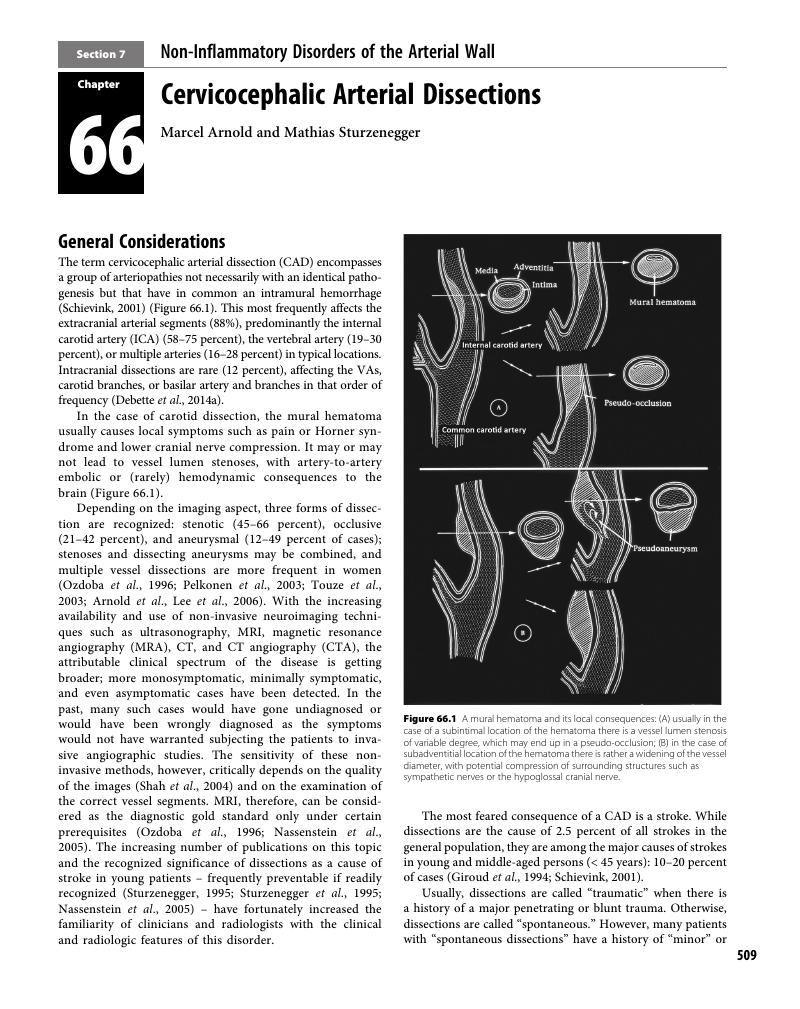
Barbour, P. J., Castaldo, J. E., Rae-Grant, A. D., et al. 1994. Internal carotid artery redundancy is significantly associated with dissection. Stroke, 25, 1201–6.Google Scholar
Bassetti, C., Carruzzo, A., Sturzenegger, M., and Tuncdogan, E. 1996. Recurrence of cervical artery dissection. A prospective study of 81 patients. Stroke, 27, 1804–7.Google Scholar
Baumgartner, R. W., Arnold, M., Baumgartner, I., et al. 2001. Carotid dissection with and without ischemic events: Local symptoms and cerebral artery findings. Neurology, 57, 827–32.CrossRefGoogle ScholarPubMed
Béjot, Y., Aboa-Eboulé, C., Debette, S., et al. 2014. Characteristics and outcomes of patients with multiple cervical artery dissection. Stroke, 45, 37–41.Google Scholar
Beletsky, V., Nadareishvili, Z., Lynch, J., et al. 2003. Cervical arterial dissection: Time for a therapeutic trial? Stroke, 34, 2856–60.Google Scholar
Benninger, D. H., Georgiadis, D., Kremer, C., et al. 2004. Mechanism of ischemic infarct in spontaneous carotid dissection. Stroke, 35, 482–5.CrossRefGoogle ScholarPubMed
Benninger, D. H., Georgiadis, D., Gandjour, J., and Baumgartner, R. W. 2006. Accuracy of color duplex ultrasound diagnosis of spontaneous carotid dissection causing ischemia. Stroke, 37, 377–81.CrossRefGoogle ScholarPubMed
Biller, J., Sacco, R. L., Albuquerque, F. C. et al. 2014. Cervical arterial dissections and association with cervical manipulative therapy. A statement for healthcare professionals from the American Heart Association/American Stroke Association. Stroke, 45, 3155–74.Google Scholar
Biousse, V., D’Anglejan-Chatillon, J., Touboul, P. J., Amarenco, P., and Bousser, M. G. 1995. Time course of symptoms in extracranial carotid artery dissections. A series of 80 patients. Stroke, 26, 235–9.CrossRefGoogle ScholarPubMed
Biousse, V., Schaison, M., Touboul, P. J., D’Anglejan-Chatillon, J., and Bousser, M. G. 1998. Ischemic optic neuropathy associated with internal carotid artery dissection. Arch Neurol, 55, 715–9.Google Scholar
Blanco Pampin, J., Morte Tamayo, N., Hinojal Fonseca, R., Payne-James, J. J., and Jerreat, P. 2002. Delayed presentation of carotid dissection, cerebral ischemia, and infarction following blunt trauma: Two cases. J Clin Forensic Med, 9, 136–40.Google Scholar
Brandt, T. and Grond-Ginsbach, C. 2002. Spontaneous cervical artery dissection: From risk factors toward pathogenesis. Stroke, 33, 657–8.Google Scholar
Brandt, T., Hausser, I., Orberk, E., et al. 1998. Ultrastructural connective tissue abnormalities in patients with spontaneous cervicocerebral artery dissections. Ann Neurol, 44, 281–5.Google Scholar
Brandt, T., Orberk, E., Weber, R., et al. 2001. Pathogenesis of cervical artery dissections: Association with connective tissue abnormalities. Neurology, 57, 24–30.Google Scholar
Calvet, D., Boutouyrie, P., Touze, E., et al. 2004. Increased stiffness of the carotid wall material in patients with spontaneous cervical artery dissection. Stroke, 35, 2078–82.CrossRefGoogle ScholarPubMed
Caprio, F. Z., Bernstein, R. A., Alberts, M. J. et al. 2014. Efficacy and safety of novel oral anticoagulants in patients with cervical artery dissection. Cerebrovasc Dis, 38, 247–53.CrossRefGoogle Scholar
Choi, Y. J., Jung, S. C., Lee, D. H. 2015. Vessel wall imaging of intracranial and cervical carotid arteries. Journal of Stroke, 17, 238–55.Google Scholar
Cohen, J. E., Ben-Hur, T., Rajz, G., Umansky, F., and Gomori, J. M. 2005. Endovascular stent-assisted angioplasty in the management of traumatic internal carotid artery dissections. Stroke, 36, e45–7.CrossRefGoogle ScholarPubMed
Cothren, C. C., Moore, E. E., Ray, C. E. Jr., et al. 2005. Carotid artery stents for blunt cerebrovascular injury: Risks exceed benefits. Arch Surg, 140, 480–5; discussion 485–6.CrossRefGoogle ScholarPubMed
Coulter, I. D., Hurwitz, E. L., Adams, A. H. et al. 2002. Patients using chiropractors in North America: Who are they, and why are they in chiropractic care? Spine, 27, 291–96.Google Scholar
Davis, J. W., Holbrook, T. L., Hoyt, D. B., et al. 1990. Blunt carotid artery dissection: Incidence, associated injuries, screening, and treatment. J Trauma, 30, 1514–7.Google Scholar
Debette, S., Metso, T., Pezzini, A., et al. 2011. Cervical Artery Dissection and Ischemic Stroke Patients (CADISP) group. Association of vascular risk factors with cervical artery dissection and ischemic stroke in young adults. Circulation, 123, 1537–44.Google Scholar
Debette, S., Goeggel-Simonetti, B., Schilling, S., et al. 2014a. Familial occurrence and heritable connective tissue disorders in cervical artery dissection. Neurology, 83, 2023–31.Google Scholar
Debette, S., Kamatani, Y., Metso, T.M., et al. 2014b. Common variation in PHACTR1 is associated with susceptibility to cervical artery dissection. Nature Genetics, 47, 78–83.Google Scholar
Debette, S., Compte, A., Labeyrie, M.A., et al. 2015. Epidemiology, pathophysiology, diagnosis, and management of intracranial artery dissection. Lancet Neurol, 14, 640–54.Google Scholar
Dreier, J. P., Lurtzing, F., Kappmeier, M., et al. 2004. Delayed occlusion after internal carotid artery dissection under heparin. Cerebrovasc Dis, 18, 296–303.Google Scholar
Droste, D. W., Junker, K., Stogbauer, F., et al. 2001. Clinically silent circulating microemboli in 20 patients with carotid or vertebral artery dissection. Cerebrovasc Dis, 12, 181–5.CrossRefGoogle ScholarPubMed
Engelter, S. T., Dallongeville, J., Kloss, M., et al. 2012. Thrombolysis in cervical artery dissection: Data from the Cervical Artery Dissection and Ischaemic Stroke Patients (CADISP) database. Eur J Neurol, 19, 1199–206.Google Scholar
Engelter, S. T., Grond-Ginsbach, C., Metso, T. M. et al. (Cervical Artery Dissection and Ischemic Stroke Patients Study Group). 2013. Cervical artery dissection: Trauma and other potential mechanical trigger events. Neurology, 80, 1950–57.Google Scholar
Fullerton, H. J., Johnston, S. C., and Smith, W. S. 2001. Arterial dissection and stroke in children. Neurology, 57, 1155–60.Google Scholar
Gao, P. H., Yang, L., Wang, G. et al. 2016. Symptomatic unruptured isolated middle cerebral artery dissection: Clinical and magnetic resonance imaging features. Clin Neuroradiol, 26, 81–91.Google Scholar
Georgiadis, D., Caso, V., and Baumgartner, R. W. 2006. Acute therapy and prevention of stroke in spontaneous carotid dissection. Clin Exp Hypertens, 28, 365–70.Google Scholar
Georgiadis, D., Arnold, M., von Buedingen, H. C. 2009. Aspirin vs. anticoagulation in carotid artery dissection: A study of 298 patients. Neurology, 72, 1810–15.CrossRefGoogle ScholarPubMed
Giossi, A., Ritelli, M., Costa, P., et al. 2014. Connective tissue anomalies in patients with spontaneous cervical artery dissection. Neurology, 83, 2032–7.Google Scholar
Giroud, M., Fayolle, H., Andre, N., et al. 1994. Incidence of internal carotid artery dissection in the community of Dijon. J Neurol Neurosurg Psychiatry, 57, 1443.Google Scholar
Grau, A. J., Brandt, T., Buggle, F., et al. 1999. Association of cervical artery dissection with recent infection. Arch Neurol, 56, 851–6.Google Scholar
Grond-Ginsbach, C., Schnippering, H., Hausser, I., et al. 2002. Ultrastructural connective tissue aberrations in patients with intracranial aneurysms. Stroke, 33, 2192–6.Google Scholar
Grond-Ginsbach, C., Engelter, S., Werner, I., et al. 2004. Alpha-1-antitrypsin deficiency alleles are not associated with cervical artery dissections. Neurology, 62, 1190–2.Google Scholar
Guillon, B. 2001. Is it necessary to treat persistent aneurysms after dissection of the cervical arteries?. Rev Neurol (Paris), 157, 1304–8.Google Scholar
Guillon, B., Brunereau, L., Biousse, V., et al. 1999. Long-term follow-up of aneurysms developed during extracranial internal carotid artery dissection. Neurology, 53, 117–22.Google Scholar
Guillon, B., Tzourio, C., Biousse, V., et al. 2000. Arterial wall properties in carotid artery dissection: An ultrasound study. Neurology, 55, 663–6.Google Scholar
Guillon, B., Berthet, K., Benslamia, L., et al. 2003. Infection and the risk of spontaneous cervical artery dissection: A case–control study. Stroke, 34, e79–81.CrossRefGoogle ScholarPubMed
Han, M., Rim, N. J., Lee, J. S., et al. 2014. Feasibility of high-resolution MR imaging for the diagnosis of intracranial vertebrobasilar artery dissection. Eur Radiol, 24, 3017–24.Google Scholar
Heldner, M. R., Nedetcheva, M., Yan, X., et al. 2015. Dynamic changes of intramural hematoma in patients with acute spontaneous internal carotid artery dissection. Int J Stroke, 10, 887–92.CrossRefGoogle ScholarPubMed
Hosoda, K., Fujita, S., Kawaguchi, T., et al. 1991. Spontaneous dissecting aneurysms of the basilar artery presenting with a subarachnoid hemorrhage. Report of two cases. J Neurosurg, 75, 628–33.Google Scholar
Hosoya, T., Adachi, M., Yamaguchi, K., et al. 1999. Clinical and neuroradiological features of intracranial vertebrobasilar artery dissection. Stroke, 30, 1083–90.Google Scholar
Hughes, K. M., Collier, B., Greene, K. A., and Kurek, S. 2000. Traumatic carotid artery dissection: A significant incidental finding. Am Surg, 66, 1023–7.Google Scholar
Kloss, M., Metso, A., Pezzini, A., et al. 2012. Towards understanding seasonal variability in cervical artery dissection (CeAD). J Neurol, 259, 1662–7.CrossRefGoogle ScholarPubMed
Kloss, M., Grond-Ginsbach, C., Pezzini, A., et al. 2014. Stroke in first-degree relatives of patients with cervical artery dissection. Eur J Neurol, 21, 1102–7.Google Scholar
Konrad, C., Muller, G. A., Langer, C., et al. 2004. Plasma homocysteine, MTHFR C677 T, CBS 844ins68bp, and MTHFD1 G1958 A polymorphisms in spontaneous cervical artery dissections. J Neurol, 251, 1242–8.CrossRefGoogle Scholar
Kremer, C., Mosso, M., Georgiadis, D., et al. 2003. Carotid dissection with permanent and transient occlusion or severe stenosis: Long-term outcome. Neurology, 60, 271–5.Google Scholar
Kwak, H. S., Hwang, S. B., Chung, G. H. et al. 2014 High-resolution magnetic resonance imaging of symptomatic middle cerebral artery dissection. J Stroke Cerebrovasc Dis, 23, 550–53.Google Scholar
Lansberg, M. G., Albers, G. W., Beaulieu, C., et al. 2000. Comparison of diffusion-weighted MRI and CT in acute stroke. Neurology, 54, 1557–61.Google Scholar
Lavallée, P. C., Mazighi, M., Saint-Maurice, J. P., et al. 2007. Stent-assisted endovascular thrombolysis versus intravenous thrombolysis in internal carotid artery dissection with tandem internal carotid and middle cerebral artery occlusion. Stroke, 38, 2270–74.CrossRefGoogle ScholarPubMed
Lee, V. H., Brown, R. D. Jr., Mandrekar, J. N., and Mokri, B. 2006. Incidence and outcome of cervical artery dissection: A population-based study. Neurology, 67, 1809–12.Google Scholar
Levy, C., Laissy, J. P., Raveau, V., et al. 1994. Carotid and vertebral artery dissections: Three-dimensional time-of-flight MR angiography and MR imaging versus conventional angiography. Radiology, 190, 97–103.Google Scholar
Leys, D. and Debette, S. 2006. Long-term outcome in patients with cervical-artery dissections: There is still a lot to know. Cerebrovasc Dis, 22, 215.Google Scholar
Lucas, C., Moulin, T., Deplanque, D., Tatu, L., and Chavot, D. 1998. Stroke patterns of internal carotid artery dissection in 40 patients. Stroke, 29, 2646–8.Google Scholar
Lucas, C., Lecroart, J. L., Gautier, C., et al. 2004. Impairment of endothelial function in patients with spontaneous cervical artery dissection: Evidence for a general arterial wall disease. Cerebrovasc Dis, 17, 170–4.Google Scholar
Lyrer, P. and Engelter, S. 2010. Antithrombotic drugs for carotid artery dissection. Cochrane Database System Review, 10, CD000255.Google Scholar
Lyrer, P. A., Brandt, T., Metso, T. M., et al. 2014. Clinical import of Horner syndrome in internal carotid and vertebral artery dissection. Neurology, 82, 1653–9.Google Scholar
Machet, A., Fonseca, A. C., Oppenheim, C., et al. 2013. Does anticoagulation promote mural hematoma growth or delayed occlusion in spontaneous cervical artery dissections? Cerebrovasc Dis, 35, 175–78.Google Scholar
Markus, H. S., Hayter, E., Levi, C., et al. (CADISS Trial Investigators). 2015. Antiplatelet treatment compared with anticoagulation treatment for cervical artery dissections (CADISS): A randomised trial. Lancet Neurol, 14, 361–67.Google Scholar
Mascalchi, M., Bianchi, M. C., Mangiafico, S., et al. 1997. MRI and MR angiography of vertebral artery dissection. Neuroradiology, 39, 329–40.Google Scholar
Metso, T. M., Metso, A. J., Salonen, O. et al., 2009. Adult cervicocerebral artery dissection: A single center study of 301 Finnish patients. Eur J Neurol, 16, 656–61.Google Scholar
Metso, A. J., Metso, T. M., Debette, S. et al. CADISP Group. 2012a. Gender and cervical artery dissection. Eur J Neurol, 19, 594–602.Google Scholar
Metso, T. M., Tatlisumak, T., Debette, S., et al. 2012b. Migraine in cervical artery dissection and ischemic stroke patients. Neurology, 78, 1221–8.Google Scholar
Mizutani, T. 2011. Natural course of intracranial arterial dissections. J Neurosurg, 114, 1037–44.Google Scholar
Mokri, B., Piepgras, D. G., and Houser, O. W. 1988. Traumatic dissections of the extracranial internal carotid artery. J Neurosurg, 68, 189–97.Google Scholar
Morel, A., Naggara, O., Touze, E., et al. 2012. Mechanism of ischemic infarct in spontaneous cervical artery dissection. Stroke, 43, 1354–61.Google Scholar
Muller, B. T., Luther, B., Hort, W., et al. 2000. Surgical treatment of 50 carotid dissections: Indications and results. J Vasc Surg, 31, 980–8.Google Scholar
Mustanoja, S., Metso, T. M., Putaala, J., et al. 2015. Helsinki experience on nonvitamin K oral anticoagulants for treating cervical artery dissection. Brain and Behavior, 5, e00349.CrossRefGoogle ScholarPubMed
Naggara, O., Morel, A., Touze, E., et al. 2012. Stroke occurrence and patterns are not influenced by the degree of stenosis in cervical artery dissection. Stroke, 43, 1150–52.Google Scholar
Nassenstein, I., Kramer, S. C., Niederstadt, T., et al. 2005. Incidence of cerebral ischemia in patients with suspected cervical artery dissection: First results of a prospective study. Rofo, 177, 1532–9.Google ScholarPubMed
Nunez, D. B. Jr., Torres-Leon, M., and Munera, F. 2004. Vascular injuries of the neck and thoracic inlet: Helical CT-angiographic correlation. Radiographics, 24, 1087–98; discussion 1099–100.Google Scholar
Okumura, A., Araki, Y., Nishimura, Y., et al. 2001. The clinical utility of contrast-enhanced 3D MR angiography for cerebrovascular disease. Neurol Res, 23, 767–71.CrossRefGoogle ScholarPubMed
Olin, J. W., Froehlich, J., Gu, X., et al. 2012. The United States Registry of Fibromuscular Dysplasia: Results of the first 447 patients. Circulation, 125, 3182–90.Google Scholar
Ozdoba, C., Sturzenegger, M., and Schroth, G. 1996. Internal carotid artery dissection: MR imaging features and clinical–radiologic correlation. Radiology, 199, 191–8.Google Scholar
Paciaroni, M., Georgiadis, D., Arnold, M., et al. 2006. Seasonal variability in spontaneous cervical artery dissection. J Neurol Neurosurg Psychiatry, 77, 677–9.Google Scholar
Pasquini, M., Trystram, D., Nokam, G. et al., 2015. Fibromuscular dysplasia of cervicocephalic arteries: Prevalence of multisite involvement and prognosis. Rev Neurol (Paris), 171, 616–23.Google Scholar
Pelkonen, O., Tikkakoski, T., Leinonen, S., et al. 2003. Extracranial internal carotid and vertebral artery dissections: Angiographic spectrum, course and prognosis. Neuroradiology, 45, 71–7.Google Scholar
Pezzini, A., Caso, V., Zanferrari, C., et al. 2006. Arterial hypertension as risk factor for spontaneous cervical artery dissection. A case–control study. J Neurol Neurosurg Psychiatry, 77, 95–7.Google Scholar
Pezzini, A., Del Zotto, E., Mazziotti, G., et al. 2006. Thyroid autoimmunity and spontaneous cervical artery dissection. Stroke, 37, 2375–7.Google Scholar
Pham, M. H., Rahme, R. J., Arnaout, O., et al. 2011. Endovascular stenting of extracranial carotid and vertebral artery dissections: A systematic review of the literature. Neurosurgery, 68, 856–66.Google Scholar
Phan, T., Huston, J. 3rd, Bernstein, M. A., et al. 2001. Contrast-enhanced magnetic resonance angiography of the cervical vessels: Experience with 422 patients. Stroke, 32, 2282–6.Google Scholar
Plouin, P. F., Perdu, J., La Batide-Alanone, A., et al. 2007. Fibromuscular dysplasia. Orphanet J Rare Dis, 2, 28–35.Google Scholar
Qureshi, A. I., Chaudhry, S. A., Hassan, A. E., et al. 2011. Thrombolytic treatment of patients with acute ischemic stroke related to underlying arterial dissection in the United States. Arch Neurol, 68, 1536–42.Google Scholar
Rothwell, D. M., Bondy, S. J., and Williams, J. I. 2001. Chiropractic manipulation and stroke: A population-based case–control study. Stroke, 32, 1054–60.Google Scholar
Rubiera, M., Ribo, M., Delgado-Mederos, R., et al. 2006. Tandem internal carotid artery/middle cerebral artery occlusion: An independent predictor of poor outcome after systemic thrombolysis. Stroke, 37, 2301–05.CrossRefGoogle ScholarPubMed
Rubinstein, S. M., Peerdeman, S. M., van Tulder, M. W., Riphagen, I., and Haldeman, S. 2005. A systematic review of the risk factors for cervical artery dissection. Stroke, 36, 1575–80.Google Scholar
Schellinger, P. D., Schwab, S., Krieger, D., et al. 2001. Masking of vertebral artery dissection by severe trauma to the cervical spine. Spine, 26, 314–9.Google Scholar
Schievink, W. I. 2001. Spontaneous dissection of the carotid and vertebral arteries. N Engl J Med, 344, 898–906.Google Scholar
Schievink, W. I., Mokri, B., and Whisnant, J. P. 1993. Internal carotid artery dissection in a community. Rochester, Minnesota, 1987–1992. Stroke, 24, 1678–80.Google Scholar
Schievink, W. I., Mokri, B., Piepgras, D. G., and Gittenberger-de Groot, A. C. 1996a. Intracranial aneurysms and cervicocephalic arterial dissections associated with congenital heart disease. Neurosurgery, 39, 685–9; discussion 689–90.Google Scholar
Schievink, W. I., Mokri, B., Piepgras, D. G., and Kuiper, J. D. 1996b. Recurrent spontaneous arterial dissections: Risk in familial versus nonfamilial disease. Stroke, 27, 622–4.Google Scholar
Shah, G. V., Quint, D. J., and Trobe, J. D. 2004. Magnetic resonance imaging of suspected cervicocranial arterial dissections. J Neuroophthalmol, 24, 315–8.Google Scholar
Sikkema, T., Uyttenboogaart, M., Eshghi, O., et al. 2014. Intracranial artery dissection. Eur J Neurol, 21, 820–26.Google Scholar
Silbert, P. L., Mokri, B., and Schievink, W. I. 1995. Headache and neck pain in spontaneous internal carotid and vertebral artery dissections. Neurology, 45, 1517–22.Google Scholar
Smith, W. S., Johnston, S. C., Skalabrin, E. J., et al. 2003. Spinal manipulative therapy is an independent risk factor for vertebral artery dissection. Neurology, 60, 1424–8.Google Scholar
Srinivasan, J., Newell, D. W., Sturzenegger, M., Mayberg, M. R., and Winn, H. R. 1996. Transcranial Doppler in the evaluation of internal carotid artery dissection. Stroke, 27, 1226–30.Google Scholar
Sturzenegger, M. 1991. Ultrasound findings in spontaneous carotid artery dissection. The value of duplex sonography. Arch Neurol, 48, 1057–63.Google Scholar
Sturzenegger, M. 1994. Headache and neck pain: The warning symptoms of vertebral artery dissection. Headache, 34, 187–93.Google Scholar
Sturzenegger, M. 1995. Spontaneous internal carotid artery dissection: Early diagnosis and management in 44 patients. J Neurol, 242, 231–8.Google Scholar
Sturzenegger, M. and Huber, P. 1993. Cranial nerve palsies in spontaneous carotid artery dissection. J Neurol Neurosurg Psychiatry, 56, 1191–9.Google Scholar
Sturzenegger, M., Mattle, H. P., Rivoir, A., Rihs, F., and Schmid, C. 1993. Ultrasound findings in spontaneous extracranial vertebral artery dissection. Stroke, 24, 1910–21.Google Scholar
Sturzenegger, M., Mattle, H. P., Rivoir, A., and Baumgartner, R. W. 1995. Ultra-sound findings in carotid artery dissection: Analysis of 43 patients. Neurology, 45, 691–8.Google Scholar
Touze, E., Gauvrit, J. Y., Moulin, T., et al. 2003. Risk of stroke and recurrent dissection after a cervical artery dissection: A multicenter study. Neurology, 61, 1347–51.Google Scholar
Tsivgoulis, G., Zahnd, R., Katsanos, A. H., et al. 2015. Safety and outcomes of intravenous thrombolysis in dissection-related ischemic stroke: An international multicenter study and comprehensive meta-analysis of reported case series. J Neurol, 262, 2135–43.Google Scholar
Tsukahara, T., Minematsu, K. 2010. Overview of spontaneous cervicocephalic arterial dissection in Japan. Acta Neurochir, 107(Suppl), 35–40.Google Scholar
Tzourio, C., Cohen, A., Lamisse, N., Biousse, V., and Bousser, M. G. 1997. Aortic root dilatation in patients with spontaneous cervical artery dissection. Circulation, 95, 2351–3.Google Scholar
Vergouwen, M. D. 2012. Intravenous thrombolysis in ischaemic stroke secondary to cervical artery dissection: Safe but not effective? Eur J Neurol, 19, 1155–56.Google Scholar
Vertinsky, A. T., Schwartz, N. E., Fischbein, N. J., et l. 2008. Comparison of multidetector CT angiography and MR imaging of cervical artery dissection. AJNR Am J Neuroradiol, 29, 1753–60.Google Scholar
Volker, W., Besselmann, M., Dittrich, R., et al. 2005. Generalized arteriopathy in patients with cervical artery dissection. Neurology, 64, 1508–13.Google Scholar
Wang, Y., Lou, X., Li, Y., et al. 2014. Imaging investigation of intracranial arterial dissecting aneurysms by using 3 T high-resolution MRI and DSA: From the interventional neuroradiologists’ view. Acta Neurochir (Wien), 156, 515–25.Google Scholar
Weimar, C., Kraywinkel, K., Hagemeister, C., et al. 2010. Recurrent stroke after cervical artery dissection. J Neurol Neurosurg Psychiatry, 81, 869–73.Google Scholar
Wiest, T., Hyrenbach, S., Bambul, P., et al. 2006. Genetic analysis of familial connective tissue alterations associated with cervical artery dissections suggests locus heterogeneity. Stroke, 37, 1697–702.Google Scholar
Willis, B. K., Greiner, F., Orrison, W. W., and Benzel, E. C. 1994. The incidence of vertebral artery injury after midcervical spine fracture or subluxation. Neurosurgery, 34, 435–41.Google Scholar
Yang, S. T., Huang, Y. C., Chuang, C. C., and Hsu, P. W. 2006. Traumatic internal carotid artery dissection. J Clin Neurosci, 13, 123–8.Google Scholar
Zhang, F.-L., Liu, Y., Xing, Y. Q., et al. 2015. Diagnosis of cervical artery dissection using 3-T magnetic resonance imaging. JAMA Neurology, 72, 600–1.Google Scholar
Zhou, M., Zheng, H., Gong, S., et al. 2015. Vertebral artery hypoplasia and vertebral artery dissection: A hospital-based cohort study., Neurology, 84, 818–24.Google Scholar
Zinkstok, S. M., Vergouwen, M. D., Engelter, S. T. et al. 2011. Safety and functional outcome of thrombolysis in dissection-related ischemic stroke: A meta-analysis of individual patient data. Stroke, 42, 2515–20.CrossRefGoogle ScholarPubMed
References
Akoudad, S., Portegies, M. L., Koudstaal, P. J., et al. (2015). Cerebral microbleeds are associated with an increased risk of stroke: The Rotterdam study. Circulation 132: 509–16.Google Scholar
Auriel, E., Charidimou, A., Gurol, M. E., et al. (2016). Validation of clinicoradiological criteria for the diagnosis of cerebral amyloid angiopathy-related inflammation. JAMA Neurol 73: 197–202.Google Scholar
Auriel, E., Gurol, M. E., Ayres, A., et al. (2012). Characteristic distributions of intracerebral hemorrhage-associated diffusion-weighted lesions. Neurology 79: 2335–41.Google Scholar
Black, S., Gao, F., and Bilbao, J. (2009). Understanding white matter disease: Imaging–pathological correlations in vascular cognitive impairment. Stroke 40: S48–52.Google Scholar
Blitstein, M. K. and Tung, G. A. (2007). MRI of cerebral microhemorrhages. AJR Am J Roentgenol 189: 720–5.Google Scholar
Boulouis, G., Charidimou, A., Auriel, E., et al. (2016). Intracranial atherosclerosis and cerebral small vessel disease in intracerebral hemorrhage patients. J Neurol Sci 369: 324–9.CrossRefGoogle ScholarPubMed
Boyle, P. A., Yu, L., Nag, S., et al. (2015). Cerebral amyloid angiopathy and cognitive outcomes in community-based older persons. Neurology 85: 1930–6.Google Scholar
Charidimou, A., Meegahage, R., Fox, Z., et al. (2013a). Enlarged perivascular spaces as a marker of underlying arteriopathy in intracerebral haemorrhage: A multicentre MRI cohort study. J Neurol Neurosurg Psychiatry 84: 624–9.CrossRefGoogle ScholarPubMed
Charidimou, A., Peeters, A. P., Jager, R., et al. (2013b). Cortical superficial siderosis and intracerebral hemorrhage risk in cerebral amyloid angiopathy. Neurology 81: 1666–73.Google Scholar
Charidimou, A., Linn, J., Vernooij, M. W., et al. (2015). Cortical superficial siderosis: detection and clinical significance in cerebral amyloid angiopathy and related conditions. Brain 138: 2126–39.Google Scholar
Charidimou, A., Boulouis, G., Haley, K., et al. (2016). White matter hyperintensity patterns in cerebral amyloid angiopathy and hypertensive arteriopathy. Neurology 86: 505–11.CrossRefGoogle ScholarPubMed
De Strooper, B. (2003). Aph-1, Pen-2, and nicastrin with presenilin generate an active gamma-secretase complex. Neuron 38: 9–12.Google Scholar
Dierksen, G. A., Skehan, M. E., Khan, M. A., et al. (2010). Spatial relation between microbleeds and amyloid deposits in amyloid angiopathy. Ann Neurol 68: 545–8.Google Scholar
Dumas, A., Dierksen, G. A., Gurol, M. E., et al. (2012). Functional magnetic resonance imaging detection of vascular reactivity in cerebral amyloid angiopathy. Ann Neurol 72: 76–81.CrossRefGoogle ScholarPubMed
Eckman, M. H., Rosand, J., Knudsen, K. A., Singer, D. E., and Greenberg, S. M. (2003). Can patients be anticoagulated after intracerebral hemorrhage? A decision analysis. Stroke 34: 1710–16.Google Scholar
Eckman, M. H., Wong, L. K., Soo, Y. O., et al. (2008). Patient-specific decision-making for warfarin therapy in nonvalvular atrial fibrillation: How will screening with genetics and imaging help? Stroke 39: 3308–15.Google Scholar
Eng, J. A., Frosch, M. P., Choi, K., Rebeck, G. W., and Greenberg, S. M. (2004). Clinical manifestations of cerebral amyloid angiopathy-related inflammation. Ann Neurol 55: 250–6.Google Scholar
Fotiadis, P., van Rooden, S., van der Grond, J., et al. (2016). Cortical atrophy in patients with cerebral amyloid angiopathy: A case–control study. Lancet Neurol 15: 811–19.Google Scholar
Glenner, G. G. and Wong, C. W. (1984). Alzheimer’s disease: Initial report of the purification and characterization of a novel cerebrovascular amyloid protein. Biochem Biophys Res Commun 120: 885–90.Google Scholar
Goldstein, L. B., Amarenco, P., Szarek, M., et al. (2008). Hemorrhagic stroke in the Stroke Prevention by Aggressive Reduction in Cholesterol Levels study. Neurology 70: 2364–70.Google Scholar
Greenberg, S. M., Vonsattel, J. P., Stakes, J. W., Gruber, M., and Finklestein, S. P. (1993). The clinical spectrum of cerebral amyloid angiopathy: Presentations without lobar hemorrhage. Neurology 43: 2073–9.Google Scholar
Greenberg, S. M., Briggs, M. E., Hyman, B. T., et al. (1996a). Apolipoprotein E epsilon 4 is associated with the presence and earlier onset of hemorrhage in cerebral amyloid angiopathy. Stroke 27: 1333–7.Google Scholar
Greenberg, S. M., Finklestein, S. P., and Schaefer, P. W. (1996b). Petechial hemorrhages accompanying lobar hemorrhage: Detection by gradient-echo MRI. Neurology 46: 1751–4.Google Scholar
Greenberg, S. M., Vonsattel, J. P., Segal, A. Z., et al. (1998). Association of apolipoprotein E epsilon2 and vasculopathy in cerebral amyloid angiopathy. Neurology 50: 961–5.CrossRefGoogle ScholarPubMed
Greenberg, S. M., Eng, J. A., Ning, M., Smith, E. E., and Rosand, J. (2004a). Hemorrhage burden predicts recurrent intracerebral hemorrhage after lobar hemorrhage. Stroke 35: 1415–20.Google Scholar
Greenberg, S. M., Gurol, M. E., Rosand, J., and Smith, E. E. (2004b). Amyloid angiopathy-related vascular cognitive impairment. Stroke 35(Suppl 1): 2616–19.Google Scholar
Greenberg, S. M., Rosand, J., Schneider, A. T., et al. (2006). A phase 2 study of tramiprosate for cerebral amyloid angiopathy. Alzheimer Dis Assoc Disord 20: 269–74.Google Scholar
Greenberg, S. M., Grabowski, T., Gurol, M. E., et al. (2008). Detection of isolated cerebrovascular beta-amyloid with Pittsburgh compound B. Ann Neurol 64: 587–91.Google Scholar
Greenberg, S. M., Vernooij, M. W., Cordonnier, C., et al. (2009). Cerebral microbleeds: A guide to detection and interpretation. Lancet Neurology 8: 165–74.Google Scholar
Gurol, M. E. (2016). Molecular neuroimaging in vascular cognitive impairment. Stroke 47: 1146–52.Google Scholar
Gurol, M. E. and Greenberg, S. M. (2008). Management of intracerebral hemorrhage. Curr Atheroscler Rep 10: 324–31.Google Scholar
Gurol, M. E., Irizarry, M. C., Smith, E. E., et al. (2006). Plasma beta-amyloid and white matter lesions in AD, MCI, and cerebral amyloid angiopathy. Neurology 66: 23–9.Google Scholar
Gurol, M. E., Dierksen, G., Betensky, R., et al. (2012). Predicting sites of new hemorrhage with amyloid imaging in cerebral amyloid angiopathy. Neurology 79: 320–6.Google Scholar
Gurol, M. E., Viswanathan, A., Gidicsin, C., et al. (2013). Cerebral amyloid angiopathy burden associated with leukoaraiosis: A positron emission tomography/magnetic resonance imaging study. Ann Neurol 73: 529–36.Google Scholar
Gurol, M. E., Becker, J. A., Fotiadis, P., et al. (2016). Florbetapir-PET to diagnose cerebral amyloid angiopathy: A prospective study. Neurology 87: 2043–9.Google Scholar
Hackam, D. G. and Mrkobrada, M. (2012). Selective serotonin reuptake inhibitors and brain hemorrhage: A meta-analysis. Neurology 79: 1862–5.Google Scholar
Haley, K. E., Greenberg, S. M., and Gurol, M. E. (2013). Cerebral microbleeds and macrobleeds: Should they influence our recommendations for antithrombotic therapies? Curr Cardiol Rep 15: 425–32.Google Scholar
Hemphill, J. C. III, Greenberg, S. M., Anderson, C. S., et al. (2015). Guidelines for the management of spontaneous intracerebral hemorrhage: A Guideline for Healthcare Professionals from the American Heart Association/American Stroke Association. Stroke 46: 2032–60.Google Scholar
Hermann, D. M. and Bassetti, C. L. (2016). Role of sleep-disordered breathing and sleep–wake disturbances for stroke and stroke recovery. Neurology 87: 1407–16.Google Scholar
Herzig, M. C., Van Nostrand, W. E., and Jucker, M. (2006). Mechanism of cerebral beta-amyloid angiopathy: Murine and cellular models. Brain Pathol 16: 40–54.Google Scholar
January, C. T., Wann, L. S., Alpert, J. S., et al. (2014). 2014 AHA/ACC/HRS guideline for the management of patients with atrial fibrillation: A report of the American College of Cardiology/American Heart Association Task Force on practice guidelines and the Heart Rhythm Society. Circulation 130: e199–267.Google Scholar
Johnson, K. A., Gregas, M., Becker, J. A., et al. (2007). Imaging of amyloid burden and distribution in cerebral amyloid angiopathy. Ann Neurol 62: 229–34.Google Scholar
Kimberly, W. T., Gilson, A., Rost, N. S., et al. (2009). Silent ischemic infarcts are associated with hemorrhage burden in cerebral amyloid angiopathy. Neurology 72: 1230–5.CrossRefGoogle ScholarPubMed
Klunk, W. E., Engler, H., Nordberg, A., et al. (2004). Imaging brain amyloid in Alzheimer’s disease with Pittsburgh compound-B. Ann Neurol 55: 306–19.Google Scholar
Knudsen, K. A., Rosand, J., Karluk, D., and Greenberg, S. M. (2001). Clinical diagnosis of cerebral amyloid angiopathy: Validation of the Boston criteria. Neurology 56: 537–9.Google Scholar
Lauer, A., Greenberg, S. M., and Gurol, M. E. (2015). Statins in intracerebral hemorrhage. Curr Atheroscler Rep 17: 46.Google Scholar
Linn, J., Halpin, A., Demaerel, P., et al. (2010). Prevalence of superficial siderosis in patients with cerebral amyloid angiopathy. Neurology 74: 1346–50.Google Scholar
Lovelock, C. E., Molyneux, A. J., and Rothwell, P. M. (2007). Change in incidence and aetiology of intracerebral haemorrhage in Oxfordshire, UK, between 1981 and 2006: A population-based study. Lancet Neurol 6: 487–93.CrossRefGoogle ScholarPubMed
Ly, J. V., Donnan, G. A., Villemagne, V. L., et al. (2010). 11 C-PIB binding is increased in patients with cerebral amyloid angiopathy-related hemorrhage. Neurology 74: 487–93.Google Scholar
Martinez-Ramirez, S., Pontes-Neto, O. M., Dumas, A. P., et al. (2013). Topography of dilated perivascular spaces in subjects from a memory clinic cohort. Neurology 80: 1551–6.Google Scholar
Martinez-Ramirez, S., Romero, J. R., Shoamanesh, A., et al. (2015). Diagnostic value of lobar microbleeds in individuals without intracerebral hemorrhage. Alzheimers Dement 11: 1480–8.Google Scholar
Masoudi, F. A., Calkins, H., Kavinsky, C. J., et al. (2015). 2015 ACC/HRS/SCAI Left atrial appendage occlusion device societal overview. J Am Coll Cardiol 66: 1497–513.Google Scholar
Masters, C. L., Simms, G., Weinman, N. A., et al. (1985). Amyloid plaque core protein in Alzheimer disease and Down syndrome. Proc Natl Acad Sci USA 82: 4245–9.Google Scholar
Masuda, J., Tanaka, K., Ueda, K., and Omae, T. (1988). Autopsy study of incidence and distribution of cerebral amyloid angiopathy in Hisayama, Japan. Stroke 19: 205–10.Google Scholar
Nicoll, J. A., Burnett, C., Love, S., et al. (1996). High frequency of apolipoprotein E epsilon 2 in patients with cerebral hemorrhage due to cerebral amyloid angiopathy. Ann Neurol 39: 682–3.Google Scholar
O’Donnell, H. C., Rosand, J., Knudsen, K. A., et al. (2000). Apolipoprotein E genotype and the risk of recurrent lobar intracerebral hemorrhage. N Engl J Med 342: 240–5.Google Scholar
Okazaki, H., Reagan, T. J., and Campbell, R. J. (1979). Clinicopathologic studies of primary cerebral amyloid angiopathy. Mayo Clin Proc 54: 22–31.Google Scholar
Oppenheim, G. (1909). Uber “drusige Nekrosen” in der Großhirnrinde. Neurol Centralbl 28: 410–13.Google Scholar
Pantelakis, S. (1954). A particular type of senile angiopathy of the central nervous system: Congophilic angiopathy, topography and frequency [in French]. Monatsschr Psychiatr Neurol 128: 219–56.Google Scholar
Peca, S., McCreary, C. R., Donaldson, E., et al. (2013). Neurovascular decoupling is associated with severity of cerebral amyloid angiopathy. Neurology 81: 1659–65.Google Scholar
Poels, M. M., Vernooij, M. W., Ikram, M. A., et al. (2010). Prevalence and risk factors of cerebral microbleeds: An update of the Rotterdam scan study. Stroke 41(Suppl): S103–6.Google Scholar
Purrucker, J. C., Haas, K., Rizos, T., et al. (2016). Early clinical and radiological course, management, and outcome of intracerebral hemorrhage related to new oral anticoagulants. JAMA Neurol 73: 169–77.Google Scholar
Reijmer, Y. D., Fotiadis, P., Martinez-Ramirez, S., et al. (2015). Structural network alterations and neurological dysfunction in cerebral amyloid angiopathy. Brain 138(Pt 1): 179–88.Google Scholar
Reijmer, Y. D., van Veluw, S. J., and Greenberg, S. M. (2016). Ischemic brain injury in cerebral amyloid angiopathy. J Cereb Blood Flow Metab 36: 40–54.Google Scholar
Revesz, T., Holton, J. L., Lashley, T., et al. (2002). Sporadic and familial cerebral amyloid angiopathies. Brain Pathol 12: 343–57.Google Scholar
Rosand, J., Hylek, E. M., O’Donnell, H. C., and Greenberg, S. M. (2000). Warfarin-associated hemorrhage and cerebral amyloid angiopathy: A genetic and pathologic study. Neurology 55: 947–51.Google Scholar
Rosand, J., Muzikansky, A., Kumar, A., et al. (2005). Spatial clustering of hemorrhages in probable cerebral amyloid angiopathy. Ann Neurol 58: 459–62.Google Scholar
Schmechel, D. E., Saunders, A. M., Strittmatter, W. J., et al. (1993). Increased amyloid beta-peptide deposition in cerebral cortex as a consequence of apolipoprotein E genotype in late-onset Alzheimer disease. Proc Natl Acad Sci USA 90: 9649–53.Google Scholar
Scholz, W. (1938). Studien zur pathologie der hirngefabe II: Die drusige entartung der hirnarterien und Capillaren. Zeit. Gesamte Neurol Psychiatr 162: 694–715.Google Scholar
Scolding, N. J., Joseph, F., Kirby, P. A., et al. (2005). Abeta-related angiitis: Primary angiitis of the central nervous system associated with cerebral amyloid angiopathy. Brain 128: 500–15.Google Scholar
Selkoe, D. J., Abraham, C. R., Podlisny, M. B., and Duffy, L. K. (1986). Isolation of low-molecular-weight proteins from amyloid plaque fibers in Alzheimer’s disease. J Neurochem 46: 1820–34.Google Scholar
Smith, E. E. and Eichler, F. (2006). Cerebral amyloid angiopathy and lobar intracerebral hemorrhage. Arch Neurol 63: 148–51.Google Scholar
Smith, E. E., Gurol, M. E., Eng, J. A., et al. (2004). White matter lesions, cognition, and recurrent hemorrhage in lobar intracerebral hemorrhage. Neurology 63: 1606–12.Google Scholar
Smith, E. E., Vijayappa, M., Lima, F., et al. (2008). Impaired visual evoked flow velocity response in cerebral amyloid angiopathy. Neurology 71: 1424–30.Google Scholar
Smith, E. E., Schneider, J. A., Wardlaw, J. M., and Greenberg, S. M. (2012). Cerebral microinfarcts: The invisible lesions. Lancet Neurol 11: 272–82.Google Scholar
Stone, N. J., Robinson, J. G., Lichtenstein, A. H., et al. (2014). 2013 ACC/AHA guideline on the treatment of blood cholesterol to reduce atherosclerotic cardiovascular risk in adults: A report of the American College of Cardiology/American Heart Association Task Force on Practice Guidelines. Circulation 129(Suppl 2): S1–45.Google Scholar
Thanprasertsuk, S., Martinez-Ramirez, S., Pontes-Neto, O. M., et al. (2014). Posterior white matter disease distribution as a predictor of amyloid angiopathy. Neurology 83: 794–800.Google Scholar
Thon, J. M. and Gurol, M. E. (2016). Intracranial hemorrhage risk in the era of antithrombotic therapies for ischemic stroke. Curr Treat Options Cardiovasc Med 18: 29.Google Scholar
van Asch, C. J., Luitse, M. J., Rinkel, G. J., van der Tweel, I., Algra, A., and Klijn, C. J. (2010). Incidence, case fatality, and functional outcome of intracerebral haemorrhage over time, according to age, sex, and ethnic origin: A systematic review and meta-analysis. Lancet Neurol 9: 167–76.Google Scholar
van Etten, E. S., Auriel, E., Haley, K. E., et al. (2014). Incidence of symptomatic hemorrhage in patients with lobar microbleeds. Stroke 45: 2280–5.Google Scholar
van Veluw, S. J., Biessels, G. J., Bouvy, W. H., et al. (2016). Cerebral amyloid angiopathy severity is linked to dilation of juxtacortical perivascular spaces. J Cereb Blood Flow Metab 36: 576–80.Google Scholar
Verbeek, M. M., De Waal, R. M. W., and Vinters, H. V. (2000). Cerebral Amyloid Angiopathy in Alzheimer’s Disease and Related Disorders. Dordrecht: Kluwer Academic.Google Scholar
Verbeek, M. M., Kremer, B. P., Rikkert, M. O., et al. (2009). Cerebrospinal fluid amyloid beta(40) is decreased in cerebral amyloid angiopathy. Ann Neurol 66: 245–49.Google Scholar
Vinters, H. V. (1987). Cerebral amyloid angiopathy: A critical review. Stroke 18: 311–24.Google Scholar
Zhang-Nunes, S. X., Maat-Schieman, M. L., van Duinen, S. G., et al. (2006). The cerebral beta-amyloid angiopathies: Hereditary and sporadic. Brain Pathol 16: 30–9.Google Scholar
References
Suzuki, J, Takaku, A. Cerebrovascular “moyamoya” disease. Disease showing abnormal net-like vessels in base of brain. Arch Neurol. 1969;20:288–99.Google Scholar
Hayashi, K, Horie, N, Izumo, T, Nagata, I. A nationwide survey on unilateral moyamoya disease in Japan. Clin Neurol Neurosurg. 2014;124:1–5.Google Scholar
Kuroda, S, Ishikawa, T, Houkin, K, et al. Incidence and clinical features of disease progression in adult moyamoya disease. Stroke. 2005;36:2148–53.Google Scholar
Natori, Y, Ikezaki, K, Matsushima, T, Fukui, M. “Angiographic moyamoya”: Its definition, classification, and therapy. Clin Neurol Neurosurg. 1997;99:S168–72.Google Scholar
Wakai, K, Tamakoshi, A, Ikezaki, K, et al. Epidemiological features of moyamoya disease in Japan: Findings from a nationwide survey. Clin Neurol Neurosurg. 1997;99 Suppl 2:S1–5.Google Scholar
Kuriyama, S, Kusaka, Y, Fujimura, M, et al. Prevalence and clinicoepidemiological features of moyamoya disease in Japan: Findings from a nationwide epidemiological survey. Stroke. 2008;39:42–7.Google Scholar
Baba, T, Houkin, K, Kuroda, S. Novel epidemiological features of moyamoya disease. J Neurol Neurosurg Psychiatry. 2008;79:900–4.Google Scholar
Im, SH, Cho, CB, Joo, WI, et al. Prevalence and epidemiological features of moyamoya disease in Korea. J Cerebrovasc Endovasc Neurosurg. 2012;14:75–8.Google Scholar
Ahn, IM, Park, DH, Hann, HJ, et al. Incidence, prevalence, and survival of moyamoya disease in Korea: A nationwide, population-based study. Stroke. 2014;45:1090–5.Google Scholar
Chen, PC, Yang, SH, Chien, KL, Tsai, IJ, Kuo, MF. Epidemiology of moyamoya disease in Taiwan: A nationwide population-based study. Stroke. 2014;45:1258–63.Google Scholar
Miao, W, Zhao, PL, Zhang, YS, Liu, HY, Chang, Y, Ma, J, et al. Epidemiological and clinical features of moyamoya disease in Nanjing, China. Clin Neurol Neurosurg. 2010;112:199–203.Google Scholar
Duan, L, Bao, XY, Yang, WZ et al. Moyamoya disease in China: Its clinical features and outcomes. Stroke. 2012;43:56–60.Google Scholar
Cho, WS, Kim, JE, Kim, CH, et al. Long-term outcomes after combined revascularization surgery in adult moyamoya disease. Stroke. 2014;45:3025–31.Google Scholar
Jo, KI, Yeon, JY, Hong, SC, Kim, JS. Clinical course of asymptomatic adult moyamoya disease. Cerebrovasc Dis. 2014;37:94–101.Google Scholar
Cho, WS, Chung, YS, Kim, JE, et al. The natural clinical course of hemodynamically stable adult moyamoya disease. J Neurosurg. 2015;122:82–9.Google Scholar
Han, C, Feng, H, Han, YQ, et al. Prospective screening of family members with moyamoya disease patients. PLoS One. 2014;9:e88765.Google Scholar
Uchino, K, Johnston, SC, Becker, KJ, Tirschwell, DL. Moyamoya disease in Washington State and California. Neurology. 2005;65:956–8.Google Scholar
Kainth, D, Chaudhry, SA, Kainth, H, Suri, FK, Qureshi, AI. Epidemiological and clinical features of moyamoya disease in the USA. Neuroepidemiology. 2013;40:282–7.Google Scholar
Takagi, Y, Kikuta, K, Nozaki, K, Hashimoto, N. Histological features of middle cerebral arteries from patients treated for moyamoya disease. Neurol Med Chir (Tokyo). 2007;47:1–4.Google Scholar
Kuroda, S, Houkin, K. Moyamoya disease: Current concepts and future perspectives. Lancet Neurol. 2008;7:1056–66.Google Scholar
Chmelova, J, Kolar, Z, Prochazka, V, Curik, R, et al. Moyamoya disease is associated with endothelial activity detected by anti-nestin antibody. Biomed Pap Med Fac Univ Palacky Olomouc Czech Repub. 2010;154:159–62.Google Scholar
Fukui, M, Kono, S, Sueishi, K, Ikezaki, K. Moyamoya disease. Neuropathology. 2000;20:S61–4.Google Scholar
Takagi, Y, Kikuta, K, Nozaki, K, et al. Expression of hypoxia-inducing factor-1 alpha and endoglin in intimal hyperplasia of the middle cerebral artery of patients with moyamoya disease. Neurosurgery. 2007;60:338–45.Google Scholar
Guo, DC, Papke, CL, Tran-Fadulu, V, et al. Mutations in smooth muscle alpha-actin (ACTA2) cause coronary artery disease, stroke, and moyamoya disease, along with thoracic aortic disease. Am J Hum Genet. 2009;84:617–27.Google Scholar
Czabanka, M, Pena-Tapia, P, Schubert, GA, Woitzik, J, Vajkoczy, P, Schmiedek, P. Characterization of cortical microvascularization in adult moyamoya disease. Stroke. 2008;39:1703–9.Google Scholar
Kim, SJ, Son, TO, Kim, KH, et al. Neovascularization precedes occlusion in moyamoya disease: Angiographic findings in 172 pediatric patients. Eur Neurol. 2014;72:299–305.Google Scholar
Ikeda, H, Sasaki, T, Yoshimoto, T, Fukui, M, Arinami, T. Mapping of a familial moyamoya disease gene to chromosome 3p24.2-p26. Am J Hum Genet. 1999;64:533–7.Google Scholar
Inoue, TK, Ikezaki, K, Sasazuki, T, Matsushima, T, Fukui, M. Linkage analysis of moyamoya disease on chromosome 6. J Child Neurol. 2000;15:179–82.Google Scholar
Yamauchi, T, Tada, M, Houkin, K et al. Linkage of familial moyamoya disease (spontaneous occlusion of the circle of Willis) to chromosome 17q25. Stroke. 2000;31:930–5.Google Scholar
Sakurai, K, Horiuchi, Y, Ikeda, H, et al. A novel susceptibility locus for moyamoya disease on chromosome 8q23. J Human Genet. 2004;49:278–81.Google Scholar
Kamada, F, Aoki, Y, Narisawa, A, et al. A genome-wide association study identifies RNF213 as the first moyamoya disease gene. J Human Genet. 2011;56:34–40.Google Scholar
Miyatake, S, Miyake, N, Touho, H, et al. Homozygous c.14576 G>A variant of RNF213 predicts early-onset and severe form of moyamoya disease. Neurology. 2012;78:803–10.Google Scholar
Kim, EH, Yum, MS, Ra, YS, et al. Importance of RNF213 polymorphism on clinical features and long-term outcome in moyamoya disease. J Neurosurg. 2016;124:1221–7.Google Scholar
Sonobe, S, Fujimura, M, Niizuma, K, et al. Temporal profile of the vascular anatomy evaluated by 9.4-T magnetic resonance angiography and histopathological analysis in mice lacking Rnf213: A susceptibility gene for moyamoya disease. Brain Res. 2014;1552:64–71.Google Scholar
Kanoke, A, Fujimura, M, Niizuma, K, et al. Temporal profile of the vascular anatomy evaluated by 9.4-tesla magnetic resonance angiography and histological analysis in mice with the R4859 K mutation of Rnf213, the susceptibility gene for moyamoya disease. Brain Res. 2015;1624:497–505.Google Scholar
Ito, A, Fujimura, M, Niizuma, K, et al. Enhanced post-ischemic angiogenesis in mice lacking Rnf213: A susceptibility gene for moyamoya disease. Brain Research. 2015;1594:310–20.Google Scholar
Fujimura, M, Sonobe, S, Nishijima, Y, et al. Genetics and biomarkers of moyamoya disease: Significance of RNF213 as a susceptibility gene. J Stroke. 2014;16:65–72.Google Scholar
Koizumi, A, Kobayashi, H, Liu, W, et al. P.R4810 K, a polymorphism of RNF213, the susceptibility gene for moyamoya disease, is associated with blood pressure. Environ Health Prev Med. 2013;18:121–9.Google Scholar
Liu, W, Morito, D, Takashima, S, et al. Identification of RNF213 as a susceptibility gene for moyamoya disease and its possible role in vascular development. PLoS One. 2011;6:e22542.Google Scholar
Cecchi, AC, Guo, D, Ren, Z, et al. RNF213 rare variants in an ethnically diverse population with moyamoya disease. Stroke. 2014;45:3200–7.Google Scholar
Research Committee on the Pathology, Treatment of Spontaneous Occlusion of the Circle of Willis, Health Labour Sciences Research Grant for Research on Measures for Infractable D. Guidelines for diagnosis and treatment of moyamoya disease (spontaneous occlusion of the circle of willis). Neurol Med Chir. 2012;52:245–66.Google Scholar
Kim, SJ, Heo, KG, Shin, HY, et al. Association of thyroid autoantibodies with moyamoya-type cerebrovascular disease: A prospective study. Stroke. 2010;41:173–6.Google Scholar
Bower, RS, Mallory, GW, Nwojo, M, et al. Moyamoya disease in a primarily white, midwestern US population: Increased prevalence of autoimmune disease. Stroke. 2013;44:1997–9.Google Scholar
Czartoski, T, Hallam, D, Lacy, JM, Chun, MR, Becker, K. Postinfectious vasculopathy with evolution to moyamoya syndrome. J Neurol Neurosurg Psychiatry. 2005;76:256–9.Google Scholar
Kumar, AH, Caplice, NM. Clinical potential of adult vascular progenitor cells. Arterioscler Thromb Vasc Biol. 2010;30:1080–7.Google Scholar
Yoshihara, T, Taguchi, A, Matsuyama, T, et al. Increase in circulating CD34-positive cells in patients with angiographic evidence of moyamoya-like vessels. J Cereb Blood Flow Metab. 2008;28:1086–9.Google Scholar
Rafat, N, Beck, G, Pena-Tapia, PG, Schmiedek, P, Vajkoczy, P. Increased levels of circulating endothelial progenitor cells in patients with moyamoya disease. Stroke. 2009;40:432–8.Google Scholar
Kim, JH, Jung, JH, Phi, JH, et al. Decreased level and defective function of circulating endothelial progenitor cells in children with moyamoya disease. J Neurosci Res. 2010;88:510–18.Google Scholar
Jung, KH, Chu, K, Lee, ST, et al. Circulating endothelial progenitor cells as a pathogenetic marker of moyamoya disease. J Cereb Blood Flow Metab. 2008;28:1795–803.Google Scholar
Kang, HS, Moon, YJ, Kim, YY, et al. Smooth-muscle progenitor cells isolated from patients with moyamoya disease: Novel experimental cell model. J Neurosurg. 2014;120:415–25.Google Scholar
Lee, JY, Moon, YJ, Lee, HO, et al. Deregulation of retinaldehyde dehydrogenase 2 leads to defective angiogenic function of endothelial colony-forming cells in pediatric moyamoya disease. Arterioscler Thromb Vasc Biol. 2015;35:1670–7.Google Scholar
Kang, HS, Kim, JH, Phi, JH, et al. Plasma matrix metalloproteinases, cytokines and angiogenic factors in moyamoya disease. J Neurol Neurosurg Psychiatry. 2010;81:673–8.Google Scholar
Nanba, R, Kuroda, S, Ishikawa, T, Houkin, K, Iwasaki, Y. Increased expression of hepatocyte growth factor in cerebrospinal fluid and intracranial artery in moyamoya disease. Stroke. 2004;35:2837–42.Google Scholar
Park, YS, Jeon, YJ, Kim, HS, et al. The role of VEGF and KDR polymorphisms in moyamoya disease and collateral revascularization. PloS One. 2012;7:e47158.Google Scholar
Kim, SK, Yoo, JI, Cho, BK, et al. Elevation of CRABP-I in the cerebrospinal fluid of patients with moyamoya disease. Stroke. 2003;34:2835–41.Google Scholar
Fujimura, M, Watanabe, M, Narisawa, A, Shimizu, H, Tominaga, T. Increased expression of serum matrix metalloproteinase-9 in patients with moyamoya disease. Surg Neurol. 2009;72:476–80.Google Scholar
Kang, HS, Kim, SK, Cho, BK, et al. Single nucleotide polymorphisms of tissue inhibitor of metalloproteinase genes in familial moyamoya disease. Neurosurgery. 2006;58:1074–80.Google Scholar
He, J, Wang, R, Zhang, D, et al. Expression of circulating vascular endothelial growth factor-antagonizing cytokines and vascular stabilizing factors prior to and following bypass surgery in patients with moyamoya disease. Exp Ther Med. 2014;8:302–8.Google Scholar
Hoshimaru, M, Takahashi, JA, Kikuchi, H, Nagata, I, Hatanaka, M. Possible roles of basic fibroblast growth factor in the pathogenesis of moyamoya disease: An immunohistochemical study. J Neurosurg. 1991;75:267–70.Google Scholar
Park, YS, Jeon, YJ, Kim, HS, et al. The role of VEGF and KDR polymorphisms in moyamoya disease and collateral revascularization. PloS One. 2012;7:e47158.Google Scholar
Wang, X, Zhang, Z, Liu, W, et al. Impacts and interactions of PDGFRB, MMP-3, TIMP-2, and RNF213 polymorphisms on the risk of moyamoya disease in Han Chinese human subjects. Gene. 2013;526:437–42.Google Scholar
Young, AM, Karri, SK, Ogilvy, CS, Zhao, N. Is there a role for treating inflammation in moyamoya disease?: A review of histopathology, genetics, and signaling cascades. Front Neurol. 2013;4:105.Google Scholar
Kim, SK, Cho, BK, Phi, JH, et al. Pediatric moyamoya disease: An analysis of 410 consecutive cases. Ann Neurol. 2010;68:92–101.Google Scholar
Horn, P, Bueltmann, E, Buch, CV, Schmiedek, P. Arterio-embolic ischemic stroke in children with moyamoya disease. Childs Nerv Syst. 2005;21:104–7.Google Scholar
Kim, JM, Lee, SH, Roh, JK. Changing ischaemic lesion patterns in adult moyamoya disease. J Neurol Neurosurg Psychiatry. 2009;80:36–40.Google Scholar
Hishikawa, T, Tokunaga, K, Sugiu, K, Date, I. Assessment of the difference in posterior circulation involvement between pediatric and adult patients with moyamoya disease. J Neurosurg. 2013;119:961–5.Google Scholar
Cho, HJ, Jung, YH, Kim, YD, et al. The different infarct patterns between adulthood-onset and childhood-onset moyamoya disease. J Neurol Neurosurg Psychiatry. 2011;82:38–40.Google Scholar
Nah, HW, Kwon, SU, Kang, DW, Ahn, JS, Kwun, BD, Kim, JS. Moyamoya disease-related versus primary intracerebral hemorrhage: [corrected] Location and outcomes are different. Stroke. 2012;43:1947–50.Google Scholar
Kikuta, K, Takagi, Y, Nozaki, K, et al. The presence of multiple microbleeds as a predictor of subsequent cerebral hemorrhage in patients with moyamoya disease. Neurosurgery. 2008;62:104–111.Google Scholar
Mori, N, Miki, Y, Kikuta, K, et al. Microbleeds in moyamoya disease: Susceptibility-weighted imaging versus T2*-weighted imaging at 3 tesla. Invest Radiol. 2008;43:574–9.Google Scholar
Sun, W, Yuan, C, Liu, W, et al. Asymptomatic cerebral microbleeds in adult patients with moyamoya disease: A prospective cohort study with 2 years of follow-up. Cerebrovasc Dis. 2013;35:469–75.Google Scholar
Ikezaki, K, Matsushima, T, Kuwabara, Y, et al. Cerebral circulation and oxygen metabolism in childhood moyamoya disease: A perioperative positron emission tomography study. J Neurosurg. 1994;81:843–50.Google Scholar
Hogan, AM, Kirkham, FJ, Isaacs, EB, Wade, AM, Vargha-Khadem, F. Intellectual decline in children with moyamoya and sickle cell anaemia. Dev Med Child Neurol. 2005;47:824–9.Google Scholar
Imaizumi, C, Imaizumi, T, Osawa, M, Fukuyama, Y, Takeshita, M. Serial intelligence test scores in pediatric moyamoya disease. Neuropediatrics. 1999;30:294–9.Google Scholar
Seol, HJ, Wang, KC, Kim, SK, Hwang, YS, Kim, KJ, Cho, BK. Headache in pediatric moyamoya disease: Review of 204 consecutive cases. J Neurosurg. 2005;103:439–42.Google Scholar
Park-Matsumoto, YC, Tazawa, T, Shimizu, J. Migraine with aura-like headache associated with moyamoya disease. Acta Neurol Scand. 1999;100:119–21.Google Scholar
Baik, JS, Lee, MS. Movement disorders associated with moyamoya disease: A report of 4 new cases and a review of literatures. Mov Disord. 2010;25:1482–6.Google Scholar
Acker, G, Goerdes, S, Schneider, UC, et al. Distinct clinical and radiographic characteristics of moyamoya disease amongst European Caucasians. Eur J Neurol. 2015;22:1012–17.Google Scholar
Kim, JE, Jeon, JS. An update on the diagnosis and treatment of adult moyamoya disease taking into consideration of controversial issues. Neurol Res. 2014;36:407–16.Google Scholar
Yamada, I, Suzuki, S, Matsushima, Y. Moyamoya disease: Comparison of assessment with MR angiography and MR imaging versus conventional angiography. Radiology. 1995;196:211–18.Google Scholar
Kaku, Y, Morioka, M, Ohmori, Y, et al. Outer-diameter narrowing of the internal carotid and middle cerebral arteries in moyamoya disease detected on 3D constructive interference in steady-state MR image: Is arterial constrictive remodeling a major pathogenesis? Acta Neurochir (Wien). 2012;154:2151–7.Google Scholar
Yuan, M, Liu, ZQ, Wang, ZQ, et al. High-resolution MR imaging of the arterial wall in moyamoya disease. Neurosci Lett. 2015;584:77–82.Google Scholar
Ryoo, S, Cha, J, Kim, SJ, et al. High-resolution magnetic resonance wall imaging findings of moyamoya disease. Stroke. 2014;45:2457–60.Google Scholar
Kurokawa, T, Tomita, S, Ueda, K, et al. Prognosis of occlusive disease of the circle of Willis (moyamoya disease) in children. Pediatr Neurol. 1985;1:274–7.Google Scholar
Hallemeier, CL, Rich, KM, Grubb, RL Jr., et al. Clinical features and outcome in North American adults with moyamoya phenomenon. Stroke. 2006;37:1490–6.Google Scholar
Kim, T, Oh, CW, Kwon, OK, et al. Stroke prevention by direct revascularization for patients with adult-onset moyamoya disease presenting with ischemia. J Neurosurg. 2016;124:1788–93.Google Scholar
Fukui, M. Guidelines for the diagnosis and treatment of spontaneous occlusion of the circle of Willis (“moyamoya” disease). Research Committee on Spontaneous Occlusion of the Circle of Willis (Moyamoya Disease) of the Ministry of Health and Welfare, Japan. Clin Neurol Neurosurg. 1997;99:S238–240.Google Scholar
Karasawa, J, Kikuchi, H, Furuse, S, Kawamura, J, Sakaki, T. Treatment of moyamoya disease with STA–MCA anastomosis. J Neurosurg. 1978;49:679–88.Google Scholar
Fujimura, M, Tominaga, T. Lessons learned from moyamoya disease: Outcome of direct/indirect revascularization surgery for 150 affected hemispheres. Neurol Med Chir. 2012;52:327–32.Google Scholar
Bang, JS, Kwon, OK, Kim, JE, et al. Quantitative angiographic comparison with the OSIRIS program between the direct and indirect revascularization modalities in adult moyamoya disease. Neurosurgery. 2012;70:625–32.Google Scholar
Guzman, R, Lee, M, Achrol, A, et al. Clinical outcome after 450 revascularization procedures for moyamoya disease. Clinical article. J Neurosurg. 2009;111:927–35.Google Scholar
Mesiwala, AH, Sviri, G, Fatemi, N, Britz, GW, Newell, DW. Long-term outcome of superficial temporal artery–middle cerebral artery bypass for patients with moyamoya disease in the US. Neurosurg Focus. 2008;24:E15.Google Scholar
Miyamoto, S, Akiyama, Y, Nagata, I, Karasawa, J, Nozaki, K, Hashimoto, N, et al. Long-term outcome after STA–MCA anastomosis for moyamoya disease. Neurosurg Focus. 1998;5:e5.Google Scholar
Amin-Hanjani, S, Du, X, Mlinarevich, N, Meglio, G, Zhao, M, Charbel, FT. The cut flow index: An intraoperative predictor of the success of extracranial–intracranial bypass for occlusive cerebrovascular disease. Neurosurgery. 2005;56:75–85.Google Scholar
Han, JS, Abou-Hamden, A, Mandell, DM, et al. Impact of extracranial–intracranial bypass on cerebrovascular reactivity and clinical outcome in patients with symptomatic moyamoya vasculopathy. Stroke. 2011;42:3047–54.Google Scholar
Matsushima, T, Fukui, M, Kitamura, K, et al. Encephalo-duro-arterio synangiosis in children with moyamoya disease. Acta Neurochir. 1990;104:96–102.Google Scholar
Bao, XY, Duan, L, Yang, WZ, et al. Clinical features, surgical treatment, and long-term outcome in pediatric patients with moyamoya disease in China. Cerebrovascular Dis. 2015;39:75–81.Google Scholar
Scott, RM, Smith, JL, Robertson, RL, et al. Long-term outcome in children with moyamoya syndrome after cranial revascularization by pial synangiosis. J Neurosurg. 2004;100:142–9.Google Scholar
Bao, XY, Duan, L, Li, DS, et al. Clinical features, surgical treatment and long-term outcome in adult patients with moyamoya disease in China. Cerebrovasc Dis. 2012;34:305–13.Google Scholar
Aoki, N. Cerebrovascular bypass surgery for the treatment of moyamoya disease: Unsatisfactory outcome in the patients presenting with intracranial hemorrhage. Surg Neurol. 1993;40:372–7.Google Scholar
Liu, X, Zhang, D, Shuo, W, et al. Long term outcome after conservative and surgical treatment of haemorrhagic moyamoya disease. J Neurol Neurosurg Psychiatry. 2013;84:258–65.Google Scholar
Ahn, JH, Wang, KC, Phi, JH, et al. Hemorrhagic moyamoya disease in children: Clinical features and surgical outcome. Childs Nerv Syst. 2012;28:237–45.Google Scholar
Miyamoto, S, Yoshimoto, T, Hashimoto, N, et al. Effects of extracranial–intracranial bypass for patients with hemorrhagic moyamoya disease: Results of the Japan adult moyamoya trial. Stroke. 2014;45:1415–21.Google Scholar
Kazumata, K, Ito, M, Tokairin, K, et al. The frequency of postoperative stroke in moyamoya disease following combined revascularization: A single-university series and systematic review. J Neurosurg. 2014;121:432–40.Google Scholar
Funaki, T, Takahashi, JC, Takagi, Y, et al. Unstable moyamoya disease: Clinical features and impact on perioperative ischemic complications. J Neurosurg. 2015;122:400–7.Google Scholar
Choi, H, Lee, JY, Phi, JH, et al. Postoperative epidural hematoma covering the galeal flap in pediatric patients with moyamoya disease: Clinical manifestation, risk factors, and outcomes. J Neurosurg Pediatr. 2013;12:181–6.Google Scholar
Fujimura, M, Kaneta, T, Mugikura, S, Shimizu, H, Tominaga, T. Temporary neurologic deterioration due to cerebral hyperperfusion after superficial temporal artery–middle cerebral artery anastomosis in patients with adult-onset moyamoya disease. Surg Neurol. 2007;67:273–82.Google Scholar
Fujimura, M, Shimizu, H, Inoue, T, Mugikura, S, Saito, A, Tominaga, T. Significance of focal cerebral hyperperfusion as a cause of transient neurologic deterioration after extracranial–intracranial bypass for moyamoya disease: Comparative study with non-moyamoya patients using n-isopropyl-p-[(123)i]iodoamphetamine single-photon emission computed tomography. Neurosurgery. 2011;68:957–64.Google Scholar
Uchino, H, Kuroda, S, Hirata, K, et al. Predictors and clinical features of postoperative hyperperfusion after surgical revascularization for moyamoya disease: A serial single photon emission CT/positron emission tomography study. Stroke. 2012;43:2610–16.Google Scholar
Hashikata, H, Liu, W, Mineharu, Y, et al. Current knowledge on the genetic factors involved in moyamoya disease. Brain Nerve. 2008;60:1261–9.Google Scholar
Fujimura, M, Mugikura, S, Kaneta, T, Shimizu, H, Tominaga, T. Incidence and risk factors for symptomatic cerebral hyperperfusion after superficial temporal artery–middle cerebral artery anastomosis in patients with moyamoya disease. Surg Neurol. 2009;71:442–7.Google Scholar
Katsuta, T, Inoue, T, Arakawa, S, Uda, K. Cutaneous necrosis after superficial temporal artery-to-middle cerebral artery anastomosis: Is it predictable or avoidable? Neurosurgery. 2001;49:879–82.Google Scholar
Takanari, K, Araki, Y, Okamoto, S, Sato, H, Yagi, S, Toriyama, K, et al. Operative wound-related complications after cranial revascularization surgeries. J Neurosurg. 2015;123:1145–50.Google Scholar
Kim, SH, Kwon, OK, Jung, CK, et al. Endovascular treatment of ruptured aneurysms or pseudoaneurysms on the collateral vessels in patients with moyamoya disease. Neurosurgery. 2009;65:1000–4.Google Scholar
Daou, B, Chalouhi, N, Tjoumakaris, S, Rosenwasser, RH, Jabbour, P. Onyx embolization of a ruptured aneurysm in a patient with moyamoya disease. J Clin Neurosci. 2015;22:1693–6.Google Scholar
Chalouhi, N, Tjoumakaris, S, Gonzalez, LF, et al. Onyx embolization of a ruptured lenticulostriate artery aneurysm in a patient with moyamoya disease. World Neurosurg. 2013;80:e437-410.Google Scholar
Khan, N, Dodd, R, Marks, MP, et al. Failure of primary percutaneous angioplasty and stenting in the prevention of ischemia in moyamoya angiopathy. Cerebrovasc Dis. 2011;31:147–53.Google Scholar
Kim, T, Kwon, OK, Oh, CW, et al. Intracranial stenting using a drug-eluting stent for moyamoya disease involving supraclinoid ICA: A case report. Neurol Med Chir (Tokyo). 2014;54:136–8.Google Scholar
References
Baccin, C. E., Krings, T., Alvarez, H., et al., 2007. A report of two cases with dolichosegmental intracranial arteries as a new feature of PHACES syndrome. Childs Nerv Syst, 23, 559–67.Google Scholar
Borota, L, Jonasson, P. 2006. Basilar and bilateral carotid dolichoectasia with spontaneous dissection of C2 segment of the internal carotid artery. AJNR Am J Neuroradio, 27, 1241–4.Google Scholar
Castelnovo, G., Jomir, L., Le Bayon, A., et al. 2003. Lingual atrophy and dolichoectatic artery. Neurology, 61, 1121.Google Scholar
Cosar, M., Yaman, M., Eser, O., et al. 2008. Basilar artery angulation and vertigo due to the hemodynamic effect of dominant vertebral artery. Med Hypotheses, 70, 941–3.Google Scholar
De Georgia, M., Belden, J., Pao, L., et al. 1999. Thrombus in vertebrobasilar dolichoectatic artery treated with intravenous urokinase. Cerebrovasc Dis, 9, 28–33.Google Scholar
De Pablo-Fernández, E., Correas-Callero, E., Sierra-Hidalgo, F., et al. 2012. Hemifacial spasm, vertebrobasilar dolichoectasia and neurofibromatosis type 1. J Clin Neurosci, 19, 1046–7.Google Scholar
Forrest, K. M., Siddiqui, A., Lim, M., et al. 2011. Basilar artery dolichoectasia in childhood: evidence of vascular compromise. Childs Nerv Syst, 27, 193–6.Google Scholar
Garibaldi, D. C. and Miller, N. R. 2003. Tortuous basilar artery as cause of hemifacial spasm. Arch Neurol, 60, 626–7.Google Scholar
Gutierrez, J., Elkind, M. S., Gomez-Schneider, M., et al. 2015a. Compensatory intracranial arterial dilatation in extracranial carotid atherosclerosis: the Northern Manhattan study. Int J Stroke, 10, 843–8.Google Scholar
Gutierrez, J., Goldman, J., Dwork, A. J., et al. 2015b. Brain arterial remodeling contribution to nonembolic brain infarcts in patients with HIV. Neurology, 85, 1139–45.Google Scholar
Hennerici, M., Rautenberg, W., and Schwartz, A. 1987. Transcranial Doppler ultrasound for the assessment of intracranial arterial flow velocity: Part 2. Evaluation of intracranial arterial disease. Surg Neurol, 27, 523–32.Google Scholar
Hirsch, C. S. and Roessmann, U. 1975. Arterial dysplasia with ruptured basilar artery aneurysm: Report of a case. Hum Pathol, 6, 749–58.Google Scholar
Horita, Y., Mikami, T., Houkin, K., and Mikuni, N. 2015. Cerebral aneurysms associated with segmental dilative arteriopathy of the circle of Willis. Surg Neurol Int, 6, S291–4.Google Scholar
Huang, L., Yu, C. Y., Wang, B. N., et al., 2013. Vertebrobasilar dolichoectasia causing a presentation resembling basilar-type migraine. Clin Neurol Neurosurg, 115, 784–6.Google Scholar
Ikeda, K., Kashihara, H., Hosozawa, K. I., et al. 2006. Concurrent dolichoectasia of basilar and coronary arteries. Neurology, 66, 1457.Google Scholar
Jha, A., Gupta, P., Haroon, M., et al., 2015. Trigeminal hypoplasia due to vertebrobasilar dolichoectasia: A new entity. J Pediatr Neurosci, 10, 153–5.Google Scholar
Kubis, N., Mikol, J., Von Langsdorff, D., et al. 2003. Dolichoectatic basilar artery: Subarachnoid hemorrhage is not so rare. Cerebrovasc Dis, 16, 292–5.Google Scholar
Kwon, H. M., Kim, J. H., Lim, J. S., et al. 2009. Basilar artery dolichoectasia is associated with paramedian pontine infarction. Cerebrovasc Dis, 2009, 27, 114–8.Google Scholar
Makos, M. M., McComb, R. D., Hart, M. N., and Bennett, D. R. 1987. Alpha-glucosidase deficiency and basilar artery aneurysm: Report of a sibship. Ann Neurol, 22, 629–33.Google Scholar
Mitsias, P. and Levine, S. R. 1996. Cerebrovascular complications of Fabry’s disease. Ann Neurol, 40, 8–17.Google Scholar
Moreira, I., Mendonça, T., Monteiro, J. P., and Santos, E. 2015. Hypnic headache and basilar artery dolichoectasia. Neurologist, 20, 106–7.Google Scholar
Mortzos, P. and Sørensen, T. L. 2013. Visual loss, homonymous hemianopia, and unilateral optic neuropathy as the presenting symptoms of vertebrobasilar dolichoectasia. Case Rep Ophthalmol Med, 2013, 562397.Google Scholar
Nagel, M. A., Gilden, D. 2014. Update on varicella zoster virus vasculopathy. Curr Infect Dis Rep, 16, 407.Google Scholar
Nakagawa, E., Hoffmann, M. 2013. Young women’s stroke etiology differs from that in young men: An analysis of 511 patients. Neurol Int, 5(3), e12.Google Scholar
Passero, S. and Filosomi, G. 1998. Posterior circulation infarcts in patients with vertebrobasilar dolichoectasia. Stroke, 29, 653–9.Google Scholar
Perrone, R. D., Malek, A. M., and Watnick, T. 2015. Vascular complications in autosomal dominant polycystic kidney disease. Nat Rev Nephrol, 11, 589–98.Google Scholar
Pessin, M. S., Chimowitz, M. I., Levine, S. R., et al. 1989. Stroke in patients with fusiform vertebrobasilar aneurysms. Neurology, 39, 16–21.Google Scholar
Pfefferkorn, T. and Rosenberg, G. A. 2003. Closure of the blood–brain barrier by matrix metalloproteinase inhibition reduces rtPA-mediated mortality in cerebral ischemia with delayed reperfusion. Stroke, 34, 2025–30.Google Scholar
Pico, F., Labreuche, J., Touboul, P. J., and Amarenco, P. 2003. Intracranial arterial dolichoectasia and its relation with atherosclerosis and stroke subtype. Neurology, 61, 1736–42.Google Scholar
Pico, F., Labreuche, J., Cohen, A., Touboul, P. J., and Amarenco, P. 2004. Intracranial arterial dolichoectasia is associated with enlarged descending thoracic aorta. Neurology, 63, 2016–21.Google Scholar
Pico, F., Labreuche, J., Touboul, P. J., Leys, D., and Amarenco, P. 2005. Intracranial arterial dolichoectasia and small-vessel disease in stroke patients. Ann Neurol, 57, 472–9.Google Scholar
Pico, F., Labreuche, J., Seilhean, D., et al. 2007. Association of small-vessel disease with dilatative arteriopathy of the brain: Neuropathologic evidence. Stroke, 38, 1197–202.Google Scholar
Read, D. and Esiri, M. M. 1979. Fusiform basilar artery aneurysm in a child. Neurology, 29, 1045–9.Google Scholar
Rosenberg, G. A., Sullivan, N., and Esiri, M. M. 2001. White matter damage is associated with matrix metalloproteinases in vascular dementia. Stroke, 32, 1162–8.Google Scholar
Savitz, S. I., Ronthal, M., and Caplan, L. R. 2006. Vertebral artery compression of the medulla. Arch Neurol, 63, 234–41.Google Scholar
Schwartz, A., Rautenberg, W., and Hennerici, M. 1993. Dolichoectatic intracranial arteries: review of selected aspects. Cerebrovasc Dis, 3, 273–9.Google Scholar
Shokunbi, M. T., Vinters, H. V., and Kaufmann, J. C. 1988. Fusiform intracranial aneurysms. Clinicopathologic features. Surg Neurol, 29, 263–70.Google Scholar
Silverman, I. E., Dike, G. L., et al. 2000. Vertebrobasilar dolichoectasia associated with Marfan syndrome. J Stroke Cerebrovasc Dis, 9, 196–8.Google Scholar
Smoker, W. R., Price, M. J., Keyes, W. D., Corbett, J. J., and Gentry, L. R. 1986. High-resolution computed tomography of the basilar artery: 1. Normal size and position. AJNR Am J Neuroradiol, 7, 55–60.Google Scholar
Stoyanov, D., Boshnjakovich, P., and Zivkovic, M. 2001. Dolichoectasia and dissection of the intracranial vertebrobasilar artery. Roentgen Radiolog, 40, 197–202.Google Scholar
Toyoshima, Y., Emura, I., Umeda, Y., et al. 2012. Vertebral basilar system dolichoectasia with marked infiltration of IgG4-containing plasma cells: A manifestation of IgG4-related disease? Neuropathology, 32, 100–4.Google Scholar
Vanikieti, K., Cheecharoen, P., Jindahra, P., et al., 2016. Atypical oculopalatal tremor as the presentation of vertebral artery dolichoectasia. Int Med Case Rep J, Sep 6, 273–7.Google Scholar
Wardlaw, J. M., Sandercock, P. A., Dennis, M. S., and Starr, J. 2003. Is breakdown of the blood–brain barrier responsible for lacunar stroke, leukoaraiosis, and dementia? Stroke, 34, 806–12.Google Scholar
Zambrino, C. A., Berardinelli, A., Martelli, A., et al. 1999. Dolichovertebrobasilar abnormality and migraine-like attacks. Eur Neurol, 41, 10–4.Google Scholar
Zis, P., Fragkis, S., Lykouri, M., et al., 2015. From basilar artery dolichoectasia to basilar artery aneurysm: Natural history in images. J Stroke Cerebrovasc Dis, 24, e117–9.Google Scholar
References
Adams, H., Bendixen, B., Kappelle, L., et al., 1993. Classification of subtype of acute ischemic stroke. Definitions for use in a multicenter clinical trial. TOAST. Trial of Org 10172 in acute stroke treatment. Stroke, 24, 35–41.Google Scholar
Aggarwal, K., Jayam, V. K., Meyer, M. A., Nayak, A. K., and Nathan, S. 2002. Thrombus-in-transit and paradoxical embolism. J Am Soc Echocardiogr, 15, 1021–2.Google Scholar
Albers, G., Amarenco, P., Easton, J., Sacco, R., and Teal, P. 2001. Antithrombotic and thrombolytic therapy for ischemic stroke. Chest, 119, S300–20.Google Scholar
Alsheikh-Ali, A., Thaler, D., and Kent, D. 2009. Patent foramen ovale in cryptogenic stroke: incidental or pathogenic? Stroke, 40, 2349–55.Google Scholar
Au, V., Walsh, G., and Fon, G. 2001. Computed tomography pulmonary angiography with pelvic venography in the evaluation of thrombo-embolic disease. Australas Radiol, 45, 141–5.Google Scholar
Ay, H., Buonanno, F., Abraham, S., Kistler, J., and Koroshetz, W. 1998. An electrocardiographic criterion for diagnosis of patent foramen ovale associated with ischemic stroke. Stroke, 29, 1393–7.Google Scholar
Bendixen, B., Posner, J., and Lango, R. 2001. Stroke in young adults and children. Curr Neurol Neurosci Rep, 1, 54–66.Google Scholar
Bergqvist, D. and Bergentz, S. E. 1990. Diagnosis of deep vein thrombosis. World J Surg, 14, 679–87.Google Scholar
Berthet, K., Lavergne, T., Cohen, A., et al. 2000. Significant association of atrial vulnerability with atrial septal abnormalities in young patients with ischemic stroke of unknown cause. Stroke, 31, 398–403.Google Scholar
Cabanes, L., Mas, J., Cohen, A., et al. 1993. Atrial septal aneurysm and patent foramen ovale as risk factors for CS in patients less than 55 years of age. A study using transesophageal echocardiography. Stroke, 24, 1865–73.Google Scholar
Cakmak, S., Nighoghossian, N., Desestret, V., et al. 2005. Pulmonary embolism: An unusual complication of cerebral venous thrombosis. Neurology, 65, 1136–7.Google Scholar
Calabro, R., La Spina, P., Serra, S., et al. 2009. Prevalence of prothrombotic polymorphisms in a selected cohort of cryptogenic and noncryptogenic ischemic stroke patients. Neurol India, 57, 636–7.Google Scholar
Carroll, J., Saver, J., Thaler, D., et al. 2013. Closure of patent foramen ovale versus medical therapy after cryptogenic stroke. N Engl J Med, 368, 1092–1100.Google Scholar
Chant, H. and McCollum, C. 2001. Stroke in young adults: The role of paradoxical embolism. Thromb Haemost, 85, 22–9.Google Scholar
Chaturvedi, S. 1998. Coagulation abnormalities in adults with CS and patent foramen ovale. J Neurol Sci, 160, 158–60.Google Scholar
Cohnheim, J. 1877. Thrombose und Embolie, Vorlesungen über Allgemeine Pathologie; ein Handbuch für Aertze und Studierende. Berlin: Hirschwald.Google Scholar
Cramer, S., Rordorf, G., Kaufman, J., et al. 1998. Clinically occult pelvic-vein thrombosis in CS. Lancet, 351, 1927–8.Google Scholar
Cramer, S., Maki, J., Waitches, G., et al. 2003. Paradoxical emboli from calf and pelvic veins in CS. J Neuroimaging, 13, 218–23.Google Scholar
Cramer, S., Rordorf, G., Maki, J., et al. 2004. Increased pelvic vein thrombi in CS: results of the Paradoxical Emboli from Large Veins in Ischemic Stroke (PELVIS) study. Stroke, 35, 46–50.Google Scholar
Dahabreh, I. and Kent, D. 2011. Index event bias as an explanation for the paradoxes of recurrence risk research. JAMA, 305, 822–3.Google Scholar
de Belder, M., Tourikis, L., Leech, G., and Camm, A. 1992. Risk of patent foramen ovale for thromboembolic events in all age groups. Am J Cardiol, 69, 1316–20.Google Scholar
De Castro, S., Cartoni, D., Fiorelli, M., et al. 2000. Morphological and functional characteristics of patent foramen ovale and their embolic implications. Stroke, 31, 2407–13.Google Scholar
Di Tullio, M., Sacco, R., Gopal, A., Mohr, J., and Homma, S. 1992. Patent foramen ovale as a risk factor for CS. Ann Intern Med, 117, 461–5.Google Scholar
Di Tullio, M., Jin, Z., Russo, C., et al. 2013. Patent foramen ovale, subclinical cerebrovascular disease, and ischemic stroke in a population-based cohort. J Am Coll Cardiol Img, 62, 35–41.Google Scholar
Dodge, S. M., Hassell, K., Anderson, C. A., et al. 2004. Antiphospholipid antibodies are common in patients referred for percutaneous patent foramen ovale closure. Catheter Cardiovasc Interv, 61, 123–7.Google Scholar
Droste, D., Lakemeier, S., Wichter, T., et al. 2002. Optimizing the technique of contrast transcranial Doppler ultrasound in the detection of right-to-left shunts. Stroke, 33, 2211–6.Google Scholar
Dubourg, O., Rigaud, M., and Bardet, J. 1984. Contrast echocardiographic visualization of cough-induced right-to-left shunt through a patent foramen ovale. J Am Coll Cardiol, 4, 587–94.Google Scholar
Fedullo, P. F. and Tapson, V. F. 2003. Clinical practice. The evaluation of suspected pulmonary embolism. N Engl J Med, 349, 1247–56.Google Scholar
Foster, P. P., Boriek, A. M., Butler, B. D., Gernhardt, M. L., and Bove, A. A. 2003. Patent foramen ovale and paradoxical systemic embolism: A bibliographic review. Aviat Space Environ Med, 74, B1–64.Google Scholar
Foulkes, M., Wolf, P., Price, T., et al., 1988. The Stroke Data Bank: Design, methods, and baseline characteristics. Stroke, 19, 547–54.Google Scholar
Fraser, D., Moody, A., Morgan, P., Martel, A., and Davidson, I. 2002. Diagnosis of lower-limb deep venous thrombosis: A prospective blinded study of magnetic resonance direct thrombus imaging. Ann Intern Med, 136, 89–98.Google Scholar
Fraser, D., Moody, A., Davidson, I., Martel, A., and Morgan, P. 2003. Deep venous thrombosis: Diagnosis by using venous enhanced subtracted peak arterial MR venography versus conventional venography. Radiology, 226, 812–20.Google Scholar
Furlan, A., Reisman, M., Massaro, J., et al. 2012. Closure or medical therapy for cryptogenic stroke with patent foramen ovale. N Engl J Med, 366, 991–9.Google Scholar
Gautier, J., Durr, A., Koussa, S., Lasault, G., and Grosgogeat, T. 1991. Paradoxical cerebral embolism with a patent foramen ovale. Cerebrovasc Dis, 1, 193–202.Google Scholar
Georgopoulos, S., Chronopoulos, A., Dervisis, K., and Arvanitis, D. 2001. Paradoxical embolism. An old but, paradoxically, under-estimated problem. J Cardiovasc Surg (Torino), 42, 675–7.Google Scholar
Gin, K., Huckell, V., and Pollick, C. 1993. Femoral vein delivery of contrast medium enhances transthoracic echocardiographic detection of patent foramen ovale. J Am Coll Cardiol, 22, 1994–2000.Google Scholar
Greer, D. and Buonanno, F. 2001. Cerebral infarction in conjunction with patent foramen ovale and May–Thurner syndrome. J Neuroimaging, 11, 432–4.Google Scholar
Ha, J., Shin, M., Kang, S., et al. 2001. Enhanced detection of right-to-left shunt through patent foramen ovale by transthoracic contrast echocardiography using harmonic imaging. Am J Cardiol, 87, 669–71, A11.Google Scholar
Haeusler, K., Herm, J., Hoppe, B., et al. 2012. Thrombophilia screening in young patients with cryptogenic stroke. Prevalence of gene polymorphisms compared to healthy blood donors and impact on secondary stroke prevention. Hamostaseologie 32, 147–152.Google Scholar
Hagen, P., Scholz, D., and Edwards, W. 1984. Incidence and size of patent foramen ovale during the first 10 decades of life: An autopsy study of 965 normal hearts. Mayo Clin Proc, 59, 17–20.Google Scholar
Hamann, G., Schatzer-Klotz, D., Frohlig, G., et al. 1998. Femoral injection of echo contrast medium may increase the sensitivity of testing for a patent foramen ovale. Neurology, 50, 1423–8.Google Scholar
Hart, R., Diener, H., Coutts, S., et al. 2014. Embolic strokes of undetermined source: The case for a new clinical construct. Lancet Neurol, 13, 429–38.Google Scholar
Hausmann, D., Mugge, A., Becht, I., and Daniel, W. 1992. Diagnosis of patent foramen ovale by transesophageal echocardiography and association with cerebral and peripheral embolic events. Am J Cardiol, 70, 668–72.Google Scholar
Hausmann, D., Mugge, A., and Daniel, W. 1995. Identification of patent foramen ovale permitting paradoxic embolism. J Am Coll Cardiol, 26, 1030–8.Google Scholar
Hirsh, J. and Hoak, J. 1996. Management of deep vein thrombosis and pulmonary embolism. A statement for healthcare professionals. Council on Thrombosis (in consultation with the Council on Cardiovascular Radiology), American Heart Association. Circulation, 93, 2212–45.Google Scholar
Homma, S., Di Tullio, M., Sacco, R., et al. 1994. Characteristics of patent foramen ovale associated with CS. A biplane transesophageal echocardiographic study. Stroke, 25, 582–6.Google Scholar
Homma, S., Sacco, R., Di Tullio, M., et al. 2002. Effect of medical treatment in stroke patients with patent foramen ovale: patent foramen ovale in the CS Study. Circulation, 105, 2625–31.Google Scholar
Jacobs, B., Boden-Albala, B., Lin, I., and Sacco, R. 2002. Stroke in the young in the northern Manhattan stroke study. Stroke, 33, 2789–93.Google Scholar
Jeanrenaud, X., Bogousslavsky, J., Payot, M., Regli, F., and Kappenberger, L. 1990. Patent foramen ovale and cerebral infarct in young patients. Schweiz Med Wochenschr, 120, 823–9.Google Scholar
Job, F., Ringelstein, E., Grafen, Y., et al. 1994. Comparison of transcranial contrast Doppler sonography and transesophageal contrast echocardiography for the detection of patent foramen ovale in young stroke patients. Am J Cardiol, 74, 381–4.Google Scholar
Jones, E., Calafiore, P., Donnan, G., and Tonkin, A. 1994. Evidence that patent foramen ovale is not a risk factor for cerebral ischemia in the elderly. Am J Cardiol, 74, 596–9.Google Scholar
Kearon, C. and Hirsh, J. 1994. Factors influencing the reported sensitivity and specificity of impedance plethysmography for proximal deep vein thrombosis. Thrombosis Haemost, 72, 652–8.Google Scholar
Kearon, C., Akl, E., Comerota, A., et al. 2012. Antithrombotic therapy for VTE disease: Antithrombotic Therapy and Prevention of Thrombosis, 9th edn: American College of Chest Physicians Evidence-Based Clinical Practice Guidelines. Chest, 141(2 Suppl): e419S-494S.Google Scholar
Kelly, J., Rudd, A., Lewis, R., and Hunt, B. 2001. Venous thromboembolism after acute stroke. Stroke, 32, 262–7.Google Scholar
Kent, D., Ruthazer, R., Weimer, C., et al. 2013. An index to identify stroke-related vs incidental patent foramen ovale in cryptogenic stroke. Neurology, 81, 619–25.Google Scholar
Kent, D., Dahabreh, I., Ruthazer, R., et al. 2015. Anticoagulant vs. antiplatelet therapy in patients with cryptogenic stroke and patent foramen ovale: An individual participant data meta-analysis. Eur Heart J, 36, 2381–9.Google Scholar
Kent, D. M., Dahabreh, I. J., Ruthazer, R., et al. 2016. Device closure of patent foramen ovale after stroke: Pooled analysis of completed randomized trials. J Am Coll Cardiol, 67, 907–17.Google Scholar
Kerut, E., Norfleet, W., Plotnick, G., and Giles, T. 2001. Patent foramen ovale: A review of associated conditions and the impact of physiological size. J Am Coll Cardiol, 38, 613–23.Google Scholar
Kiernan, T., Yan, B., Cubeddu, R., et al. 2009. May–Thurner syndrome in patients with cryptogenic stroke and patent foramen ovale: An important clinical association. Stroke, 40, 1502–4.Google Scholar
Kitsios, G., Dahabreh, I., Abu, D., et al. 2012. Patent foramen ovale closure and medical treatments for secondary stroke prevention: A systematic review of observational and randomized evidence. Stroke, 43, 422–31.Google Scholar
Kitsios, G. D., Thaler, D. E., and Kent, D. M. 2013. Potentially large yet uncertain benefits: A meta-analysis of patent foramen ovale closure trials. Stroke, 44, 2640–3.Google Scholar
Klotzsch, C., Janssen, G., and Berlit, P. 1994. Transesophageal echocardiography and contrast-TCD in the detection of a patent foramen ovale. Neurology, 44, 1603–6.Google Scholar
Koessler, M. J. and Pitto, R. P. 2002. Fat embolism and cerebral function in total hip arthroplasty. Int Orthop, 26, 259–62.Google Scholar
Konstantinides, S., Geibel, A., Kasper, W., et al. 1998. Patent foramen ovale is an important predictor of adverse outcome in patients with major pulmonary embolism. Circulation, 97, 1946–51.Google Scholar
Kunitz, S., Gross, C., Heyman, A., et al., 1984. The pilot Stroke Data Bank: Definition, design, and data. Stroke, 15, 740.Google Scholar
Lamy, C., Giannesini, C., Zuber, M., et al. 2002. Clinical and imaging findings in CS patients with and without patent foramen ovale: The PFO–ASA Study. Atrial septal aneurysm. Stroke, 33, 706–11.Google Scholar
Landi, G., D’Angelo, A., Boccardi, E., et al. 1992. Venous thromboembolism in acute stroke: Prognostic importance of hypercoagulability. Arch Neurol, 49, 279–83.Google Scholar
Lapergue, B., Decroix, J., Evrard, S., et al. 2015. Diagnostic yield of venous thrombosis and pulmonary embolism by combined CT venography and pulmonary angiography in patients with cryptogenic stroke and patent foramen ovale. Eur Neurol, 74, 69–72.Google Scholar
Lechat, P., Mas, J., and Lascault, G. 1988. Prevalence of patent foramen ovale in patients with stroke. N Engl J Med, 318, 1148–52.Google Scholar
Leon, L., Giannoukas, A. D., Dodd, D., Chan, P., and Labropoulos, N. 2005. Clinical significance of superficial vein thrombosis. Eur J Vasc Endovasc Surg, 29, 10–7.Google Scholar
Lethen, H., Flachskampf, F., Schneider, R., et al. 1997. Frequency of deep vein thrombosis in patients with patent foramen ovale and ischemic stroke or transient ischemic attack. Am J Cardiol, 80, 1066–9.Google Scholar
Liberman, A., Daruwalla, V., Collins, J., et al. 2014. Diagnostic yield of pelvic magnetic resonance venography in patients with cryptogenic stroke and patent foramen ovale. Stroke, 45, 2324–9.Google Scholar
Lip, P. and Lip, G. 2014. Patent foramen ovale and migraine attacks: A systematic review. Am J Med, 127, 411–20.Google Scholar
Lohr, J., James, K., Deshmukh, R., et al. 1995. Karmody Award. Calf vein thrombi are not a benign finding. Am J Surg, 170, 86–90.Google Scholar
Loscalzo, J. 1986. Paradoxical embolism: Clinical presentation, diagnostic strategies, and therapeutic options. Am Heart J, 112, 141–5.Google Scholar
Loud, P., Katz, D., Bruce, D., Klippenstein, D., and Grossman, Z. 2001. Deep venous thrombosis with suspected pulmonary embolism: Detection with combined CT venography and pulmonary angiography. Radiology, 219, 498–502.Google Scholar
Maldjian, P. D., Anis, A., and Saric, M. 2006. Radiological reasoning: Pulmonary embolism – thinking beyond the clots. AJR Am J Roentgenol, 186, S219–23.Google Scholar
Mas, J.-L., Arquizan, C., Lamy, C., et al. 2001. Recurrent cerebrovascular events associated with patent foramen ovale, atrial septal aneurysm, or both. N Engl J Med, 345, 1740–6.Google Scholar
Mas, J. L, Derumeaux, G., Guillon, B., et al. 2017. Patent foramen ovale closure or anticoagulation vs. antiplatelets after stroke. N Engl J Med, 377, 1011–21.Google Scholar
Meier, B., Kalesan, B., Heinrich, M., et al. 2013. Percutaneous closure of patent foramen ovale in cryptogenic embolism. N Engl J Med, 368, 1083–91.Google Scholar
Meissner, I., Khandheria, B. K., Heit, J. A., et al. 2006. Patent foramen ovale: Innocent or guilty? Evidence from a prospective population-based study. J Am Coll Cardiol, 47, 440–5.Google Scholar
Messe, S., Silverman, I., Kizer, J., et al. 2004. Practice parameter: Recurrent stroke with patent foramen ovale and atrial septal aneurysm. Neurology, 62, 1042–50.Google Scholar
Miller, R. L., Das, S., Anandarangam, T., et al. 1997. Relation between patent foramen ovale and perfusion abnormalities in acute pulmonary embolism. Am J Cardiol, 80, 377–8.Google Scholar
Modan, B., Sharon, E., and Jelin, N. 1972. Factors contributing to the incorrect diagnosis of pulmonary embolic disease. Chest, 62, 388–93.Google Scholar
Mohr, J., Caplan, L., Melski, J., et al. 1978. The Harvard Cooperative Stroke Registry: A prospective registry of patients hospitalized with stroke. Neurology, 28, 754–62.Google Scholar
Mohr, J., Thompson, J., Lazar, R., et al. 2001. A comparison of warfarin and aspirin for the prevention of recurrent ischemic stroke. N Engl J Med, 345, 1444–51.Google Scholar
Ning, M., Lo, E., Ning, P., et al. 2013. The brain’s heart: Therapeutic opportunities for patent foramen ovale (PFO) and neurovascular disease. Pharmacol Ther, 139, 111–123.Google Scholar
Noser, E., Felberg, R., and Alexandrov, A. 2001. Thrombolytic therapy in an adolescent ischemic stroke. J Child Neurol, 16, 286–8.Google Scholar
Obernosterer, A., Aschauer, M., Schnedl, W., and Lipp, R. 2002. Anomalies of the inferior vena cava in patients with iliac venous thrombosis. Ann Intern Med, 136, 37–41.Google Scholar
Offelli, P., Zanchetta, M., Pedon, L., et al. 2007. Thrombophilia in young patients with cryptogenic stroke and patent foramen ovale (PFO). Thromb Haemost, 98, 906–7.Google Scholar
Osgood, M., Budman, E., Carandang, R., Goddeau, R. P. Jr., and Henninger, N. 2015. Prevalence of pelvic vein pathology in patients with cryptogenic stroke and patent foramen ovale undergoing MRV pelvis. Cerebrovasc Dis, 39, 216–223.Google Scholar
Overell, J., Bone, I., and Lees, K. 2000. Interatrial septal abnormalities and stroke: A meta-analysis of case–control studies. Neurology, 55, 1172–9.Google Scholar
Parisi, D. M., Koval, K., and Egol, K. 2002. Fat embolism syndrome. Am J Orthop, 31, 507–12.Google Scholar
Petty, G., Khandheria, B., Chu, C.-P., Sicks, J., and Whisnant, J. 1997. Patent foramen ovale in patients with cerebral infarction. Arch Neurol, 54, 819–22.Google Scholar
Pezzini, A., Del Zotto, E., Magoni, M., et al. 2003. Inherited thrombophilic disorders in young adults with ischemic stroke and patent foramen ovale. Stroke, 34, 28–33.Google Scholar
Philbrick, J. and Becker, D. 1988. Calf deep venous thrombosis. A wolf in sheep’s clothing? Arch Intern Med, 148, 2131–8.Google Scholar
Puls, R., Hosten, N., Bock, J., et al. 1999. Signal-enhanced color Doppler sonography of deep venous thrombosis in the lower limbs and pelvis. J Ultrasound Med, 18, 185–90.Google Scholar
Ranoux, D., Cohen, A., Cabanes, L., et al. 1993. Patent foramen ovale: Is stroke due to paradoxical embolism? Stroke, 24, 31–4.Google Scholar
Roth, C. and Alli, O. (2015) Role of occlusive devices to prevent thromboembolism among persons with a patent foramen ovale and prior stroke. Curr Treat Options Neurol 17, 337.Google Scholar
Sacco, R., Ellenberg, J., Mohr, J., et al. 1989. Infarcts of undetermined cause: The NINCDS Stroke Data Bank. Ann Neurol, 25, 382–90.Google Scholar
Salem, D. and Thaler, D. 2013. Patent foramen ovale science: Keeping the horse in front of the cart. J Am Coll Cardiol Img, 62, 42–43.Google Scholar
Saver, J. L., Carroll, J. D., Thaler, D. E., et al. 2017. Long-term outcomes of patent foramen ovale closure or medical therapy after stroke. N Engl J Med, 377, 1022–32.Google Scholar
Schneider, B., Hofmann, T., Justen, M. H., and Meinertz, T. 1995. Chiari’s network: normal anatomic variant or risk factor for arterial embolic events? J Am Coll Cardiol, 26, 203–10.Google Scholar
Schuchlenz, H., Weihs, W., Horner, S., and Quehenberger, F. 2000. The association between the diameter of a patent foramen ovale and the risk of embolic cerebrovascular events. Am J Med, 109, 456–62.Google Scholar
Schuchlenz, H., Saurer, G., and Weihs, W. 2002a. Patent foramen ovale, atrial septal aneurysm, and recurrent stroke. N Engl J Med, 346, 1331–2; author reply 1331–2.Google Scholar
Schuchlenz, H., Weihs, W., Beitzke, A., et al. 2002b. Transesophageal echocardiography for quantifying size of patent foramen ovale in patients with cryptogenic cerebrovascular events. Stroke, 33, 293–6.Google Scholar
Sondergaard, L., Kasner, S. E., Rhodes, J. F., et al. 2017. Patent foramen ovale closure or antiplatelet therapy for cryptogenic stroke. N Engl J Med, 377, 1033–42.Google Scholar
Spritzer, C., Arata, M., and Freed, K. 2001. Isolated pelvic deep venous thrombosis: Relative frequency as detected with MR imaging. Radiology, 219, 521–5.Google Scholar
Steiner, M., Di Tullio, M., Rundek, T., et al. 1998. Patent foramen ovale size and embolic brain imaging findings among patients with ischemic stroke. Stroke, 29, 944–8.Google Scholar
Stendel, R., Gramm, H., Schroder, K., Lober, C., and Brock, M. 2000. Transcranial Doppler ultrasonography as a screening technique for detection of a patent foramen ovale before surgery in the sitting position. Anesthesiology, 93, 971–5.Google Scholar
Stern, J., Abehsera, M., Grenet, D., et al. 2002. Detection of pelvic vein thrombosis by magnetic resonance angiography in patients with acute pulmonary embolism and normal lower limb compression ultrasonography. Chest, 122, 115–21.Google Scholar
Stollberger, C., Slany, J., Schuster, I., et al. 1993. The prevalence of deep vein thrombosis in patients with suspected paradoxical embolism. Ann Intern Med, 119, 461–5.Google Scholar
Tatlisumak, T. and Fisher, M. 1996. Hematologic disorders associated with ischemic stroke. J Neurol Sci, 140, 1–11.Google Scholar
Thaler, D., Di Angelantonio, D., Di Tullio, M., et al. 2013a. The Risk of Paradoxical Embolism (RoPE) Study: Initial description of the completed database. Int J Stroke, 8, 612–9.Google Scholar
Thaler, D., Ruthazer, R., Di Angelantonio, E., et al. 2013b. Neuroimaging findings in cryptogenic stroke patients with and without patent foramen ovale. Stroke, 44, 675–80.Google Scholar
Thaler, D., Ruthazer, R., Weimer, C., et al. 2014a. Recurrent stroke predictors differ in medically treated patients with pathogenic vs. other PFOs. Neurology, 83, 221–6.Google Scholar
Thaler, D., Ruthazer, R., Weimer, C., et al. 2014b. Determinants of antithrombotic choice for patent foramen ovale in cryptogenic stroke. Neurology, 83, 1954–7.Google Scholar
The National Institute of Neurological Disorders and Stroke rt-PA Stroke Study Group. 1995. Tissue plasminogen activator for acute ischemic stroke. N Engl J Med, 333, 1581–7.Google Scholar
The Publications Committee for the Trial of ORG 10172 in Acute Stroke Treatment (TOAST) Investigators. 1998. Low molecular weight heparinoid, ORG 10172 (Danaparoid), and outcome after acute ischemic stroke. JAMA, 279, 1265–72.Google Scholar
Unno, N., Mitsuoka, H., Uchiyama, T., et al. 2002. Superficial thrombophlebitis of the lower limbs in patients with varicose veins. Surg Today, 32, 397–401.Google Scholar
Vaidya, V., DeSimone, C., Asirvatham, S., et al. 2014. Implanted endocardial lead characteristics and risk of stroke or transient ischemic attack. J Interv Card Electrophysiol, 41, 31–8.Google Scholar
Verlato, F., Zucchetta, P., Prandoni, P., et al. 1999. An unexpectedly high rate of pulmonary embolism in patients with superficial thrombophlebitis of the thigh. J Vasc Surg, 30, 1113–5.Google Scholar
Warlow, C., Ogston, D., and Douglas, A. 1976. Deep venous thrombosis of the legs after strokes. Br Med J, 1, 1178–83.Google Scholar
Webster, M., Chancellor, A., Smith, H., et al. 1988. Patent foramen ovale in young stroke patients. Lancet, 2, 11–2.Google Scholar
Wessler, B., Thaler, D., Ruthazer, R., et al. 2014. Transesophageal echocardiography in cryptogenic stroke and patent foramen ovale: Analysis of putative high-risk features from the risk of paradoxical embolism database. Circ Cardiovasc Imaging, 7, 125–31.Google Scholar
Wheeler, H. and Anderson, F. 1995. Diagnostic methods for deep vein thrombosis. Haemostasis, 25, 6–26.Google Scholar
References
Ashleigh, RJ, Weller, JM, Leggate, JRS (1992) Fibromuscular hyperplasia of the internal carotid artery. A further cause of the “moya-moya” collateral circulation. British Journal of Neurosurgery, 6, 269–73.Google Scholar
Assadian, A, Senekowitsch, C, Assadian, O, et al. (2005) Combined open and endovascular stent grafting of internal carotid artery fibromuscular dysplasia: Long term results. European Journal of Vascular and Endovascular Surgery, 29, 345–9.Google Scholar
Baumgartner, RW, Waespe, W (1993) Behandelbare Erkrankungen des Nervensystems mit Kataraktbildung. Klinik Monatsblatter für Augenheilkunde, 202, 89–93.Google Scholar
Bour, P, Taghavi, I, Bracard, S, Frisch, N, Fieve, G (1992) Aneurysms of the extracranial internal carotid artery due to fibromuscular dysplasia: Results of surgical management. Annals of Vascular Surgery, 6, 205–8.Google Scholar
Chiche, L, Bahnini, A, Koskas, F, et al. (1997) Occlusive fibromuscular disease of arteries supplying the brain: Results of surgical treatment. Annals of Vascular Surgery, 11, 496–504.Google Scholar
Choi, PM, Singh, D, Trivedi, A, et al. (2015) AJNR American Journal of Neuroradiology, 36, 2134–9.Google Scholar
Connett, MC, Lansche, JM (1965) Fibromuscular hyperplasia of the internal carotid artery: Report of a case. Annals of Surgery, 162, 59–61.Google Scholar
Corrin, LS, Sandok, BA, Houser, OW (1981) Cerebral ischemic events in patients with carotid artery fibromuscular dysplasia. Archives of Neurology, 38, 616–18.Google Scholar
Corwin, H, Geller, S, (1990) Case records of the Massachusetts General Hospital: Case 9–1990. The New England Journal of Medicine, 322, 612–22.Google Scholar
Currie, ADM, Bentley, CR, Bloom, PA (2001) Retinal hemorrhage and fatal stroke in an infant with fibromuscular dysplasia. Archives of Diseases of Childhood, 84, 263–4.CrossRefGoogle Scholar
DiFazio, M, Hinds, SR II, Depper, M, Tom, B, David, R (2000) Intracranial fibromuscular dysplasia in a six-year old child: A rare cause of stroke. Journal of Child Neurology, 15, 559–62.Google Scholar
Edell, SL, Huang, P (1981) Sonographic demonstration of fibromuscular hyperplasia of the cervical internal carotid artery. Stroke, 12, 518–20.Google Scholar
Ehrenfeld, WK, Wylie, EJ (1974) Fibromuscular dysplasia of the internal carotid artery: Surgical management. Archives of Surgery, 109, 161–6.Google Scholar
Eskenasy-Cottier, AC, Leu, HJ, Bassetti, C et al (1994) A case of dissection of intracranial cerebral arteries with segmental mediolytic “arteritis”. Clinical Neuropathology, 13, 329–37.Google Scholar
Finsterer, J, Strassegger, J, Haymerle, A, Hagmuller, G (2000) Bilateral stenting and asymptomatic internal carotid artery stenosis due to fibromuscular dysplasia. Journal of Neurology, Neurosurgery and Psychiatry, 69, 683–6.Google Scholar
Gatalica, Z, Gibas, Z, Martinez-Hernandez, A (1992). Dissecting aneurysm as a complication of generalized fibromuscular dysplasia. Human Pathology, 23, 586–8.Google Scholar
Gavin, J, Gu, X, Olin, J, et al (2013) Prevalence of arterial aneurysms in patients with fibromuscular dysplasia: A report of the United States Registry for fibromuscular dysplasia. Journal of the American College of Cardiology, 61, e2078.Google Scholar
Goldstein, SJ (1982) Dissecting hematoma of the cervical vertebral artery: Case report. Journal of Neurosurgery, 56, 451–4.Google Scholar
Harrington, OB, Crosby, VG, Nicholas, L (1970) Fibromuscular hyperplasia of the internal carotid arteries. Annals of Thoracic Surgery, 9, 516–24.Google Scholar
Heiserman, JE, Drayer, BP, Fram, EK, Keller, PJ (1992) MR angiography of cervical fibromuscular dysplasia. AJNR American Journal of Neuroradiology, 13, 1454–7.Google Scholar
Hill, SF, Sheppard, MN (2010) Non-atherosclerotic coronary artery disease associated with sudden cardiac death. Heart, 96, 1119–25.Google Scholar
Huizar, JF, Awasthi, A, Kozman, H (2006) Fibromuscular dysplasia and acute myocardial infarction: Evidence for a unique clinical and angiographic pattern. Journal of Invasive Cardiology, 18, E99–101.Google Scholar
Hugenholtz, H, Pokrupa, R, Montpetit, VJA, et al. (1982) Spontaneous dissecting aneurym of the extracranial vertebral artery. Neurosurgery, 10, 96–100.Google Scholar
Javid, H (1965) Discussion of Hill LD, Antonius JL. Arterial dysplasia. Archives of Surgery, 90, 595.Google Scholar
Josien, E (1992) Extracranial vertebral artery dissection: Nine cases. Journal of Neurology, 239, 327–30.Google Scholar
Kelly, TF Jr, Morris, GC Jr (1982) Arterial fibromuscular disease: Observations on pathogenesis and surgical management. American Journal of Surgery, 143, 232–6.Google Scholar
Kaneko, K, Someya, T, Ohtaki, R et al. (2004) Congenital fibromuscular dysplasia involving multivessels in an infant with fatal outcome. European Journal of Pediatrics, 163, 241–4.Google Scholar
Kim, ESH, Olin, JW, Froehlich, JB, et al (2013) Clinical manifestations of fibromuscular dysplasia vary by patient sex: A report of the United States Registry for fibromuscular dysplasia. Journal of the American College of Cardiology, 61, 2026–8.Google Scholar
Kubis, N, von Langsdorrff, D, Petitjean, C, et al (1999) Thrombotic carotid megabulb: Fibromuscular dysplasia, septae, and ischemic stroke. Neurology, 52, 883–6.Google Scholar
Leadbetter, WF, Burkland, CE (1938) Hypertension in unilateral renal disease. Journal of Urology, 39, 611–26.Google Scholar
Lemahieu, SF, Marchau, MMB (1979) Intracranial fibromuscular dysplasia and stroke in children. Neuroradiology, 18, 99–102.Google Scholar
Lie, JT, Berg, KK (1987) Isolated fibromuscular dysplasia of the coronary arteries with spontaneous dissection and myocardial infarction. Human Pathology, 18, 654–6.Google Scholar
Luscher, TF, Lie, JT, Stanson, AW, et al (1987) Arterial fibromuscular dysplasia. Mayo Clinic Proceedings, 62, 931–52.Google Scholar
McCormack, LJ, Hazard, JB, Poutasse, EF (1958) Obstructive lesions of the renal artery associated with remediable hypertension. American Journal of Pathology, 34, 582.Google Scholar
McCormack, LJ, Poutasse, EF, Meaney, TF, Noto, TJ Jr, Dunstan, HP (1966) A pathologic–arteriographic correlation of renal arterial disease. American Heart Journal, 72, 188–98.Google Scholar
Mettinger, K, Ericson, K (1982) Fibromuscular dysplasia and the brain. Stroke, 13, 46–52.Google Scholar
Momose, KJ, New, PF (1973) Non-atheromatous stenosis and occlusion of the internal carotid artery and its main branches. American Journal of Roentgenology Radium Therapy and Nuclear Medicine, 118, 550–6.Google Scholar
Moreau, P, Albat, B, Thevenet, A (1993) Fibromuscular dysplasia of the internal carotid artery: Long term surgical results. Journal of Cardiovascular Surgery, 34, 465–72.Google Scholar
Meyers, PM, Schumacher, HC, Higashida, RT, Leary, MC, Caplan, LR (2006) Use of stents to treat extracranial cerebrovascular disease. Annual Review of Medicine, 57, 437–54.Google Scholar
Nishiyama, K, Fuse, S, Shimizu, J, Takeda, K, Sakuta, M (1992) A case of fibromuscular dysplasia presenting with Wallenberg syndrome, and developing a giant aneurysm of the internal carotid artery in the cavernous sinus. Rhinsho Shinkeigaku, 32, 1117–20.Google Scholar
O’Connor, SC, Gornik, HL (2014) Recent developments in the understanding and management of fibromuscular dysplasia. Journal of the American Heart Association, 3, e001259.Google Scholar
Olin, JW, Sealove, BA (2011) Diagnosis, management and future developments of fibromuscular dysplasia. Journal of Vascular Surgery, 53, 826–36.Google Scholar
Olin, JW, Froehlich, J, Gu, X, et al (2012) The United States Registry for fibromuscular dysplasia: Results in the first 447 patients. Circulation, 125, 3182–90.Google Scholar
Olin, JW, Gornik, HL, Bacharach, JM, et al (2014) Fibromuscular dysplasia: state of the science and critical unanswered questions: A scientific statement from the American Heart Association. Circulation, 129, 1048–78.Google Scholar
Osborn, AG, Anderson, RE (1977) Angiographic spectrum of cervical and intracranial fibromuscular dysplasia. Stroke, 8, 617–26.Google Scholar
Palubinskas, AJ, Ripley, HR (1964) Fibromuscular hyperplasia in extrarenal arteries. Radiology, 82, 451–5.Google Scholar
Pasquini, M, Trystram, D, Nokam, G, et al (2015) Fibromuscular dysplasia of cervicocephalic arteries: Prevalence of multisite involvement and prognosis. Revue Neurologique (Paris), 171, 616–23.Google Scholar
Patman, RD, Thompson, JE, Talkington, CM, Garrett, WV (1980) Natural history of fibromuscular dysplasia of the internal carotid artery. Stroke, 11, 135.Google Scholar
Perren, F, Urbano, L, Rossetti, AO et al (2004) Ultrasound image of a single symptomatic carotid artery stenosis disclosed as fibromuscular dysplasia. Neurology, 62, 1023–4.Google Scholar
Sandok, BA (1983) Fibromuscular dysplasia of the internal carotid artery. In Barnett, HJM (ed), Neurologic Clinics, Volume 1. Philadelphia: Saunders, pp. 17–26.Google Scholar
Sandok, BA (1989) Fibromuscular dysplasia of the cephalic arterial system In Toole, JF (ed), Handbook of Clinical Neurology, Volume 11. Amsterdam: Elsevier Science Publishers, pp. 283–92.Google Scholar
Shulze, HE, Ebner, A, Besinger, UA (1992) Report of dissection of the internal carotid artery in three cases. Neurosurgical Review, 15, 61–4.Google Scholar
Slavin, RE, Gonzalez-Vitale, JC (1976) Segmental mediolytic arteritis: A clinical pathologic study. Laboratory Investigations, 35, 23–9.Google Scholar
Slavin, RE, Cafferty, L, Cartwright, J (1989) Segmental mediolytic arteritis: A clinicopathologic and ultrastructural study of two cases. American Journal of Surgical Pathology, 13, 558–68.Google Scholar
Slavin, RE, Saeki, K, Bhagavan, B, Maas, AE (1995) Segmental arterial mediolysis: A precursor to fibromuscular dysplasia. Modern Pathology, 8, 287–94.Google Scholar
So, EL, Toole, JF, Moody, DM, Challa, VR (1979) Cerebral embolism from septal fibromuscular dysplasia of the common carotid artery. Annals of Neurology, 6, 75–8.Google Scholar
So, EL, Toole, JF, Dalal, P, et al. (1981) Cephalic fibromuscular dysplasia in 32 patients. Archives of Neurology, 38, 619–22.Google Scholar
Slovut, DP, Olin, JW (2004) Fibromuscular dysplasia. New England Journal of Medicine, 350, 1862–71.Google Scholar
Starr, DS, Lawrie, GM, Morris, GC Jr (1981) Fibromuscular disease of carotid arteries: Long term results of graduated internal dilatation. Stroke, 12, 196–199.Google Scholar
Stewart, MT, Moritz, MW, Smith, RB lll, Fulenwider, JT, Perdue, GD (1986) The natural history of carotid fibromuscular dysplasia. Journal of Vascular Surgery, 3, 305–10.Google Scholar
Trinquart, L, Mounier-Vehier, C, Sapoval, M, Gagnon, N, Plouin, P (2010) Efficacy of revascularization for renal artery stenosis caused by fibromuscular dysplasia: A systemic review and meta-analysis. Hypertension, 56, 525–32.Google Scholar
Watanabe, S, Tanaka, K, Nakayama, T, Kazneko, M (1993) Fibromuscular dysplasia at the internal carotid origin: A case of carotid web. No Shinkei Geka, 21, 449–52.Google Scholar
Weinberg, I, Gu, X, Giri, J, et al (2015) Anti-platelet and anti-hypertension medication use in patients with fibromuscular dysplasia. Results from the United States Registry for fibromuscular dysplasia. Vascular Medicine, 20, 447–53.Google Scholar
Wells, RP, Smith, RR (1982) Fibromuscular dysplasia of the internal carotid artery: A long term follow-up. Neurosurgery, 10, 39–43.Google Scholar
Wesen, CA, Elliott, BM (1986) Fibromuscular dysplasia of the carotid arteries. American Journal of Surgery, 151, 448–51.Google Scholar
Woldenberg, R, Holz, A, Black, K et al (1997) Fibromuscular dysplasia: A comparison of 2D time-of-flight magnetic resonance angiography, 3D time-of-flight magnetic resonance angiography and catheter angiography. Journal of Neurovascular Disease, 2, 74–8.Google Scholar
Zardkoohi, O, Haupert, GT Jr (2007) Scarce among men. American Journal of Medicine, 120, 136–9.Google Scholar
References
Aktas, C., Cinar, O., Ay, D. et al. 2008. Acute aortic dissection with painless paraplegia: Report of 2 cases. Am J Emerg Med, 26, 631.e3–631.e5.Google Scholar
April, M. D., Fossum, K., Hounshell, C. et al. 2015. A sinister cause of anterograde amnesia: Painless aortic dissection. Am J Emerg Med, 33, 989.e5–989.e7.Google Scholar
Asha, S. E. and Miers, J. W. 2015. A systematic review and meta-analysis of D-dimer as a rule-out test for suspected acute aortic dissection. Am Coll Emerg Physicians, 66, 368–78.Google Scholar
Bossone, E., Rampoldi, V., Nienaber, C. A., et al. 2002. Usefulness of pulse deficit to predict in-hospital complications and mortality in patients with acute type A aortic dissection. Am J Cardiol, 89, 851–5.Google Scholar
Bossone, E., Corteville, D. C., Harris, K. M., et al. 2013. Stroke and outcomes in patients with acute type A aortic dissection. Circulation, 128, S175–9.Google Scholar
Chase, T. N., Rosman, N. P., and Price, D. L. 1968. The cerebral syndromes associated with dissecting aneurysm of the aorta. A clinicopathological study. Brain, 91, 173–90.Google Scholar
Chua, C., Lien, L., Lin, C., and Hung, C. 2005. Emergency surgical intervention in a patient with delayed diagnosis of aortic dissection presenting with acute ischemic stroke and undergoing thrombolytic therapy. J Thorac Cardiovasc Surg, 130, 1222–4.Google Scholar
Crawford, E. S. 1990. The diagnosis and management of aortic dissection. JAMA, 264, 2537–41.Google Scholar
Daily, P. O., Trueblood, H. W., Stinson, E. B., et al. 1970. Management of acute aortic dissections. Ann Thorac Surg, 3, 237–24.Google Scholar
DeBakey, M. E., Henly, W. S., Cooley, D. A., et al., 1965. Surgical mangement of dissecting aneurysms of the aorta. J Thorac Cardiovasc Surg, 49, 130–49.Google Scholar
Demaerschalk, B. M., Kleindorfer, D. O., Opeolu, A. M., et al. 2015. Scientific rationale for the inclusion and exclusion criteria for intravenous alteplase in acute ischemic stroke. Stroke, 47, 581–641.Google Scholar
DeSanctis, R. W., Doroghazi, R. M., Austen, W. G., and Buckley, M. J. 1987. Aortic dissection. N Engl J Med, 317, 1060–7.Google Scholar
Di Eusanio, M., Patel, H. J., Nienaber, C. A. et al. 2013. Patients with type A acute aortic dissection presenting with major brain injury: Should we operate on them? J Thorac Cardiovasc Surg, 145, S213–21.Google Scholar
Dudzinski, D. M. and Isselbacher, E. M. 2015. Diagnosis and management of thoracic aortic disease. Curr Cardiol Rep, 17, 106.Google Scholar
Fessler, A. J. and Alberts, M. J. 2000. Stroke treatment with tissue plasminogen activator in the setting of aortic dissection. Neurology, 54, 1010.Google Scholar
Gaul, C., Dietrich, W., Friedrich, I., Sirch, J., and Erbguth, F. J. 2007. Neurological symptoms in type A aortic dissections. Stroke, 38, 292–7.Google Scholar
Gaul, C., Dietrich, W., and Erbguth, F. J. 2008. Neurological Symptoms in Aortic Dissection: A Challenge for Neurologists. Cerebrovasc Dis. 26. 1–8.Google Scholar
Gerber, O., Heyer, E. J., and Vieux, U. 1986. Painless dissections of the aorta presenting as acute neurologic syndromes. Stroke, 17, 644–7.Google Scholar
Hagan, P. G., Nienaber, C. A., Isselbacher, E. M. et al. 2000. The International Registry of Acute Aortic Dissection (IRAD). New insights into an old disease (2000). JAMA, 283, 897–903.Google Scholar
Hirst, A. E. Jr., Johns, V. J., and Kime, S. W. 1958. Dissecting aneurysm of the aorta: A review of 505 cases. Medicine, 37, 217–79.Google Scholar
Januzzi, J. L., Isselbacher, E. M., Fattori, R. et al. 2004. Characterizing the young patient with aortic dissection: Results from the International Registry of Aortic Dissection (IRAD). J Am Coll Cardiol, 43, 665–9.Google Scholar
Kaul, P. 2011. Spontaneous retrograde dissection of ascending aorta from descending thoracic aorta: A case review. Perfusion, 26, 215–22.Google Scholar
Khan, I. A. and Nair, C. K. 2002. Clinical, diagnostic, and management perspectives of aortic dissection. Chest, 122, 311–28.Google Scholar
Kruger, T., Conzelmann, L. O., Bonser, R. S., et al. 2012. Acute aortic dissection type A. British J Surg, 99, 1331–44.Google Scholar
Kruger, T., Hoffmann, I., Blettner, M., et al. 2013. Intraoperative neuroprotective drugs without beneficial effects? Results of the German Registry for Acute Aortic Dissection Type A (GERAADA). Eur J Cardiothorac Surg, 44, 939–46.Google Scholar
Kumar, V., Sandhu, H. K., Meyer, A. L., et al. 2015. Pearls & oysters: Ophthalmic artery malperfusion in aortic dissection with common carotid artery involvement. Neurology, 84, e27–9.Google Scholar
Lee, C., Lee, M. G., Chen, Y., et al. 2015. Risk of aortic dissection and aortic aneurysm in patients taking oral fluoroquinolone. JAMA Intern Med, 11, 1839–47.Google Scholar
Mehta, R. H., Manfredini, R., Hassan, F., et al. 2002a. Chronobiological patterns of acute aortic dissection. Circulation, 106, 1110–15.Google Scholar
Mehta, R. H., O’Gara, P. T., Bossone, E. et al. 2002b. Acute type A aortic dissection in the elderly: Clinical characteristics, management, and outcomes in the current era. J Am Coll Cardiol, 40, 685–92.Google Scholar
Mehta, R. H., Manfredini, R., Bossone, E. et al. 2005. The winter peak in the occurrence of acute aortic dissection is independent of climate. Chronobiol Int, 22, 723–9.Google Scholar
Meszaros, I., Morocz, J., Szlavi, J., et al. 2000. Epidemiology and clinicopathology of aortic dissection. Chest, 117, 1271–8.Google Scholar
Moersch, F. P. and Sayre, G. P. 1950. Neurologic manifestations associated with dissecting aneurysm of the aorta. JAMA, 144, 1141–8.Google Scholar
Nagamine, H., Miyazaki, M., Wakabayashi, N., et al. 2015. Clinical significance of a false lumen pathway through the arch in acute type A aortic dissection and its influence on cervical branch compromise. Eur J Cardiothorac Surg, 48, 671–8.Google Scholar
Nicholls, F. 1761. Observations concerning the body of His Late Majesty. Phil Trans, 52, 265–75.Google Scholar
Nienaber, C. A. and Eagle, K. A. 2003. Aortic dissection: New frontiers in diagnosis and management. Part I: From etiology to diagnostic strategies. Circulation, 108, 628–35.Google Scholar
Noel, M., Short, J., and Farooq, M. U. 2010. Thrombolytic therapy in a patient with acute ischemic stroke caused by aortic dissection. Clin Neurol Neurosurg, 112, 695–6.Google Scholar
Olsson, C., Thelin, S., Stahle, E. et al. 2006. Thoracic aortic aneurysm and dissection: Increasing prevalence and improved outcomes reported in a nationwide population-based study of more than 14,000 cases from 1987–2002. Circulation, 114, 2611–18.Google Scholar
Pape, L. A., Awais, M., Woznicki, E. M. et al. 2015. Presentation, diagnosis, and outcomes of acute aortic dissection: 17-year trends from the International Registry of Acute Aortic Dissection. J Am Coll Cardiol, 66, 350–8.Google Scholar
Rampoldi, V., Trimarchi, S., Eagle, K. A. et al. 2007. Simple risk models to predict surgical mortality in acute type A aortic dissection: The International Registry of Acute Aortic Dissection score. Ann Thorac Surg, 83, 55–61.Google Scholar
Roberts, W. C. 1981. Aortic dissection: Anatomy, consequences, and causes. Am Heart J, 101, 195–214.Google Scholar
Rosenberg, G. A. 1979. Transient global amnesia with a dissecting aortic aneurysm. Arch Neurol, 36, 255.Google Scholar
Rylski, B., Suedkamp, M., Beyersdorf, F. et al. 2011. Outcome after surgery for acute aortic dissection type A in patients over 70 years: Data analysis from the German Registry for Acute Aortic Dissection Type A (GERAADA). Eur J Cardiothorac Surg, 40, 435–40.Google Scholar
Sheikh, A. S., Kamran, A., and Mazhar, S. 2013. Acute aortic syndrome. Circulation, 128, 1122–7.Google Scholar
Spittell, P. C., Spittell, J. A., Joyce, J. W. et al. 1993. Clinical features and differential diagnosis of aortic dissection: Experience with 236 cases (1980 through 1990). Mayo Clin Proc, 68, 642–51.Google Scholar
Stanley, I., Sharma, V. K., Tsivgoulis, G. et al. 2007. Painless aortic dissection with unusual extension into intracranial internal carotid arteries. Cerebrovasc Dis, 24, 314–15.Google Scholar
Stecker, M. M., Bavaria, J. E., Barclay, D. K., et al. 1997. Carotid dissection with acute aortic dissection. J Neurovasc Dis, 2, 166–71.Google Scholar
Thrumurthy, S. G., Karthikesalingam, A., Patterson, B. O. et al. 2011. The diagnosis and management of aortic dissection. BMJ, 344, d8920.Google Scholar
Uchino, K., Estrera, A., Calleja, S. et al. 2005. Acute dissection presenting as an acute ischemic stroke for thrombolysis. J Neuroimaging, 15, 281–3.Google Scholar
von Kodolitsch, Y., Schwartz, A. G., and Nienaber, C. A. 2000. Clinical prediction of acute aortic dissection. Arch Intern Med, 160, 2977–82.Google Scholar
Wang, J., Chen, H., Su, X., and Zhang, Z. 2016. Aortic dissection manifesting as dysphagia and hoarseness: Ortner’s syndrome. Am J Emerg Med, In Press.Google Scholar
Williams, D. M., Lee, D. Y., Hamilton, B. H., et al. 1997. The dissected aorta: Part III. Anatomy and radiologic diagnosis of branch-vessel compromise. Radiology, 203, 37–44.Google Scholar
Zurbrugg, H. R., Leupi, F., Schupbach, P., and Althaus, U. 1988. Duplex scanner study of carotid artery dissection following surgical treatment of aortic dissection type A. Stroke, 19, 970–6.Google Scholar