Skip to main content Accessibility help
×
Hostname: page-component-8448b6f56d-wq2xx Total loading time: 0 Render date: 2024-04-25T04:51:22.469Z Has data issue: false hasContentIssue false

Chapter 9 - The Cavus Foot

Published online by Cambridge University Press:  29 March 2018

Andrew Robinson
Affiliation:
Addenbrooke’s Hospital, Cambridge
James W. Brodsky
Affiliation:
Baylor College of Medicine, Texas
John P. Negrine
Affiliation:
Orthosports, Sydney
Get access

Summary

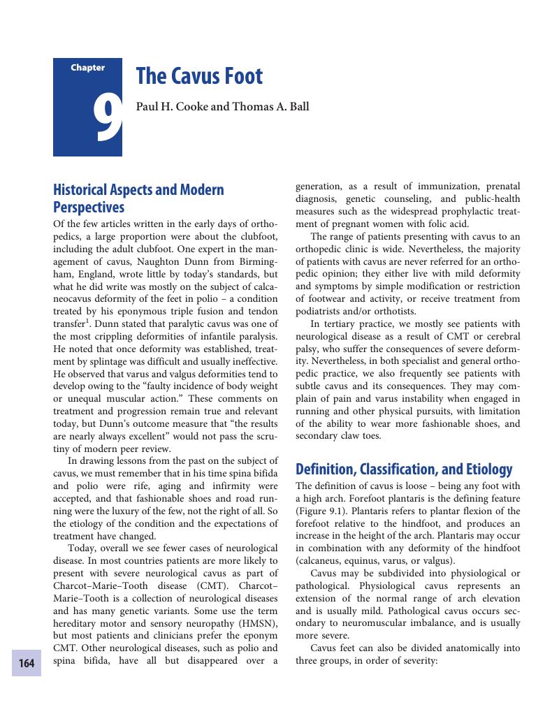
Image of the first page of this content. For PDF version, please use the ‘Save PDF’ preceeding this image.'
Type
Chapter
Information
Publisher: Cambridge University Press
Print publication year: 2018

Access options

Get access to the full version of this content by using one of the access options below. (Log in options will check for institutional or personal access. Content may require purchase if you do not have access.)

References

Dunn, N. Calcaneo cavus and its treatment. J Bone Joint Surg. 1919; 1, 711–21.Google Scholar
Aminian, A, Sangeorzan, BJ. The anatomy of cavus foot deformity. Foot Ankle Clin. 2008; 13, 191–8, v.Google Scholar
Burns, J, Crosbie, J, Ouvrier, R, Hunt, A. Effective orthotic therapy for the painful cavus foot: a randomized controlled trial. J Am Podiatr Med Assoc. 2006; 96, 205–11.Google Scholar
Scherer, PR. Orthotic management of the pes cavus foot. Lower Extremity Review Magazine. 2011.Google Scholar
McCluskey, WP, Lovell, WW, Cummings, RJ. The cavovarus foot deformity. Etiology and management. Clin Orthop Relat Res. 1989; 27–37.CrossRefGoogle Scholar
Duchenne, GB. Physiologie des mouvements démontrée à l’aide de l’expérimentation électrique et de l’observation clinique, et applicable à l’étude des paralysies et des déformations. https://archive.org/stream/physiologiedesm00duch#page/449: (J.-B. Bailliere et Fils, 1867) pp. 526–35.Google Scholar
Sabir, M, Lyttle, D. Pathogenesis of Charcot–Marie–Tooth disease. Gait analysis and electrophysiologic, genetic, histopathologic, and enzyme studies in a kinship. Clin Orthop Relat Res. 1984; 223–35.Google Scholar
Price, AE, Maisel, R, Drennan, JC. Computed tomographic analysis of pes cavus. J Pediatr Orthop. 1993; 13, 646–53.CrossRefGoogle ScholarPubMed
Helliwell, TR, Tynan, M, Hayward, M, Klenerman, L, Whitehouse, G, Edwards, RH. The pathology of the lower leg muscles in pure forefoot pes cavus. Acta Neuropathol. 1995; 89, 552–9.CrossRefGoogle ScholarPubMed
Garceau, GJ, Brahms, MA. A preliminary study of selective plantar-muscle denervation for pes cavus. J Bone Joint Surg Am. 1956; 38, 553–62.Google Scholar
Ledoux, WR, Shofer, JB, Ahroni, JH, Smith, DG, Sangeorzan, BJ, Boyko, EJ. Biomechanical differences among pes cavus, neutrally aligned, and pes planus feet in subjects with diabetes. Foot Ankle Int. 2003; 24, 845–50.CrossRefGoogle ScholarPubMed
Burns, J, Crosbie, J, Hunt, A, Ouvrier, R. The effect of pes cavus on foot pain and plantar pressure. Clin Biomech (Bristol, Avon). 2005; 20, 877–82.Google Scholar
Crosbie, J, Burns, J, Ouvrier, RA. Pressure characteristics in painful pes cavus feet resulting from Charcot–Marie–Tooth disease. Gait Posture. 2008; 28, 545–51.CrossRefGoogle ScholarPubMed
Taylor, A, Porter, D, Cooke, PL. Leg, foot and ankle pain in Charcot–Marie–Tooth disease. J Bone Joint Surg Br. 2003; 85-B, proceedings, 128.Google Scholar
LoPicollo, Mea. Effectiveness of the cavus foot orthosis. J Surg Orthop Adv. 2010; 19, 166–9.Google Scholar
Molloy, JM, Christie, DS, Teyhen, DS, et al. Effect of running shoe type on the distribution and magnitude of plantar pressures in individuals with low- or high-arched feet. J Am Podiatr Med Assoc. 2009; 99, 330–8.Google Scholar
Vlachou, M, Beris, A, Dimitriadis, D. Modified Chuinard–Baskin procedure for managing mild-to-moderate cavus and claw foot deformity in children and adolescents. J Foot Ankle Surg. 2008; 47, 313–20.CrossRefGoogle ScholarPubMed
Swanson, AB, Braune, HS, Coleman, JA. The cavus foot concept of production and treatment by metatarsal osteotomy. J Bone Joint Surg Am. 1966; 48, 1019.Google Scholar
Jahss, MH. Tarsometatarsal truncated-wedge arthrodesis for pes cavus and equinovarus deformity of the fore part of the foot. J Bone Joint Surg Am. 1980; 62, 713–22.Google Scholar
Japas, LM. Surgical treatment of pes cavus by tarsal V-osteotomy. Preliminary report. J Bone Joint Surg Am. 1968; 50, 927–44.Google Scholar
Wilcox, PG, Weiner, DS. The Akron midtarsal dome osteotomy in the treatment of rigid pes cavus: a preliminary review. J Pediatr Orthop. 1985; 5, 333–8.CrossRefGoogle ScholarPubMed
Weiner, DS, Morscher, M, Junko, JT, Jacoby, J, Weiner, B. The Akron dome midfoot osteotomy as a salvage procedure for the treatment of rigid pes cavus: a retrospective review. J Pediatr Orthop. 2008; 28, 6880.Google Scholar
Wülker, N, Hurschler, C. Cavus foot correction in adults by dorsal closing wedge osteotomy. Foot Ankle Int. 2002; 23, 344–7.Google Scholar
Naudi, S, Dauplat, G, Staquet, V, Parent, S, Mehdi, N, Maynou, C. Anterior tarsectomy long-term results in adult pes cavus. Orthop Traumatol Surg Res. 2009; 95, 293300.CrossRefGoogle ScholarPubMed
Dwyer, FC. Osteotomy of the calcaneum for pes cavus. J Bone Joint Surg Br. 1959; 41-B, 80–6.Google ScholarPubMed
Kendal, A, Ball, T, Rogers, M, Cooke, P, Sharp, R. Minimally invasive calcaneal osteotomy: a safe alternative to open calcaneal osteotomy with fewer complications. Foot Ankle Int. 2015; 36, 685–90.Google Scholar
Wetmore, RS, Drennan, JC. Long term results of triple arthrodesis in CMT disease. J Bone Joint Surg Am. 1989; 71, 417–22.CrossRefGoogle Scholar
Roper, BA, Tibrewal, SB. Soft tissue surgery in Charcot–Marie–Tooth disease. J Bone Joint Surg Br. 1989; 71, 1720.Google Scholar
Gould, N. Surgery in advanced Charcot–Marie–Tooth disease. Foot Ankle. 1984; 4, 267273.Google Scholar
Ward, CM, Dolan, LA, Bennett, DL, Morcuende, JA, Cooper, RR. Long-term results of reconstruction for treatment of a flexible cavovarus foot in Charcot–Marie–Tooth disease. J Bone Joint Surg Am. 2008; 90, 2631–42.Google Scholar
Leeuwesteijn, AEEPM, de Visser, E, Louwerens, JWK. Flexible cavovarus feet in Charcot–Marie–Tooth disease treated with first ray proximal dorsiflexion osteotomy combined with soft tissue surgery: a short-term to mid-term outcome study. Foot Ankle Surg. 2010; 16, 142–7.Google Scholar

Save book to Kindle

To save this book to your Kindle, first ensure coreplatform@cambridge.org is added to your Approved Personal Document E-mail List under your Personal Document Settings on the Manage Your Content and Devices page of your Amazon account. Then enter the ‘name’ part of your Kindle email address below. Find out more about saving to your Kindle.

Note you can select to save to either the @free.kindle.com or @kindle.com variations. ‘@free.kindle.com’ emails are free but can only be saved to your device when it is connected to wi-fi. ‘@kindle.com’ emails can be delivered even when you are not connected to wi-fi, but note that service fees apply.

Find out more about the Kindle Personal Document Service.

Available formats
×

Save book to Dropbox

To save content items to your account, please confirm that you agree to abide by our usage policies. If this is the first time you use this feature, you will be asked to authorise Cambridge Core to connect with your account. Find out more about saving content to Dropbox.

Available formats
×

Save book to Google Drive

To save content items to your account, please confirm that you agree to abide by our usage policies. If this is the first time you use this feature, you will be asked to authorise Cambridge Core to connect with your account. Find out more about saving content to Google Drive.

Available formats
×