


Introduction
After their crystallization from magmatic melt, minerals are incorporated as major or minor constituents in the texture of intrusive or extrusive rocks. In the solid state, many mineral phases undergo various processes of chemical transformation, which may result in partial or complete destruction of the crystal structure [Reference Ewing1,Reference Weber2]. Concerning the accessory mineral zircon, which contributes about 0.05–0.1% to the rock bulk mass, two different types of chemical destruction can be observed: (1) disintegration of the crystal structure by radioactivity and (2) crystal dissolution by metamorphic fluid phases [Reference Ewing1–Reference Sturm4].
The first phenomenon, which is commonly known as “metamictization,” causes the continuous transformation of the regular crystal structure into an amorphous mass (Figure 1a). Thereby, three self-contained processes play an essential role: the first process represents the radiation-induced crack of chemical compounds within different structural units, resulting in a permanent destabilization of the crystal lattice; the second process describes the collision of α-particles (He2+) with various components of the crystal structure, which additionally stimulates structural disintegration; and the third process includes the recoil of radioactive atomic nuclei due to the emission of α-particles. Small dislocations resulting from this effect may produce point defects or defect clusters in the crystal structure [Reference Hanchar and Hoskin5–Reference Nasdala7].

Figure 1: Graphic illustrating the two processes of chemical crystal destruction outlined in this contribution.
Concerning the dissolution of zircon crystals due to chemical reaction with the fluid phases, two self-contained processes may be distinguished (Figure 1b). The first process describes the chemical reaction between the solid and fluid phases causing the generation of deep corrosion pits. The intensity of crystal dissolution depends on the temperature and pressure acting upon the host rock and the amount of fluid phases. The second process includes refilling of the corrosion pits due to the recrystallization of dissolved material, which is highly incompatible with other minerals [Reference Sturm3,Reference Sturm4].
In the contribution presented here, the phenomena of metamictization and fluid-induced crystal dissolution in accessory zircon are demonstrated by using cross sections of single mineral grains. Visualization of crystal destruction was carried out by application of backscattered-electron imaging (BSEI), representing an appropriate technique in material sciences.
Materials and Methods
Preparation of the zircon crystals was conducted according to a well-defined standard procedure [Reference Benisek and Finger8–Reference Sturm10]. In the first step, selected mineral grains were fixed on the central line of a glass slide (4 × 2 cm) by using a 2-component adhesive (for example, Köropax 431). The crystal axes were aligned parallel to each other and parallel to the longer side of the slide. After fixation of the grains, the whole preparation was covered with a 2 mm thick layer of the adhesive. The second step included the halving of the preparation (and crystals) by application of an extensive process of polishing. The halved preparation was mounted on another glass slide in erect position, again using the 2-component adhesive. In the last step, the erected part of the preparation was polished from the other side to a final thickness of 35 μm (Figure 2). Working progress was permanently controlled using a stereomicroscope.

Figure 2: Single steps of crystal preparation required to obtain a cross section: (1) fixation of the crystals on a glass slide; (2) covering with a 2-component adhesive; (3) halving of the preparation; (4) fixation of the halved preparation in erect position on another slide; (5) coarse polishing; and (6) fine polishing and documentation.
For microscopy documentation of the cross sections of single zircon crystals, the preparations had to be covered with a thin layer of carbon and inserted into the vacuum chamber of an electron microprobe (JEOL JXA-6800). Visualization of various phenomena of destruction was achieved by using the BSEI mode and a standard setup (accelerating voltage: 15 kV, beam current: 30–40 nA, beam diameter: 1 μm). Photographs of the crystal sections were made with a digital camera (NIKON D-60) integrated in the microprobe.
Results and Discussion
The electron micrographs clearly show that metamictization of accessory zircon represents a multi-stage process starting with small point defects and defect clusters in the crystal structure and ending with a completely amorphous core area (Figure 3a). The initial defect clusters are subject to continuous concentric expansion, whereby radiation-induced transformation of the crystal lattice often begins in the central part of the mineral grains, where an accumulation of incompatible (radioactive) elements can be observed [Reference Ewing1,Reference Weber2]. Single stages of this structural modification can be appropriately analyzed in the BSE image by respective variation of the contrast setup. The results obtained from electron microscopy correspond with descriptions reported in the literature [Reference Hanchar and Hoskin5–Reference Nasdala7]. According to previous studies, zircon crystals affected by extensive metamictization are additionally characterized by dark discoloration and significant modification of several physical and chemical properties.

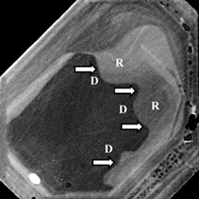
Figure 3: BSE images of zircon cross sections for the documentation of chemical destruction affecting the internal structure: (a) zircon grain affected by extensive metamictization (area within the dashed line; MZ = metamictic zone; arrows mark point defects); (b) zircon crystal affected by remarkable chemical dissolution (D, arrows) and subsequent recrystallization (R). Scale bars = 20 μm.
Chemical dissolution of accessory zircon is seen in the BSE images as a complex phenomenon with significant effects on the crystal structure (Figure 3b). Corrosion pits commonly occur in the rim areas of the crystals and are mostly marked by refilling with recrystallized matter. In most cases, zircon grains exhibiting extreme traces of chemical dissolution were subjected to high-temperature metamorphism (anatexis) causing the partial melting of the host rock and the generation of high amounts of fluid phases. These phases were responsible for both the dissolution and subsequent recrystallization process, whereby the first phenomenon took place during the heating period and the second during the cooling-off period [Reference Sturm3–Reference Hanchar and Hoskin5].
Conclusion
Metamictization and fluid-induced chemical dissolution represent two processes of crystal destruction, which can be most efficiently studied by application of (1) appropriate crystal preparation methods and (2) specific electron microscopy visualization techniques. In the exemplary case of accessory zircon, radiation and fluid-induced destruction mainly affect the internal crystal structure, which is best exposed by the production of cross sections. Both phenomena represent multi-stage transformations, which may remarkably change the physical and chemical properties of the mineral.


