

The study of low-frequency brain signals, which is important for the diagnosis of many medical conditions, is limited by the microelectrode materials currently available. Now, researchers have shown that these infraslow brain waves, that is, brain activity that occurs at frequencies less than 0.1 Hz, can be monitored with high spatial resolution using graphene transistors.
From the outside, the brain looks like a colony of neurons that communicate with each other in some sort of bizarre electrical language. Chemical conditions inside and around a neuron decide whether it will respond to or ignore an incoming signal from another neuron. If the conditions are right, a traveling electric potential is generated that moves across the brain. Most neural activity that has been studied so far is fast—greater than 1 Hz. However, infraslow activity (ISA) is becoming recognized for its involvement in a number of electrophysiological states such as sleep, coma, wakefulness, and anesthesia. Among these slow-moving brain murmurs are such ominously named entities as “spreading depolarizations” and “cortical spreading depressions” or CSDs. These slow-moving waves are often called brain tsunamis, which depolarize neurons and shut down large sections of the brain. CSDs are often triggered in cases of stroke or brain injury as well as during migraines and epileptic seizures.
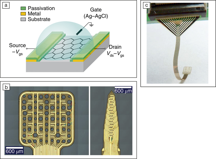
(a) Schematic of a flexible graphene solution-gated field-effect transistor (gSGFET) array technology; (b) microscope images of the active area of a 4 × 4 gSGFET array and a 15-channel intracortical array; and (c) photograph of the neural probe after peeling from the wafer and being introduced into a zero insertion force connector to interface with recording electronics. Credit: Nature Materials.
“In humans, terminal spreading depolarization (SD) has been measured within minutes following circulatory arrest and during the development of brain death. Transient SDs have been recorded in 90–100% of patients with severe stroke, 60–80% of patients with brain hemorrhage, and about 50% of patients with severe traumatic brain injury,” Jens P. Dreier of the Center for Stroke Research Berlin told MRS Bulletin. The study of these low-frequency brain signals are therefore important for diagnosis and cure in neurocritical care.
In a recent issue of Nature Materials (doi:10.1038/s41563-018-0249-4), Eduard Masvidal-Codina and colleagues, led by Anton Guimerà-Brunet at the Institute of Microelectronics of Barcelona (IMB-CNM, CSIC) and Jose A. Garrido at the Catalan Institution for Research and Advanced Studies (ICREA), reported their study of graphene microtransistors for studying low-frequency brain signals. The unique geometry of these devices allows good integration with the tissue of the brain and can cover a significant area, allowing for comprehensive monitoring. The research team developed a graph-ene solution-gated field-effect transistor (gSGFET) that used a graphene sheet to mediate the conduction between the source and the drain. The potential of the neural tissue changes the resistance of the graphene channel that is then read from the current variation. These graphene transistors not only have a high surface-to-volume ratio—enabling greater coverage of the brain tissue than was possible previously—but also function as signal amplifiers, boosting the signal-to-noise ratio.
“Mapping infraslow activity with high-fidelity and spatial resolution simultaneously with local field potentials with non-cytotoxic materials was not previously possible and is a technological advance provided by graphene microtransistors,” the researchers commented in a communication to MRS Bulletin; “Combined with the mapping capabilities, this could allow determining the role of different brain structures on infraslow signals.”
The use of transistors also removes another fundamental bottleneck in high fidelity measurements of brain signals. Current technology requires each electrode to be attached to a wire—limiting the space available for exploration. Due to the multiplexing capability of transistor technology, this limitation can be overcome. As the recording sensor is an active electronic device, it can be easily arranged in an array to implement multiplexing strategies that ultimately lead to a significant reduction in the number of connectors. “We envision that if the results of these studies are positive, this technology could be applied by neurosurgeons in the clinic for a more precise detection of the onset of certain epileptic seizures,” the research team says. The device is pending patent approval.
Other researchers in the field have reacted positively to this study. Robert Wykes, in the Department of Clinical and Experimental Epilepsy at University College London, says, “I believe that gSGFETs will aid preclinical studies aimed at understanding the importance of DC shifts and infraslow activity to seizure initiation and termination. Additionally, these devices could be used clinically during pre-surgical monitoring as studies have shown that favorable surgical outcome correlates with resection of areas generating ictal [relating to a seizure] baseline shifts.”
Dreier concurs. “The results are very promising as this material seems to have the potential to perform intracranial monitoring in humans with much higher accuracy than before. This certainly deserves further study and development.” Both Wykes and Dreier are not involved with this study.
