


Executive Summary
This executive summary will provide a brief overview of the status of international point of care ultrasound (PoCUS) curriculum development in emergency medicine and will outline the educational processes and content recommended by the International Federation for Emergency Medicine (IFEM) for PoCUS.
Definition: Point of Care Ultrasound (PoCUS; also referred to as emergency, focused, or clinician-performed ultrasound) is defined as “diagnostic or procedural guidance ultrasound that is performed by a clinician during a patient encounter to help guide the evaluation and management of the patient.”
The content of the IFEM POCUS Curriculum can be grouped into two main sections: the selection of PoCUS applications to be included, and the methodology required for training and skills maintenance in each application.
Application selection
Every curriculum should include two mandatory applications (or module): the demonstration of how to generate and optimize an ultrasound image and demonstration of good practice in PoCUS which includes image quality assurance and competency assessments for the clinician. This forms the basis for training in all further applications. Mandatory core (or basic) applications are selected to complement local emergency medicine practice. Training bodies will consider disease prevalence, impact of disease, potential for patient benefit and resources when deciding what core applications to include. Optional enhanced (or advanced) applications are selected to encourage skill development and progression of PoCUS practice. Many PoCUS applications may be used by emergency physicians, and their non-inclusion in the core applications should not diminish their importance in practice.
All applications should be defined as diagnostic or procedural. Diagnostic applications can be single area applications (e.g., assessment of the abdominal aorta) or multiple area applications (e.g., shock assessment). Procedural applications should be selected for their ability to improve patient and procedural safety and efficiency. The curriculum describes each application in terms of what it is, potential benefits, equipment requirements, required knowledge of anatomy and pathology, as well as skills required.
Training methodology
Training in each application of PoCUS is divided into four stages: introduction, gaining experience, confirmation of competency, and skills maintenance.
Each application should be introduced by either delivery of recognized course material, online learning, modular learning, or by a combination of these methods. Further experience must be gained by a combination of scanning real patients with supervision (or proctoring), simulation, or by undertaking dedicated PoCUS fellowship training. It is recognized that learning curves vary for each individual and as such it is important that this stage optimizes the skills of image generation, recognition and clinical integration. Assessment of competency for each PoCUS application should be comprehensive and structured. A template global assessment form provides an assessment structure that includes task specific checklists, unique for each module, compiled using Delphi methodology amongst experts, and a global rating scale to assess good clinical practice (standard for all modules). Skills maintenance is essential and is achieved by continuous quality improvement methods. Hiatuses in performing a particular application may result in loss of skill. Peer review and audit has an important role in demonstrating continued competency. Keeping up to date with the latest medical research in this area is essential.
This process is summarized in Figure 1.

Figure 1 Essential components of an emergency PoCUS curriculum.
Purpose
This IFEM curriculum defines the educational framework, goals and objectives, content, experience, and desired outcomes of an educational program, which will provide the foundational knowledge in point of care ultrasound required of any specialist in emergency medicine (EM).
The IFEM PoCUS Curriculum is an evidence-based consensus document drawing on regional and national guidelines already in place internationally. It presents PoCUS as a complementary diagnostic tool that can help make clinicians more efficient, more independent, and more confident in their medical decision-making.
The curriculum applies to PoCUS training during residency/specialty training and beyond into clinical practice. It is anticipated that most EM trainees should become familiar with the theoretical principles of ultrasound early in specialty training. Training and assessment for PoCUS should be included in emergency medicine curricula and specialty examinations.
Core PoCUS competency should be a universal skill for emergency physicians that will not require special or additional education and assessment outside a standard EM training program or residency.
The curriculum provides a structure to ensure educational programs achieve appropriate selection of PoCUS applications; to support the delivery of comprehensive PoCUS programs that include adequate training; consistent credentialing or certification; and outline effective governance and continuous quality improvement; and to encourage the integration of PoCUS into the practice of emergency physicians worldwide. IFEM is tasked with supporting the international development of training and practice in PoCUS regardless of region and socioeconomic issues. This document will help authors of existing curricula ensure that key components are incorporated. It will also help regions that have no existing PoCUS curriculum to establish one. Providing a common structure and theme for the content and methodology of PoCUS curricula may allow transferability of PoCUS skills internationally leading to establishment of reciprocity arrangements and a more uniform educational approach.
At the completion of PoCUS training, emergency physicians will demonstrate the Knowledge, Skills and Behaviour enabling them to:
-
∙ Generate and optimize ultrasound images;
-
∙ Interpret the images;
-
∙ Incorporate the information provided by the images into clinical decision-making;
-
∙ Maintain their skills;
-
∙ Support the training of other physicians in PoCUS.
A checklist of curriculum elements that should be included in a PoCUS curriculum can be found in Appendix A. IFEM will not accredit curricula — this should be a self-regulating step and an important part of developing a mature program — but are are happy to provide advice via the USIG Chair.
Background
The use of PoCUS as an adjunct to the practice of emergency medicine is now well established internationally. Initially, evidence to support the use of PoCUS came from the management of blunt trauma patients. The scope of practice has expanded as emergency physicians have identified further clinical problems where PoCUS is able to aid diagnosis and guide procedures.
A review of currently available PoCUS curricula and guidelines highlights that most divide applications into core or basic, and advanced or extended. Core applications cover traditional clinical questions, where there is a body of evidence of clinical relevance, and that are within the likely skill set of a novice practitioner. Common core applications include:
-
∙ Trauma Ultrasound (Extended Focused Assessment with Sonography for Trauma (E-FAST), including pericardial, peritoneal and pleural fluid detection, along with assessment for pneumothorax),
-
∙ Assessment of the Abdominal Aorta,
-
∙ Focused Cardiac Assessment (or Echo in Life Support),
-
∙ Pelvic Early Pregnancy Assessment,
-
∙ Ultrasound Guided Vascular Access (or Procedural Guidance).
Other applications that are often considered as core include:
-
∙ Thoracic assessment,
-
∙ Biliary assessment,
-
∙ Urinary Tract assessment,
-
∙ DVT assessment,
-
∙ Soft-tissue/musculoskeletal assessment,
-
∙ Ocular assessment.
Examples of other appropriate applications include:
Diagnostic
-
∙ Evaluation of left ventricular function,
-
∙ Volume depletion/IVC assessment,
-
∙ Jugular venous distension,
-
∙ Undifferentiated hypotension, shortness of breath, chest pain,
-
∙ Testicular pain,
-
∙ Joint effusion and tendon rupture;
Procedural
-
∙ Thoracentesis,
-
∙ Paracentesis,
-
∙ Pericardiocentesis,
-
∙ Lumbar puncture,
-
∙ Cutaneous and peritonsillar abscess drainage,
-
∙ Foreign body removal,
-
∙ Pediatric bladder catheterization,
-
∙ Joint aspiration,
-
∙ Temporary pacemaker placement,
-
∙ Regional anesthesia,
-
∙ Confirmation of endotracheal tube placement.
Most curricula require initial theoretical and practical education, followed by a period of supervised experience with widely varying numbers of recommended scans, followed by some form of competency assessment. With a plethora of PoCUS applications available, it is important to ensure that PoCUS can be delivered with adherence to good governance principles.
What Pocus Applications to Include?
Training programs for PoCUS in EM must include mandatory introductory and core applications based upon defined criteria. The majority of emergency physicians should be able to generate the required image, interpret the image and apply the findings to patient care for all core applications. Recommendations for optional enhanced applications are provided.
The anatomy of a PoCUS application
For each application it is important to understand what it is, when to use it, and why and how it is of benefit to EM practice. Hence the anatomy of each application needs to include details of what trainees will learn with regards to Knowledge, Skills, and Behaviour.
Demonstration of how to generate and optimize an image
It is imperative that trainees have a foundation in understanding how ultrasound works, safety issues, ergonomic considerations and how to operate and maintain the equipment. IFEM advocates that this topic is included as an application in its own right even though this is not a clinically directed module. Below is a general guide to the level of understanding expected:
Knowledge
-
∙ The basic components of an ultrasound system;
-
∙ Types of transducer and the production of ultrasound, with an emphasis on operator controlled variables;
-
∙ Use of ultrasound controls;
-
∙ Know the frequencies used in medical ultrasound and the effect on image quality and penetration;
-
∙ The interaction of ultrasound with tissue including biological effects;
-
∙ Safety issues in ultrasound;
-
∙ The basic principles of real time and Doppler ultrasound including color flow and power Doppler;
-
∙ The recognition and explanation of common artefacts;
-
∙ Image recording systems.
Skills
-
∙ Operation of the key machine controls;
-
∙ Transducer changing;
-
∙ Image manipulation and storage.
Behaviour
-
∙ Safe practice;
-
∙ Limitations of own skills;
-
∙ Integration of ultrasound findings with clinical assessment.
Demonstration of good practice in emergency ultrasound
Every user of ultrasound should embrace principles of good practice and governance or continuing quality improvement. IFEM advocates that this is included as an application in its own right even though this is not a clinically directed module. Below is a general guide to the level of understanding expected:
Knowledge
-
∙ Image recording, storing and filing;
-
∙ Reporting;
-
∙ Medico-legal aspects – outlining the responsibility to practise within specific levels of competence and the requirements for training;
-
∙ Consent issues;
-
∙ The value and role of departmental protocols;
-
∙ The resource implications of ultrasound use.
Skills
-
∙ Integrate PoCUS into departmental continuous quality improvement governance system
Behaviour
-
∙ Adherence to a rule-in philosophy (namely, that a focused ultrasound exam often rules-in a pathology but may be unable to rule it out due to a higher specificity than sensitivity of the application).
Core clinical applications
In general, core clinical PoCUS applications:
-
∙ Are simple to learn, perform and interpret;
-
∙ Are rapid to perform;
-
∙ Answer simple questions, ideally with binary (yes/no) answers;
-
∙ Allow learners to consolidate key ultrasound skills, which provide a solid foundation to their practice;
-
∙ Have significant impact in the area/region being practiced due to burden of disease, local resources or mortality/morbidity considerations.
Examples of recommended core applications include:
-
∙ Basic cardiac
-
○ Is the heart beating normally?
-
○ Is there pericardial fluid?
-
○ Is there an enlarged right ventricle?
-
○ Is the left ventricle enlarged?
-
-
∙ Basic trauma/E-FAST
-
○ Is there free fluid in the pleural/pericardial/peritoneal space?
-
○ Is there a pneumothorax?
-
-
∙ Simple assessment of fluid status
-
○ Is the inferior vena cava (IVC) full or empty?
-
-
∙ Looking for sites of bleeding
-
○ Is there an abdominal aortic aneurysm?
-
-
∙ Simple lung assessment
-
○ Is there a pneumothorax?
-
○ Is there pleural fluid?
-
-
∙ Simple early pregnancy assessment
-
○ Is there an intra-uterine pregnancy?
-
-
∙ Simple venous occlusive assessment
-
○ Is there an occlusive above-knee DVT?
-
-
∙ Simple procedure guidance
-
○ Peripheral and central line insertion
-
Enhanced clinical applications
In general, enhanced PoCUS applications are those that:
-
∙ Are more difficult to learn, perform and interpret;
-
∙ Require prior proficiency in a related more basic core application;
-
∙ Answer more complex questions;
-
∙ Or, they may be simple to learn but have less impact in the area/region being practiced due to different burdens of disease, local resources or mortality/morbidity considerations.
Examples of enhanced applications include:
-
∙ Intermediate and advanced echocardiography
-
○ Is there valvular disease?
-
○ Is there a segmental wall motion abnormality in the left ventricle?
-
-
∙ Advanced trauma
-
○ Is there solid organ injury?
-
-
∙ Advanced Lung
-
○ Are the lungs wet or dry?
-
○ Is there consolidation?
-
○ Is there pleural thickening?
-
-
∙ Comprehensive venous occlusive assessment
-
○ Is there a below-knee DVT?
-
Choosing and defining what applications should be included
Principles that can be used when deciding which applications to include as core or enhanced include:consideration for local patterns of disease (e.g., if diagnosis of pleural fluid is required frequently, ensure that this is included in the curriculum in addition to peritoneal and pericardial fluid); and complimenting the skills of the provider, and accommodating their needs (e.g., emergency physicians may not need to learn obstetric applications in some health care systems, whereas in other systems this may be the single most important application).
Applications can be divided by Organ System, Disease System and Procedure Type, and then by Core and Enhanced knowledge. Facilitating a “building-block” approach to PoCUS education by building enhanced applications on the skills learned with core applications.
As such, a beginner may become competent in scanning a particular organ system for simple but important pathology (e.g., pericardial effusion), while an advanced user may develop competency to scan the same organ system for other more complex pathology (valvular pathology). A comprehensive table of applications and suggested curriculum content is provided in Supplementary Table 1.
Training Requirements
Training
This section of the curriculum provides an overview of the three essential training steps required for PoCUS for any specific application, namely: initial introduction, gaining experience and achieving competency (see Figure 2). Formalizing these steps into a well-structured training programme improves long term learning outcomes. The final make up of a specific region’s training programme and curriculum will be influenced by local factors/logistics and should therefore be flexible in order to optimally incorporate the three basic learning steps.
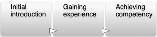
Figure 2 Essential Steps in PoCUS Training
Initial introduction
The initial introduction is the first contact session between trainer and trainee, which should focus on the providing the core knowledge of the application, demonstration of the skill and the first time practice under trainer supervision. The formal introductory course is most commonly used but should include reading materials such as online learning, video resources. Simulation or other variations may be used due to local resource and logistical challenges. Recommended teaching formats include:
-
∙ Short lectures,
-
∙ Demonstrations,
-
∙ Hands on skills teaching,
-
∙ Simulation sessions,
-
∙ Open and closed discussions,
-
∙ Passive and interactive web based learning,
-
∙ Practical scanning on real patients.
Gaining experience
Gaining experience should be based on the use of an ultrasound machine on actual patients if possible. The key features of this phase are focused around optimizing the trainee’s skill in:
-
∙ Image acquisition,
-
∙ Image interpretation,
-
∙ Clinical integration.
Image acquisition
We recommend supervision by a clinician trained in PoCUS or by a sonographer while gaining experience in image acquisition. There is no universal number of scans required to achieve competency, as this is operator and situation dependent and may range from 10 to 50 or more scans per application. Accumulation of experience in PoCUS in general is likely to improve skills globally, in addition to gaining experience specific to each particular application. Alternatives to proctored scanning include scanning patients who are undergoing diagnostic imaging (computed tomography scan, formal echocardiogram, or any other imaging test). There are services that will review images for new clinicians for a fee and offer remote mentoring via web based image portals. The increasing wireless capabilities of ultrasound machines mean that images can be transmitted in real time and several studies have demonstrated that streaming to web based interactive portals like Skype, FaceTime, etc. can be ways of getting image feedback and mentoring even if a local expert is not available in one’s own practice environment. However, one must be mindful of not breaching patient confidentiality and adhering to local information governance arrangements.
Finally, there are increasing numbers of simulators that can provide opportunities for practice – especially for scans that are invasive and involve some discomfort to the patient (i.e. transesophageal echocardiography or transvaginal sonography) it may be to the novice scanner’s advantage to practice initially on a simulator.
Image interpretation
Gaining experience interpreting images is part of the process of learning how to obtain a good image. The immediate feedback of formal diagnostic imaging for the patient and the new clinician sonographer can help to confirm initial image interpretation made at the bedside and a new clinician sonographer can gain experience and confidence by utilizing formal diagnostic imaging in the early phase of their training as a check. There are also increasing numbers of on-line and web-based image banks where new clinician sonographer can practice their interpretation skills [see Supplementary Table 1]. Some websites and portals have competency assessment tests that the new clinician sonographer can take to provide evidence to their practice governing board or hospital administration as evidence of their growing competency.
Clinical integration
The trainee should be able to apply the findings of the application to clinical practice. It is essential to understand the clinical question or circumstance. It is also necessary to be familiar with the quoted accuracies in the medical literature as this provides a greater understanding of the limitations or strengths of the application. For example:
-
∙ Highly sensitive tests are good at ruling out pathology (e.g., seeing a normal calibre abdominal aorta from the proximal aspect to the distal bifurcation effectively rules out an abdominal aortic aneurysm);
-
∙ Highly specific tests are good at ruling in pathology (e.g., seeing free intra-peritoneal fluid during a focused assessment with sonography scan (FAST) effectively confirms free fluid is present).
Achieving competency
The final phase is assessment to determine if the PoCUS trainee has achieved competence in performing, interpreting and incorporating the application. There are various methods available to assess competence. Current evidence supports the use of a task specific checklist (unique to each module) and a global rating scale (good clinical practice) irrespective of the assessment method chosen. The curriculum template assessment forms can be adapted and incorporated into local curricula and assessment processes.
These provide the opportunity for local facilities to adapt PoCUS assessment process to their local needs, allowing comprehensive competency assessment and quality assurance.
The assessment tools provided include 2 components (shown in Table 1 and Table 2):
-
1. Task specific checklists, unique for each module. These should be be compiled using a Delphi methodology amongst experts for a specific ultrasound module.
-
2. Global rating scale to assess good clinical practice (standard for all modules).
Table 1 Example of image demonstration tasks required for FAST examination

* Adapted with permission from College of Emergency Medicine Emergency Medicine Ultrasound Core (Level 1) competency Workplace Based Triggered Assessment – 2010, Adapted from de Cossart and Fish 2005©
Table 2 Example of global assessment for a PoCUS application

* Endorsed by the International Federation for Emergency Medicine; Adapted with permission from College of Emergency Medicine Emergency Medicine Ultrasound Core (Level 1) competency Workplace Based Triggered Assessment – 2010, Adapted from de Cossart and Fish 2005©
IFEM recognizes the variation in current methods of certification and credentialing internationally. The application-specific competency based assessment process recommended here provides a flexible yet standardized approach to skills assessment. IFEM does not provide direct certification of competency, rather it supports local assessment in keeping with agreed applications, standards and skill sets. Further examples of assessment documentation can be found in Appendix B.
Skill Maintenance and Continuous Quality Improvement
It is essential to ensure maintenance of skills and personal quality assurance after competency has been achieved. IFEM recommends continuous logging of activity (before and after assessment of competency/credentialing), quality assurance with peers and regular PoCUS continuing medical education (CME)/continuing professional development (CPD) to keep up to date.
The amount of CME/CPD required to maintain competency is related to the number of applications being utilized, the frequency of use, and other developments in emergency ultrasound and emergency medicine at large. In general, those in charge of ultrasound programs should have at least 5 to 10 hours of CME/CPD credits pertaining to ultrasound activities per year including conference attendance, online educational activities, preceptorships, teaching, research, hands-on teaching, administration, quality assurance/audit, image review, in-service examinations, textbook and journal readings, morbidity and mortality conferences inclusive of ultrasound cases, or others.
Individual credentialed physicians should have 2.5 to 5 hours of the above continuing educational ultrasound activities per year. Educational sessions that integrate ultrasound into the practice of EM are encouraged, and do not have to be didactic in nature but can be participatory.
Conclusion
The IFEM Point of Care Ultrasound (PoCUS) Curriculum provides an overview of the processes involved in the development of PoCUS training programs, resources that can be adapted for local training and competency assessment, and a statement of the principles that should guide the on-going integration of PoCUS into the practice of emergency physicians across the world. The practice of emergency medicine varies internationally, with marked differences in the spectrum of disease, available resources, and institutional structures. As a result, a one-size curriculum does not fit all, and regional processes require optimization to accommodate such differences. However, there are essential principles and processes that are required in all good curricula; the IFEM PoCUS Curriculum provides guidance on how a curriculum should be constructed and what content should be chosen. It provides a framework for good practice in PoCUS training, certification and skills maintenance, enabling institutions to develop programs that suit their needs, yet conform to accepted international standards. We encourage use of the curriculum and the accompanying assessment documents.
Acknowledgments
This article was published on behalf of the IFEM Ultrasound Special interest Group, which includes Paul Atkinson (Canada), Justin Bowra (Australia), Raoul Breitkreutz (Germany), Arif Cevik (Turkey), Toh Hong Chuen (Singapore), Jim Connolly (United Kingdom), Rip Gangahar (Chair) (United Kingdom), Bob Jarman (Vice-Chair and Project Lead) (United Kingdom), Mike Lambert (United States of America), Hein Lamprecht (South Africa), Vicki Noble (United States of America), and Ang Shiang Hu (Singapore). The Ultrasound Special Interest Group (USIG) was tasked by the Clinical Practice Committee (CPC) of the International Federation of Emergency Medicine (IFEM) to produce curriculum guidance for ultrasound training in emergency medicine (EM). The document was written by the Point-of-Care Ultrasound Curriculum Project Team (PoCUS-CPT).
Supplementary material
To view supplementary material for this article, please visit http://dx.doi.org/10.1017/cem.2015.8


