


Case report
A full-term newborn was transferred to our hospital for the cardiological evaluation because of hypoxia resistant to ventilatory therapy with the addition of nitric oxide.
At birth, the body weight was 2.770 kg, the APGAR score was 9’–10’, and peripheral oxygen saturation was 85%. Electrocardiogram was normal. Chest X-ray showed bronchial situs solitus (Fig. 1). Echocardiogram revealed situs solitus, dilated right atrium and right atrial appendage, huge ostium secundum atrial septal defect of 8 mm and right-to-left atrial shunt, and three pulmonary veins draining into the right atrium near to the opening of the superior caval vein without obstruction. The superior caval vein was not dilated. (Figs. 2 and 3).

Figure 1. Posteroanterior chest radiography shows normal lungs, bronchial situs solitus, and normal heart size.
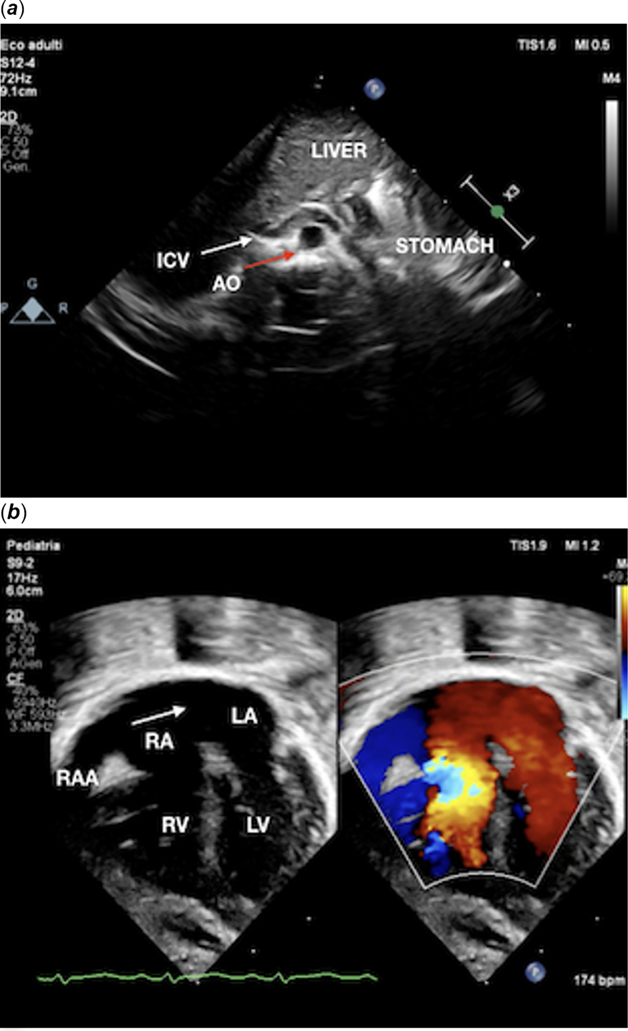
Figure 2. A. Subcostal view shows situs solitus, right-sided liver, left-sided stomach, left-sided aorta (red arrow), and right-sided inferior caval vein (white arrow). B. Four chamber view shows dilated right atrial appendage and right atrium, atrial septal defect (arrow), normal-size left atrium, mildly dilated and hypertrophied right ventricle, normal-size left ventricle, no shunts through the ventricular septum, normal left ventricle, normal left and right outflow tracts, and no pericardial effusion. No pulmonary veins are seen draining to either atrium. AO = aorta; ICV = inferior caval vein; LA = left atrium; LV = left ventricle; RA = right atrium; RAA = right atrial appendage; RV = right ventricle.
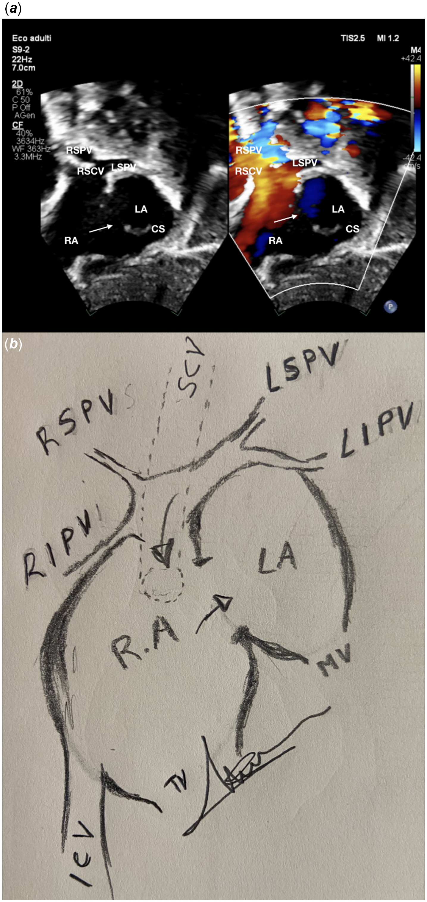
Figure 3. a . Subcostal view shows expanded right atrium and huge atrial septal defect (arrow) of 8 mm with right-to-left shunt; three pulmonary veins (the right superior and inferior veins, and the left superior one). b . Courtesy of Elio Caruso. Manual drawing of subcostal view of total anomalous pulmonary venous returns into the right superior caval vein’s ostium, huge atrial septal defect (arrow) with right-to-left shunt. CS = coronary sinus; ICV = inferior caval vein; LA = left atrium; LIPV = left inferior pulmonary vein; LSPV = left superior pulmonary vein; MV = mitral valve; RA = right atrium; RIPV = right inferior pulmonary vein; RSCV = right superior caval vein; RSPV = right superior pulmonary vein; SCV = superior caval vein; TV = tricuspid valve.
CT scan angiography (Fig. 4c) and maximum intensity projection (Fig. 4d) showed abdominal and bronchial situs solitus and confirmed the total anomalous pulmonary venous returns into the right atrium at the base of the superior caval vein ostium without vertical vein, better appreciable from the volume rendering by Horos® software (Fig. 4a,b). All four pulmonary veins appeared patent.

Figure 4. a , b . CT angiography, volume rendering by Horos® software, antero-posterior view (A) and posterior-anterior view (B), show four pulmonary veins draining at the base of the right superior caval vein’s ostium into the right atrium. c . Chest CT scan shows all four pulmonary veins draining C. CT angiography axial plane shows all four pulmonary veins draining into the right superior caval vein’s ostium in the right atrium. d . CT angiography multiplanar reconstruction with perfusion colour look up table by Horos® software, shows four pulmonary veins draining into the right atrium at the base of the right superior caval vein ostium. AO = aorta; DA = descending aorta; LA = left atrium; LIPV = left inferior pulmonary vein; LSPV = left superior pulmonary vein; LV = left ventricle; PT = pulmonary trunk; RA = right atrium; RAA = right atrial appendage; RIPV = right inferior pulmonary vein; RPA = right pulmonary artery; RIPV = right inferior pulmonary vein; RSCV= right superior caval vein; RSPV = right superior pulmonary vein; RV = right ventricle.
We believe this is an uncommon case report of total anomalous pulmonary venous return to the right atrium at the base of the superior caval vein’s ostium without a sinus venosus defect, in situs solitus, and without vertical vein or a posterior pulmonary venous confluence. Reference Gaynor, Burch, Dollery, Sullivan, Deanfield and Elliott1
The two-dimensional echocardiography with colour Doppler allowed to accurately describe the anatomy of the pulmonary venous drainage; however, advanced imaging scan may be needful for defining the anatomy and confirming the diagnosis. Reference Rocamora Salort, Ruiz González, Cano Sánchez and Fernández Tudela2
Acknowledgements
None.
Author contribution
Elio Caruso wrote article and critical revision.
Silvia Farruggio wrote article and data interpretation.
Norman H Silverman developed concept of publication, critical revision, and approval of article.
Financial support
Additional supporting information may be found in the online version of the article at the publisher’s website.
Competing interests
All authors have read and approved submission of the manuscript and have no conflict of interest to disclose.


