


Introduction
Intestinal helminth infections are among the most prevalent infections, both in humans and animals. In humans, it is estimated that more than 1.5 billion people are infected with 1 or more species of intestinal helminths, especially in Asia, Africa and Latin-America, causing a significant impact on human health, mainly in children (Hotez et al., Reference Hotez, Brindley, Bethony, King, Pearce and Jacobson2008; Weatherhead and Hotez, Reference Weatherhead and Hotez2015). Moreover, intestinal helminths cause a large amount of economic losses due to diminishing productivity and also the cost of anthelminthic treatments (Roeber et al., Reference Roeber, Jex and Gasser2013). Due to the unavailability of vaccines, the development of new control strategies is needed and, in this context, the use of experimental models has served to increase our knowledge on the factors governing the resistance to intestinal helminths.
Evidence from studies using different experimental models suggests that resistance to intestinal helminths relies on the activation of a Th2-type immunity involving the production of interleukin-4 (IL-4), IL-5, IL-9 and IL-13 (Cortés et al., Reference Cortés, Muñoz-Antoli, Esteban and Toledo2017). In this context, IL-25 has been regarded as the main regulatory cytokine responsible for the development of the protective Th2-biased immune response. IL-25 participates in the maintenance of the intestinal homoeostasis, tissue adaptation to external damage and tissue repair (Borowczyk et al., Reference Borowczyk, Shutova, Brembilla and Boehncke2021; Varyani et al., Reference Varyani, Löser, Filbey, Harcus, Drurey, Poveda, Rasid, White, Smyth, Gerbe, Jay and Maizels2022). IL-25 is mainly produced by epithelial tuft cells because of the chemosensory receptors that these cells present (Borowczyk et al., Reference Borowczyk, Shutova, Brembilla and Boehncke2021), linking with type 2 immunity by stimulating production of IL-13 by type 2 innate lymphoid cells which, in turn, induces signal transducer and activator of transcription 6-mediated intestinal alterations leading to resistance to infection (Fallon et al., Reference Fallon, Ballantyne, Mangan, Barlow, Dasvarma, Hewett, McIlgorm, Jolin and McKenzie2006; Owyang et al., Reference Owyang, Zaph, Wilson, Guild, McClanahan, Miller, Cua, Goldschmidt, Hunter and Kastelein2006; Zhao et al., Reference Zhao, Urban, Sun, Stiltz, Morimoto, Notari, Madden, Yang, Grinchuk and Ramalingam2010; Angkasekwinai et al., Reference Angkasekwinai, Srimanote, Wang, Pootong, Sakolvaree, Pattanapanyasat, Chaicumpa, Chaiyaroj and Dong2013, Reference Angkasekwinai, Sodthawon, Jeerawattanawart, Hansakon, Pattanapanyasat and Wang2017; Cortés et al., Reference Cortés, Muñoz-Antoli, Esteban and Toledo2017; Varyani et al., Reference Varyani, Löser, Filbey, Harcus, Drurey, Poveda, Rasid, White, Smyth, Gerbe, Jay and Maizels2022). In fact, emerging evidence shows that IL-25 is required for resistance to intestinal helminth infections (Muñoz-Antoli et al., Reference Muñoz-Antoli, Cortés, Santano, Sotillo, Esteban and Toledo2016a).
Our group showed that mice susceptibility to primary infection with the intestinal trematode Echinostoma caproni depends on the inability of mice to respond to IL-25 production. However, mice develop partial resistance to secondary infection in relation to the innate production of IL-25 as a consequence of the curation of the primary infection (Muñoz-Antoli et al., Reference Muñoz-Antoli, Cortés, Santano, Sotillo, Esteban and Toledo2016a, Reference Muñoz-Antoli, Cortés, Martin-Grau, Fried, Esteban and Toledo2016b). Furthermore, Álvarez-Izquierdo et al. (Reference Álvarez-Izquierdo, Pérez-Crespo, Esteban, Muñoz-Antoli and Toledo2020a) demonstrated that type 2 response to secondary E. caproni infection in mice was unable to develop resistance in the absence of IL-25, confirming the requirement of IL-25 for resistance and showing that the role of this cytokine is not limited to regulatory functions, but also has a critical effector role in the resistance. However, the factors that determine the upregulation of IL-25 are not known, but there are several lines of evidence suggesting that it can be driven by resident gut microbiota (Zaph et al., Reference Zaph, Du, Saenz, Nair, Perrigoue, Taylor, Troy, Kobuley, Kastelein and Cua2008; Sawa et al., Reference Sawa, Lochner, Satoh-Takayama, Dulauroy, Bérard, Kleinschek, Cua, Di Santo and Eberl2011; Donaldson et al., Reference Donaldson, Bradford, Artis and Mabbott2015; Watanabe et al., Reference Watanabe, Gilchrist, Uddin, Burgess, Abhyankar, Moonah, Noor, Donowitz, Schneider and Arju2017). IL-25-mediated intestinal immune regulation is impaired in mice in the absence of microbiota (Sawa et al., Reference Sawa, Lochner, Satoh-Takayama, Dulauroy, Bérard, Kleinschek, Cua, Di Santo and Eberl2011; Donaldson et al., Reference Donaldson, Bradford, Artis and Mabbott2015; Watanabe et al., Reference Watanabe, Gilchrist, Uddin, Burgess, Abhyankar, Moonah, Noor, Donowitz, Schneider and Arju2017). Expression of ileal IL-25 is reduced in germ-free mice compared to wild-type mice, but exposure to environmental microbes induced IL-25 overexpression (Sawa et al., Reference Sawa, Lochner, Satoh-Takayama, Dulauroy, Bérard, Kleinschek, Cua, Di Santo and Eberl2011; Donaldson et al., Reference Donaldson, Bradford, Artis and Mabbott2015). Furthermore, antibiotic treatment of mice significantly decreased the expression of gut IL-25 (Zaph et al., Reference Zaph, Du, Saenz, Nair, Perrigoue, Taylor, Troy, Kobuley, Kastelein and Cua2008).
To investigate the potential role of resident microbiota in the production of IL-25 and the subsequent resistance to infection we have used the experimental model of E. caproni in mice. Echinostoma caproni (Trematoda: Echinostomatidae) is an intestinal trematode with no tissue phase in the definitive host (Fried and Huffman, Reference Fried and Huffman1996; Toledo et al., Reference Toledo, Esteban and Fried2009). Although E. caproni is able to parasitize a wide range of laboratory rodent hosts, its compatibility differs considerably between rodent species. In highly compatible hosts, such as mice, primary infection becomes chronic, while pharmacological healing of the primary infection induces a sudden production of IL-25. Secondary infection in the presence of IL-25 results in partial resistance to the challenge infection (Muñoz-Antoli et al., Reference Muñoz-Antoli, Cortés, Santano, Sotillo, Esteban and Toledo2016a, Reference Muñoz-Antoli, Cortés, Martin-Grau, Fried, Esteban and Toledo2016b; Álvarez-Izquierdo et al., Reference Álvarez-Izquierdo, Pérez-Crespo, Esteban, Muñoz-Antoli and Toledo2020a, Reference Álvarez-Izquierdo, Esteban, Muñoz-Antoli and Toledo2020b). Herein, we analyse the changes in resident microbiota induced by primary E. caproni infection in mice, cure of the primary and secondary infections with the aim to gain further insight into the potential role of microbiota in the production of IL-25 and resistance to infection. This experimental model allows us to analyse 3 different situations: (1) susceptibility in primary infections associated with the lack of IL-25; (2) changes induced by healing of the primary infections and (3) resistance to secondary infection in an environment with elevated levels of IL-25.
Materials and methods
Parasites, hosts and experimental infections
The strain of E. caproni has been described previously (Fujino and Fried, Reference Fujino and Fried1993). Encysted metacercariae of E. caproni were removed from the kidney and pericardial cavity of experimentally infected Biomphalaria glabrata snails and used to infect male ICR (Institute of Cancer Research) mice of the same age and weighing 30–35 g by gastric gavage (50 metacercariae each). The positivity of the infection was determined at necropsy or detection of eggs in stools as described previously (Toledo et al., Reference Toledo, Espert, Carpena, Munoz-Antoli, Fried and Esteban2004).
Primary and secondary experimental infections
A total of 15 mice were infected and randomly allocated to 3 groups (infected, treated and secondarily infected) of 5 mice each. The characteristics of the primary infections were studied in the animals in the primary infected group. These animals were maintained as infected without additional treatment until the end of the experiment at 10 weeks post-primary infection (wppi). At 4 wppi, animals in the other 2 groups were treated with a double dose of praziquantel (pzq) of 100 mg kg−1 (pzq-treated and secondarily infected groups), orally administered on alternate days as described previously (Muñoz-Antoli et al., Reference Muñoz-Antoli, Cortés, Santano, Sotillo, Esteban and Toledo2016a, Reference Muñoz-Antoli, Cortés, Martin-Grau, Fried, Esteban and Toledo2016b). All mice belonging to these 2 groups responded to the treatment and reverted to the negative state as determined by coprological examination. Two weeks after treatment, all mice in the secondarily infected group were challenged with 50 metacercariae of E. caproni following the same procedure used in primary infections. Additionally, 5 mice were maintained uninfected and used as naïve controls. At 4, 6 and 10 wppi, stools from each individual mouse were collected to investigate the resident microbiota. The influence of the pharmacological treatment over the studied parameters was ignored since 4 additional mice were left uninfected, treated with pzq as described above and analysed as in the other animals. All the animals were individually maintained in cages under conventional conditions with food and water ad libitum. All mice were sacrificed and necropsied at the end of the experiment at 10 wppi.
The effect of the infection, pzq-treatment and reinfection on IL-25 production and its potential relation to changes in microbiota composition was studied on 45 additional mice. These animals were randomly allocated into 3 groups as described above (infected, treated and secondarily infected) of 15 each and the procedures described above were followed for each group. At 2, 4, 5, 6 and 10 wppi, 3 animals from each group were sacrificed and necropsied to study the production of IL-25.
Induction of intestinal dysbacteriosis and IL-25 production
To analyse the effect of resident microbiota on the production of IL-25 in response to secondary E. caproni infections, a total of 10 mice were primarily infected and treated with pzq at 4 wppi. In 5 of these animals, dysbacteriosis was induced using a cocktail of broad-spectrum antibiotics. Mice received drinking water containing ampicillin (Sigma, Darmstadt, Germany) (1.0 g L−), metronidazole (Guinama, Valencia, Spain) (1.0 g L−), neomycin (Sigma) (1.0 g L−) and vancomycin (Sigma) (0.5 g L−) for 2 weeks before secondary infection with 50 metacercariae of E. caproni. The remaining 5 mice were secondarily infected at 2 weeks post-pzq treatment without antibiotic treatment. All mice were necropsied at 10 wppi and expression of IL-25 was compared between both groups of mice. Additionally, 5 mice were used to evaluate the potential effect of antibiotic treatment in the expression of IL-25. These mice primarily infected and treated with pzq and antibiotics followed the same procedure but were not secondarily infected. No changes in IL-25 were observed in these control animals.
Total RNA extraction from intestinal tissues and relative quantification analysis of IL-25 by quantitative polymerase chain reaction (qPCR)
Total RNA was extracted from full-thickness sections of ileum of necropsied mice. Total RNA was isolated using a Real Total ARN Spin Plus kit (Durviz) according to the manufacturer's instructions. The cDNA was synthesized using a High Capacity cDNA Reverse Transcription kit (Applied Biosystems, Foster City, CA, USA).
For qPCR, 40 ng total RNA was reverse transcribed to cDNA and added to 10 μL TaqMan Universal PCR Master Mix, No AmpErase UNG (2×), 1 μL of the specified TaqMan gene expression assay and water to obtain a final reaction volume of 20 μL.
Reactions were performed on the Abi Prism 7000 (Applied Biosystems, Foster City, CA, USA), with the following thermal cycler conditions: initial set-up of 10 min at 95°C, and 40 cycles of 15 s denaturation at 95°C and 1 min of annealing/extension at 60°C each. Samples were amplified in a 96-well plate. In each plate, endogenous control, samples and negative controls were analysed in triplicate. IL-25 and β-actin TaqMan gene expression primers were designed by Applied Biosystems and offered as inventoried assays (IL-25 ID: Mm00499822_m1; β-actin ID: Mm01205647_g1). Each assay contains 2 unlabelled primers and 1 6-FAM dye-labelled, TaqMan MGB probe. Primer concentration was optimized by a matrix of reactions testing a range of concentrations for each primer against different concentrations of the partner primer and also negative controls were included. Cycle threshold (C t) value was calculated for each sample, housekeeping and uninfected control. To normalize for differences in efficiency of sample extraction or cDNA synthesis we used β-actin as a housekeeping gene. To estimate the influence of infection on the gene expression levels we used a comparative quantification method (2−ΔΔCT). This method is based on the fact that the difference in threshold cycles (ΔC t) between the gene of interest and the housekeeping gene is proportional to the relative expression level of the gene of interest. The fold change in the target gene was normalized to β-actin and standardized to the expression at time 0 (uninfected animals) to generate a relative quantification of the expression levels.
DNA extraction from stool samples
Total DNA was extracted from ~200 mg of fecal samples of individual mice by using the QIAamp Fast DNA Stool Mini Kit (51504, QIAGEN, Germany), according to the specifications from the manufacturer. The samples were immediately frozen and stored at −80°C until use in qPCR analysis of the gene 16S rRNA or massive sequencing.
Determination of bacterial load in feces
To determine the total bacterial load present in fecal samples of animals subjected to dysbacteriosis prior to the induction of a secondary infection with E. caproni and compare it with animals in the presence of conventional secondary infection, qPCR of the 16S rRNA gene was performed. For this, the KAPA SYBR FAST qPCR kit (Sigma) was used. For each sample, a final PCR reaction volume of 20 μL was prepared in duplicate, containing 2 μL sample DNA, 10 μL solution provided in the kit and 0.4 μL forward and reverse primers (27F-PCRrtAGAGTTTGATCMTGGCTCAG; 338R-PCRrtTGCTGCCTCCCGTAGG AGT) at a final concentration of 0.2 mm. The reaction volume was completed with the addition of 7.2 μL distilled water.
Prior to establishing the optimal conditions for qPCR, a PCR product of the 16S rRNA gene of Enterococcus faecium strain C68 was used to obtain a standard curve. This E. faecium 16S rRNA PCR was performed as follows. Briefly, a 25 μL reaction solution was prepared containing 1 μL 1 bacterial colony resuspended in phosphate-buffered saline, 2.5 μL 10× Taq standard reaction buffer (New England BioLabs, Ipswich, MA, USA), 0.25 mm deoxynucleoside triphosphates, 2 0.5 U Taq DNA polymerases (New England BioLabs) and 0.2 mm primers. The volume was made up with water. The amplification conditions were an initial cycle of 5 min at 94°C and 35 cycles of denaturation for 30 s at 94°C, annealing for 30 s at 56°C and elongation for 30 s at 68°C, and a final extension cycle of 5 min at 68°C. Amplification was confirmed by electrophoresis by loading 4 μL PCR reaction product on a 1.6% agarose gel. The remaining volume was purified using ExcelaPure™ 96-Well PCR Purification Plates (Edge Bio, San Jose, CA, USA).
The ENDMEMO program was used to determine the number of 16S rDNA molecules in the E. faecium C68 PCR product based on the 16S rRNA gene sequence and the concentration of the PCR product. A standard curve was obtained by making 5-fold dilutions of the PCR product. The qPCR cycling conditions were 94°C for 5 min and 45 cycles of 94°C for 30 s, 56°C for 30 s and 68°C for 30 s, and a final elongation cycle at 68°C for 30 s. By extrapolating the results with those obtained using the standard curve, the number of 16S rRNA genes was determined for each sample. The final number of 16S rRNA genes per g of fecal sample was calculated using the following formula:

where E represents the volume of the buffer used for DNA dilution after extraction, N represents the number of 16S rDNA molecules obtained by qPCR, 2 represents the volume of DNA used for the qPCR reaction and F represents the weight (in g) from the fecal sediment from which the DNA was extracted.
Bacterial 16S rRNA gene Illumina sequencing
Stool collection and DNA extraction was performed as described above. Afterwards, concentrations of DNA in stool samples were measured using a Qubit® 2.0 Fluorometer (Life Technology, CA, USA) and normalized to 10 ng μL−. Gut microbiota composition and diversity were determined by the V3–V4 variable region of the 16S rRNA gene sequencing. It was amplified by PCR using the Illumina protocol for the preparation of metagenomic sequencing libraries with the primers proposed by Klindworth et al. (Reference Klindworth, Pruesse, Schweer, Peplies, Quast, Horn and Glöckner2013) for detection of bacteria (forward: S-D-Bact-0564-a-S-15; reverse: S-D-Bact-0785-b-A-18; total coverage: 89%). Following the Illumina amplicon library protocol, DNA amplicon libraries were generated using a limited cycle PCR: initial denaturation at 95°C for 3 min, followed by 25 cycles of annealing (95°C for 30 s, 55°C for 30 s, 72°C for 30 s) and extension at 72°C for 5 min, using a KAPA HiFi HotStart ReadyMix (KK2602). Then, a Nextera XT index kit (Illumina, CA, USA) was used for the multiplexing step and a Bioanalyser DNA 1000 chip (Agilent Technologies, CA, USA) was used to check the PCR product quality. Libraries were sequenced using a 2× 300 pb paired-end run (MiSeq reagent kit v3) on a MiSeq-Illumina platform (FISABIO Sequencing Service, Valencia, Spain) according to the manufacturer's instructions (Illumina). For quality control, reagents employed for DNA extraction and PCR amplification were also sequenced. Taxonomy assignment was conducted using Silva138 database (Quast et al., Reference Quast, Pruesse, Yilmaz, Gerken, Schweer, Yarza, Peplies and Glöckner2013).
Bioinformatics and statistical analysis
The evaluation of the quality of the sequences was carried out using the PRINSEQ-lite program (Schmieder and Edwards, Reference Schmieder and Edwards2011) that allows establishing a quality control and pre-processing of the genomic dataset quickly and easily from raw sequences in FAST or FASTQ format. A series of filters were established defining the minimum sequence length (min_length: 50); quality of the sequence cut (trim_qual_right: 20); sequence cut quality type (trim_qual_type: mean) and sequence cut quality window (trim_qual_window: 20). R1 and R2 from Illumina sequencing were joined using fastq-join from the ea-tools suite (Aronesty, Reference Aronesty2011). Data were obtained using an ad hoc pipeline written in R Statistics environment (R Core Team, 2012) and data processing was performed using a QIIME pipeline (version 1.9.0) (Caporaso et al., Reference Caporaso, Kuczynski, Stombaugh, Bittinger, Bushman, Costello, Fierer, Peña, Goodrich, Gordon, Huttley, Kelley, Knights, Koenig, Ley, Lozupone, McDonald, Muegge, Pirrung and Knight2010). Additional filters were applied: taxa with <3 reads in at least 20% of the samples, and taxa with <0.01% of the total reads in all samples were removed. In addition, the decontam package (Davis et al., Reference Davis, Proctor, Holmes, Relman and Callahan2018) in the R environment [Rizzo, Reference Rizzo2019; RStudio Team (Reference Rstudio2020), RStudio: Integrated Development Environment for R, PBC, Boston, MA, USA] was used to find possible sequences related to contaminants. The clustered sequences were utilized to construct operational taxonomic unit tables with 97% identity and representative sequences were taxonomically classified at the taxonomic level of phylum, family and genus according to the data found in the Greengenes 16S rRNA gene database (version 13.8). Sequences that could not be classified to the domain level, or were classified as Cyanobacteria, Chloroplasts or Rhizobiales were removed from the dataset. All those sequences that could not be classified for a given taxonomic level were described at the previous taxonomic level with the prefix ‘Unclassified’. From these data, tables of relative abundance of taxa were generated for the different taxonomic levels (phylum, family and genus). Subsequently, α diversity indices (Chao1 and Shannon, species richness estimates and diversity index, respectively) and β diversity indices based on the Bray–Curtis distance (nonphylogenetic) between groups at the genus level were studied and permutational multivariate analysis of variance was used to test significance. Calypso software version 8.84 (http://cgenome.net/wiki/index.php/Calypso/) was used with total sum normalization for the statistical analysis, and also, cumulative sum scaling normalization for multivariate test (redundancy discriminant analysis – RDA) as well as principal coordinate analysis (PCoA).
Moreover, to identify taxa with differentiating abundance in the different environments the linear discriminant analysis (LDA) effect size (LEfSe) algorithm was used with the online interface Galaxy (http://huttenhower.sph.harvard.edu/lefse) (Segata et al., Reference Segata, Izard, Waldron, Gevers, Miropolsky, Garrett and Huttenhower2011). LEfSe couples robust tests for measuring statistical significance (Kruskal–Wallis test) with quantitative tests for biological consistency (Wilcoxon-rank-sum test). The differentially abundant and biologically relevant features are ranked by effect size after undergoing LDA. An effect size threshold between 2 and 3 (on a log10 scale) was used for all biomarkers discussed in this study.
Additionally, Student's t-test was used to compare worm recoveries and IL-25 expression. One-way analysis of variance with Bonferroni test as post-hoc analysis was used to compare expression levels of IL-25 and bacterial loads in feces. The P < 0.05 value was considered as significant. Prior to analyses, data were log-transformed to achieve normality and verified by using the Anderson–Darling test.
Results
Experimental infection outcomes
The results obtained herein show that ICR mice develop partial resistance against homologous challenge infection with E. caproni on the basis of worm recovery (Fig. 1). All the mice exposed to 50 metacercariae of E. caproni were infected and were positive to egg detection and necropsy at 10 wppi. Worm recovery was significantly higher in animals primarily infected than those exposed to metacercariae after pzq treatment (Fig. 1). Worm recovery in primarily infected mice ranged from 19 to 42 worms per mouse (27.1 ± 8.7), whereas a reduction of 86.8% was observed in the secondarily infected mice with 1–9 worms per mouse (3.6 ± 3.8). Application of the Student's t-test to the worm recovery showed that the values of worm recovery were significantly lower in secondary animals than in infected mice (P < 0.001).

Fig. 1. Worm recovery expressed as percentage in primarily and secondarily infected mice with metacercariae of Echinostoma caproni. a: significant differences between infected and secondarily infected mice (P < 0.001).
Effect of primary infection, treatment with pzq and secondary infection in the production of IL-25
Our results confirm that primary E. caproni infection in mice does not induce significant changes in the production of IL-25. No significant changes in IL-25 production were observed in any of the 3 groups of mice used to study IL-25 production. In fact, no change in IL-25 expression was observed during the entire experiment in that group of mice that were only subjected to a primary infection (Fig. 2). However, significant increases in IL-25 expression were detected in the pzq-treated and reinfected groups of mice as a consequence of treatment with pzq and healing of the primary infection (P < 0.05). IL-25 expression levels remained high in both groups of mice until the end of the experiment at 10 wppi (Fig. 2).

Fig. 2. Expression of IL-25 mRNA in the intestinal tissue of ICR mice infected, pzq-treated and reinfected with E. caproni. The relative quantities (RQ) of cytokine genes are shown after normalization with β-actin and standardization of the relative amount against day 0 sample. The vertical bars represent the standard deviation. a: significant differences with respect to negative controls; b: significant differences between groups at each week of the study (P < 0.05).
Effect of resident microbiota on the production of IL-25 and resistance to infection
To analyse the influence of intestinal resident microbiota in the non-specific upregulation of IL-25 after cure of primary infection, the changes in the bacterial load as a consequence of the infection was initially analysed by reverse transcription qPCR. The results obtained showed that exposure to primary E. caproni infection induced a significant quantitative reduction of bacterial load. Nevertheless, after pzq treatment and cure of the primary infection the bacterial charge was suddenly recovered (Fig. 3A).

Fig. 3. Quantitative changes in the composition of the resident microbiota in the intestine of mice subjected to primary and secondary infections with E. caproni and expression of IL-25 in the presence or absence of dysbiosis. (A) Quantitative evaluation of the resident microbiota in the intestine of mice in the presence of primary and secondary infections with E. caproni analysed by qPCR of the 16S rRNA gene of Enterococcus faecium C68 strain expressed as 16S rRNA genes per mg of fecal sample. (B) Relative expression of IL-25 mRNA in the intestinal tissue of secondary infected mice with or without previous antibiotic treatment to induce dysbiosis. The RQs of cytokine genes are shown after normalization with β-actin and relative amount of standardization against naïve mice. The vertical bars represent the standard deviation. a: significant differences with respect to naïve mice; b: significant differences between groups (P < 0.05).
To further confirm the involvement of intestinal microbiota in the regulation of IL-25 expression, we treated a group of mice at 4 wppi with a cocktail of broad-spectrum antibiotics to induce dysbacteriosis and challenged at 6 wppi. All mice were sacrificed at 10 wppi. Analysis by qPCR showed that antibiotic-treated mice did not produce IL-25 in response to reinfection with E. caproni. In contrast, non-treated mice responded with elevated levels of IL-25 gene expression to secondary exposure to metacercariae of E. caproni (Fig. 3B).
Fecal bacterial profile
After filtration and chimaera removal, our dataset contained 6 426 398 reads (min–max 106 229–227 068). The resulted phyloseq object consisted of a total of 43 different genera catalogued in the Silva138 database (Quast et al., Reference Quast, Pruesse, Yilmaz, Gerken, Schweer, Yarza, Peplies and Glöckner2013). Furthermore, the retrieved genera were distributed among 9 phyla and 36 families.
For brief characterization, the most abundant phyla, families and genera are presented in Fig. 4A–C and further information in relation to the relative abundance of bacteria in each group of animals can be found in Supplementary Tables S1–S3.

Fig. 4. Analysis of the relative abundance of the main phyla, families and genera detected in fecal samples from mice under different experimental conditions at 4, 6 and 10 wppi. (A–C) Relative abundance at the phylum, family and genera levels (expressed as a percentage at 4, 6 and 10 weeks from the beginning of the experiment) detected in naïve, primarily infected with E. caproni, pzq-treated and secondarily infected mice.
Bacteroidetes followed by Firmicutes were the most abundant phyla in all the experimental groups. At the family level, S247, Unclassified Clostridiales and Unclassified Bacteroidaceae were the most represented in all samples. Likewise, all experimental groups shared a core microbiota composed of the genera Unclassified S247, Unclassified Clostridiales, Bacteroides, Akkermansia, Unclassified Rikenellaceae, Prevotella, Unclassified Bacteroidales, Unclassified Lachnospiraceae, Lactobacillus, Oscillospira, Unclassified Ruminococcaceae, Odoribacter and Ruminococcus (Fig. 4C). In contrast, members of the genera Turicibacter and Bifidobacterium were exclusively detected in pzq-treated mice in each week of the experiment. The genera Unclassified S247 and Unclassified Clostridiales were the most prevalent in all samples and in all experimental groups.
Impact of primary infection, pzq treatment and secondary infection on gut microbiota profile
All the experimental procedures induced changes in the gut microbiota of mice. The composition of the intestinal microbiota analysed from feces obtained from mice infected with E. caproni at the different experimental times showed statistically significant differences at the phylum, family and genera levels (Fig. 4A–C). More detailed data regarding the percentage proportion of bacterial phyla and at each fecal sample are presented in Supplementary Tables S1–S3.
Differences in the α-diversity in the 4 experimental groups were determined by using the Shannon index. Mice with primary infection showed a moderately low diversity at 4 wppi compared to naïve mice in the same experimental week (P < 0.05) (Fig. 5A). Naïve, infected, pzq-treated and secondarily infected mice showed similar α-diversity values at both 6 and 10 weeks from the beginning of the experiment and no significant differences were observed in α-diversity over the course of the experiment. On the other hand, the richness of the microbiota of infected mice at 4 wppi, evaluated by the Chao1 index (Fig. 5B), was quite low compared to naïve animals at the same experimental time (P < 0.05). In contrast, no significant differences were observed in neither intra- nor inter-groups or within during different weeks of study.

Fig. 5. Analysis of α-diversity and microbial richness in fecal samples from mice in the presence and/or absence of infection with E. caproni at different experimental times. The α-diversity was measured from the microbiota detected in fecal samples using the Shannon index (A) and the richness using the Chao1 index (B) at 4, 6 and 10 weeks from the beginning of the experiment in naïve, primarily infected with E. caproni, pzq-treated and secondarily infected mice. a: significant differences between naïve and infected mice with P < 0.05.
However, in mice exposed to reinfection, a greater variability in diversity in samples within the group was observed. No significant differences were observed in α-diversity over the course of the experiment after primary infection between groups.
LEfSe) showed that high abundance of the order Bacteroidales (LDA 4.56, P < 0.05), family Rikenellaceae (LDA 4.13, P < 0.05) and the genus Prevotella (LDA 3.93, P < 0.05) were characteristics of infected mice (Fig. 6). However, bacteria belonging to the phylum Verrucomicrobia (LDA 4.11, P < 0.05), order Verrucomicrobiales, Bacillales and Bifidobacteriales (LDA 4.41, 3.75 and 3.54, respectively, P < 0.05) and families S247, Verrucomicrobiaceae, Lachnospiraceae and Bifidobacteriaceae (LDA 4.81, 4.11, 4.18 and 3.44, respectively, P < 0.05) were distinctive of the pzq-treated mice group. In the same way, genera Staphylococcus, Akkermansia, Turicibacter and Bifidobacterium (LDA 4.22, 4.12, 3.48 and 3.48, respectively, P < 0.05) were prevalent in this group of mice. The family Odoribacteriaceae (LDA 3.95 P < 0.05), the class Clostridia (LDA 4.31 P < 0.05) and genera Lactobacillus and Odoribacter (LDA 4.52 and 3.95, respectively, P < 0.05) were found to be significantly enriched in secondarily infected mice compared to the remaining groups of mice.

Fig. 6. Analysis of the bacterial taxa showing significant differences in the relative abundance of the microbial profiles obtained from fecal samples of naïve, primarily infected with E. caproni, pzq-treated and secondarily infected mice, according to LEfSe. (A) Significant differences obtained in the relative abundance of the main microbial taxa detected in feces of infected mice with E. caproni (red), pzq-treated (blue) and secondarily infected (green) with an LDA score of >2. (B) Cladogram representation of the significant differences obtained from the LEfSe analysis in the relative microbial abundance detected in the feces of infected (red), treated (blue) and secondarily infected (green) mice.
Exposure to primary E. caproni infection induced significant changes in the resident microbiota of infected mice that can be observed at different wppi. The composition of the intestinal microbiota analysed from feces obtained from infected mice with E. caproni at the different experimental time showed statistically significant differences at the phylum and family levels (Fig. 4A and B). However, the gut microbiota composition was rather homogeneous over the course of primary infection and no significant changes were detected until 10 wppi in this group. Exposure to primary E. caproni infection caused a significant increase in the relative abundance of the members of the phylum Bacteroidetes (71.72 vs 61.61%) concomitantly with a reduction of the phylum Firmicutes (22.62 vs 35.21%) at 4 wppi with respect to naïve mice (P < 0.05) (Fig. 4A).
The diversity detected in the microbiota at the family level was evident in samples collected under all experimental conditions. The 9 most abundant bacterial families in the microbiota of all experimental groups analysed are presented in Fig. 3B. More detailed data regarding the percentage proportions of bacterial families at each fecal sample are presented in Supplementary Table S2. The bacterial families S247, Unclassified Clostridiales and Bacteroidaceae were the most abundant contributing together around 49.71–67.78% of the global microbiota composition in all experimental group as mean value (Fig. 4B). Moreover, at the family level, a reduction in the relative abundance of Unclassified Clostridiales (11.75 vs 22.38%) was observed, concurrently with an increase in the abundance of Unclassified Bacteroidales (6.17 vs 1.95%) in infected mice at 4 wppi with respect to naïve mice at the same experimental time (P < 0.05) (Fig. 4B). At 6 wppi, a significant increase in the S247 family (33.64 vs 21.51%) was observed in infected mice as compared to naïve mice (P < 0.05). Likewise, in primarily infected mice, a significant increase in the relative abundance of the genera Bacteroides, Prevotella and Unclassified Bacteroidales (13.43 vs 7.47%, 6.64 vs 3.8% and 6.17 vs 1.95%, respectively) was detected, as well as a decrease in Unclassified Clostridiales (11.75 vs 25.19%) with respect to naïve mice at 4 wppi (P < 0.05). On the other hand, at 6 wppi levels of Unclassified S247 (33.66 vs 21.51%) were significantly increased in relation to naïve mice (P < 0.05). In addition, the significant increase in the genus Unclassified Bacteroidales (8.05 vs 3.05%) observed at 4 wppi in infected mice compared to naïve mice appeared again at 10 weeks of the experiment (P < 0.05). However, the gut microbiota composition was rather homogeneous over the course of primary infection.
An enrichment in the relative abundance of the phylum Verrucomicrobia was observed in pzq-treated mice at both 6 wppi (3.04 vs 0.08%) (P < 0.001) and 10 wppi (2.54 vs 0.44%) (P < 0.05) and compared to age-matched naïve mice. In addition, at the family level, a significant increase in the relative abundance of S247 (44.60 vs 21.51%) was detected at 6 wppi (P < 0.001) as well as that of the Verrucomicrobiaceae family both at 6 and 10 wppi (3.04 vs 0.08% and 2.54 vs 0.43%, respectively) from the start of the experiment with respect to naïve mice (P < 0.05). However, these animals showed a significant reduction in the relative abundance of the families Bacteroidaceae, Rikenellaceae and Unclassified Bacteroidales compared to what was observed in naïve mice at 6 wppi (6.03 vs 12.54%, 8.24 vs 13.5% and 0.26 vs 2.65%, respectively) (P < 0.05), and this reduction was constant until the end of the experiment in the Unclassified Bacteroidales family (0.27 vs 3.05%) (P < 0.05). These changes in the microbiota composition of pzq-treated mice were also observed at the genus level where a significant increase in the relative abundance of the genera Unclassified S247 and Akkermansia (44.60 vs 21.51% and 3.04 vs 0.08%, respectively) was found and at the same time Unclassified Rikenellaceae (8.24 vs 13.01%) decreased with respect to naïve mice at 6 wppi of the experiment (P < 0.05). The increase in the genus Akkermansia (2.54 vs 0.01%) was maintained at 10 wppi of the experiment, at which time there was a decrease in the genus Unclassified Bacteroidales (0.27 vs 3.05%) in relation to what was detected in naïve mice (P < 0.05).
Impact of pzq treatment
PCoA of the samples corresponding to pzq-treated mice were clearly separated and showed greater variability than that observed in the infected mice (Fig. 7A). In the RDA, no significant interaction was observed between the microbiota of primary infected and pzq-treated mice (Fig. 7B).

Fig. 7. PCoA Bray–Curtis (A) and RDA. Microbial profile obtained from fecal samples from naïve, primarily infected with E. caproni, pzq-treated and secondarily infected mice at 10 weeks of the experiment.
A comparison of the relative abundance of bacterial taxa after healing of the primary infection with the data observed in infected mice showed a significant increase of the Unclassified Lachnospiraceae (4.37 vs 2.48%) and a decrease of Lactobacillus (1.83 vs 5.46%) and Unclassified Bacteroidales (0.27 vs 8.05%) at 10 wppi (P < 0.05).
Probably, the most striking feature observed in the pzq-treated mice was the marked increase in the relative abundance of Verrucomicrobia exclusively represented by the members of genus Akkermansia (3.04 vs 0.02%) and this abundance was constant in the pzq-treated mice until the end of the experiment (2.54% vs non-detected) (Fig. 3C). A similar pattern followed the members of the genera Bifidobacterium (0.13% vs non-detected) and Turicibacter (0.5% vs non-detected), although the increase after treatment was less pronounced (Supplementary Table S3).
Impact of secondary infection
The microbial profile detected by the PCoA in the samples corresponding to secondarily infected mice clustered separately from the rest of the other experimental groups and showed greater variability than that observed in infected mice (Fig. 7A). In the RDA, no significant interaction was observed between the microbiota of infected and secondarily infected mice (Fig. 7B).
Secondary infection of the pzq-treated animals was characterized by the complete abrogation of the members of the phylum Verrucomicrobia. Moreover, significant increase of the relative abundances of the families Unclassified Bacteroidales (4.85 vs 0.27%) and Lactobacillaceae (4.52 vs 1.83%) (P < 0.05) was detected at 10 wppi. At the genus level, reinfection was characterized by a significant reduction in the relative abundance of Unclassified Lachnospiraceae (1.62 vs 4.37%) (P < 0.05) and the complete disappearance of the genera Akkermansia, Bifidobacterium and Turicibacter. Concomitantly, a significant increase of the members of Lactobacillus (8.04 vs 1.83%) was observed (P < 0.05).
These facts entailed a return to a similar profile to that observed in primary infection, characterized by predominance of the phyla Bacteroidetes and Firmicutes and the complete abrogation of the members of the phylum Verrucomicrobia. The most represented families were S247, Unclassified Clostridiales and Bacteroidaceae. Only a decrease in the relative abundance of Unclassified Bacteroidales (4.85 vs 8.05%) (P < 0.05) was observed at 10 wppi. Moreover, 32 genera out of a total of 33 were common in both groups of animals.
Discussion
There is growing evidence supporting the cross-talking among parasites, hosts and resident microbiota plays a critical role in the outcome of intestinal helminth infections by affecting mechanisms of parasite establishment or modulation of the host immune responses (Li et al., Reference Li, Li, Sun, Yu, Baldwin and Urban2016; El-Ashram et al., Reference El-Ashram, Al Nasr, Abouhajer, El-Kemary, Huang, Dinçel, Mehmood, Hu and Suo2017; Cortés et al., Reference Cortés, Rooney, Bartley, Nisbet and Cantacessi2020). In this context, the experimental model of E. caproni mouse offers the possibility to study the role of microbiota in processes such as susceptibility and resistance to infection (primary and secondary infections, respectively) and cure of infection.
Susceptibility of mice to E. caproni infection depends on the ability of the host to respond to IL-25 production (Muñoz-Antoli et al., Reference Muñoz-Antoli, Cortés, Santano, Sotillo, Esteban and Toledo2016a; Álvarez-Izquierdo et al., Reference Álvarez-Izquierdo, Pérez-Crespo, Esteban, Muñoz-Antoli and Toledo2020a). Primary infections become chronic and characterized by elevated levels of inflammation and tissue damage in relation to the lack of IL-25 production (Muñoz-Antoli et al., Reference Muñoz-Antoli, Sotillo, Monteagudo, Fried, Marcilla and Toledo2007). Pharmacological curation of the primary infection abruptly elevated the levels of IL-25 conferring resistance to homologous secondary infection (Muñoz-Antoli et al., Reference Muñoz-Antoli, Cortés, Santano, Sotillo, Esteban and Toledo2016a, Reference Muñoz-Antoli, Cortés, Martin-Grau, Fried, Esteban and Toledo2016b). Considering that several studies support that IL-25 production is mediated by signals derived from gut microbiota (Sawa et al., Reference Sawa, Lochner, Satoh-Takayama, Dulauroy, Bérard, Kleinschek, Cua, Di Santo and Eberl2011; Donaldson et al., Reference Donaldson, Bradford, Artis and Mabbott2015; Watanabe et al., Reference Watanabe, Gilchrist, Uddin, Burgess, Abhyankar, Moonah, Noor, Donowitz, Schneider and Arju2017), we analyse the changes in the resident microbiota induced by a primary E. caproni infection and curation and a secondary infection.
Treatment of mice with a cocktail of antibiotics abrogated the IL-25 response after the curation of the primary E. caproni infection concomitantly with a decrease in bacterial abundance in feces and susceptibility to challenge infection at 2 wppi. This indicates that resident microbiota may play a pivotal role in the expression of IL-25 and, consequently, in the resistance to challenge infections. This is consistent with previous studies showing that IL-25 production is dependent on microbial-derived signals (Sawa et al., Reference Sawa, Lochner, Satoh-Takayama, Dulauroy, Bérard, Kleinschek, Cua, Di Santo and Eberl2011; Donaldson et al., Reference Donaldson, Bradford, Artis and Mabbott2015; Watanabe et al., Reference Watanabe, Gilchrist, Uddin, Burgess, Abhyankar, Moonah, Noor, Donowitz, Schneider and Arju2017).
Primary E. caproni infection in mice induced a significant reduction of α-diversity, which is consistent with the fact that primary infection causes chronic infections with elevated levels of inflammation and tissue damage (Toledo et al., Reference Toledo, Monteagudo, Espert, Fried, Esteban and Marcilla2006; Muñoz-Antoli et al., Reference Muñoz-Antoli, Sotillo, Monteagudo, Fried, Marcilla and Toledo2007). α-Diversity is commonly used as a measurement to determine the health status of the microbiota. High values of α-diversity are associated with mature, homogeneous, stable and healthy intestinal microbial environment (Mosca et al., Reference Mosca, Leclerc and Hugot2016; Menni et al., Reference Menni, Jackson, Pallister, Steves, Spector and Valdes2017). Consequently, it has been proposed that the direct or immune-mediated ability of gastrointestinal helminths to restore intestinal homoeostasis by promoting increases in microbial richness and uniformity may represent a mechanism by which parasites exert therapeutic properties in individuals with chronic inflammatory disorders (Giacomin et al., Reference Giacomin, Croese, Krause, Loukas and Cantacessi2015, Reference Giacomin, Zakrzewski, Jenkins, Su, Al-Hallaf, Croese, De Vries, Grant, Mitreva and Loukas2016; Zaiss et al., Reference Zaiss, Rapin, Lebon, Dubey, Mosconi, Sarter, Piersigilli, Menin, Walker and Rougemont2015; Jenkins et al., Reference Jenkins, Rathnayaka, Perera, Peachey, Nolan, Krause, Rajakaruna and Cantacessi2017).
This reduction was confirmed by the Chao1 index. The richness of the intestinal microbiota of mice in the presence of primary infection at 4 wppi evaluated by the Chao1 index was lower than that observed in control animals at the same time point. Likewise, this index increased in all experimental groups both at 6 and 10 wppi and no significant differences were observed between groups or within the same group at different weeks of study. This fact suggests that the individual microbiota of mice with primary and secondary infections at 6 and 10 wppi differentially responded to the alteration of intestinal homoeostasis observed in the early phases of primary infection. Previous studies have detected a significant increase in microbial richness in the gut microbiota of laboratory rodents experimentally infected with Trichuris suis and Hymenolepis diminuta (Holm et al., Reference Holm, Sorobetea, Kiilerich, Ramayo-Caldas, Estellé, Ma, Madsen, Kristiansen and Svensson-Frej2015; Kreisinger et al., Reference Kreisinger, Bastien, Hauffe, Marchesi and Perkins2015), as well as in fecal samples from humans infected with various intestinal helminth species (Jenkins et al., Reference Jenkins, Rathnayaka, Perera, Peachey, Nolan, Krause, Rajakaruna and Cantacessi2017). These data suggest that a higher richness could represent a common characteristic given in the intestinal microbiota of mammals parasitized by helminths, regardless of the species of parasites and the location in the gastrointestinal tract. However, in addition to the state of infection (acute or chronic), together with the characteristics of the parasite species in general, it is likely that the parasite load also affects the changes induced by intestinal helminths in the intestinal microbial richness (Jenkins et al., Reference Jenkins, Rathnayaka, Perera, Peachey, Nolan, Krause, Rajakaruna and Cantacessi2017).
Moreover, analysis of β-diversity supports the relevant role of helminth infection in the composition of resident microbiota. The taxonomic profile of microbial communities, using PCoA and RDA, revealed strong associations between intestinal composition and infection stage (primary or secondary infection), thus providing further evidence of the modulating function of the microbiota (directly or immune-mediated) in infections caused by intestinal helminths (Jenkins et al., Reference Jenkins, Formenti, Castro, Piubelli, Perandin, Buonfrate, Otranto, Griffin, Krause and Bisoffi2018a). Moreover, LEfSe analysis showed that all of the experimental procedures markedly affected the richness of microbial communities with the major changes observed in the populations of Lachnospiraceae, Rikenellaceae, Unclassified Clostridiales, Unclassified Bacteroidales, Lactobacillus, Akkermansia, Prevotella and Odoribacter.
The overall observation of the microbiota profile in each of the 4 experimental groups shows that Bacteroidetes and Firmicutes were the most represented phyla in all the groups. However, each experimental procedure elicited significant changes that may contribute to explain the susceptibility or resistance to E. caproni infection.
Primary infection with E. caproni induced a significant increase in the relative abundance of Bacteroidetes to the detriment of the Firmicutes, affecting the Bacteroidetes/Firmicutes ratio (1.75 in naïve vs 3.17 in primarily infected mice). Growth of Bacteroidetes is mainly related to an increase of Unclassified Bacteroidales. It has been suggested that intestinal helminths, such as Trichuris spp., enhance to expansion of Bacteroidetes which may contribute to the restoration of the intestinal tissue damages by the helminth infection (Giacomin et al., Reference Giacomin, Croese, Krause, Loukas and Cantacessi2015; Myhill et al., Reference Myhill, Stolzenbach, Hansen, Skovgaard, Stensvold, Andersen, Nejsum, Mejer, Thamsborg and Williams2018).
Members of Bacteroidetes are involved in the degradation of carbohydrates and proteins, suggesting that primary E. caproni infection induces an increase of the metabolism of this type of molecules as previously described for other helminths. Increase of Bacteroidetes/Firmicutes ratio has been commonly associated with the growth of beneficial bacteria enhancing anti-inflammatory responses and may induce several metabolic changes in the intestine (Myhill et al., Reference Myhill, Stolzenbach, Hansen, Skovgaard, Stensvold, Andersen, Nejsum, Mejer, Thamsborg and Williams2018). The increase of Bacteroidetes induced by primary E. caproni infections is consistent with the metabolic changes previously observed. Primary infection of mice with E. caproni affects the carnitine biosynthetic pathway, which induces an increase of β-oxidation of fatty acids as an alternative source of energy (Cortés et al., Reference Cortés, Sotillo, Muñoz-Antoli, Fried, Esteban and Toledo2015). Bacteroidetes has the ability to use a large range of substrates to degrade carbohydrates as a source of energy (Thomas et al., Reference Thomas, Hehemann, Rebuffet, Czjzek and Michel2011). In this context, the release of fatty acids as a consequence of the anaerobic degradation of carbohydrates by members of Bacteroidetes may facilitate the procurement of additional energy to the host.
Moreover, the decrease of Firmicutes may contribute to the accumulation of short-chain fatty acids and the inflammation observed in primary E. caproni infections. Several studies have demonstrated that the members of Firmicutes are able to degrade pro-inflammatory factors, reduce cellular infiltration and reactive oxygen species levels, ameliorating the intestinal inflammation (Mannick and Udall, Reference Mannick and Udall1996; von Schillde et al., Reference von Schillde, Hörmannsperger, Weiher, Alpert, Hahne, Bäuerl, van Huynegem, Steidler, Hrncir and Pérez-Martínez2012; Hörmannsperger et al., Reference Hörmannsperger, von Schillde and Haller2013; Yin et al., Reference Yin, Yin, Wang, Lei, Wang, Guo, Aggrey, Nie and Yuan2018). Thus, reduction of Firmicutes may favour the pathology induced by primary E. caproni infections due to an increase of inflammatory responses, oxidative stress and accumulation of short-chain fatty acids which are relevant factors causing pathology (Cortés et al., Reference Cortés, Sotillo, Muñoz-Antoli, Fried, Esteban and Toledo2015).
Increased populations of Clostridia were detected in the fecal microbiota of mice during primary infection with E. caproni compared to subjects treated with pzq or subjected to secondary infection. Several strains of Clostridia have been identified as main actors in the maintenance of intestinal homoeostasis, due to their role in the protection of the intestine against the colonization of pathogens, as a mediator of the development of the host's immune system and as a modulator of the immunological tolerance (Lopetuso et al., Reference Lopetuso, Scaldaferri, Petito and Gasbarrini2013; Jenkins et al., Reference Jenkins, Peachey, Ajami, MacDonald, Hsieh, Brindley, Cantacessi and Rinaldi2018b).
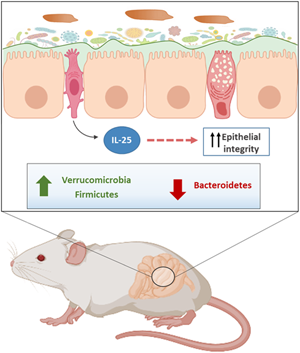
Pharmacological curation of primary E. caproni infection rapidly activates mechanisms for wound healing, including the development of a Th2 phenotype with elevated levels of IL-25 and alternative activation of macrophages and the transition from an inflammatory to anti-inflammatory milieu (Cortés et al., Reference Cortés, Sotillo, Muñoz-Antoli, Fried, Esteban and Toledo2015; Muñoz-Antoli et al., Reference Muñoz-Antoli, Cortés, Santano, Sotillo, Esteban and Toledo2016a, Reference Muñoz-Antoli, Cortés, Martin-Grau, Fried, Esteban and Toledo2016b). Strikingly, this process is associated with a marked increase of beneficial microbes such as those of the Verrucomicrobia phylum (Verrucomicrobiaceae) together with a depletion of native bacteria including Bacteroidaceae or Unclassified Bacteroidales. Increased abundance of Verrucomicrobia has been associated with the development of Th2 phenotype in response to Trichinella spiralis infections (Liu et al., Reference Liu, Chen, Xu, Wu, Tang, Chen, Ji, Peng, Jiang and Xiao2019). It has been shown that Verrucomicrobia cause a decrease in inflammation and enhanced glucose metabolism of the host (Fujio-Vejar et al., Reference Fujio-Vejar, Vasquez, Morales, Magne, Vera-Wolf, Ugalde, Navarrete and Gotteland2017; Plovier and Cani, Reference Plovier and Cani2017; Liu et al., Reference Liu, Chen, Xu, Wu, Tang, Chen, Ji, Peng, Jiang and Xiao2019; Fujisaka et al., Reference Fujisaka, Usui, Nawaz, Igarashi, Okabe, Furusawa, Watanabe, Yamamoto, Sasahara, Watanabe, Nagai, Yagi, Nakagawa and Tobe2020; Ray et al., Reference Ray, Pandey and Aich2021). Shift from the Th1 to Th2 phenotype in E. caproni infections after pharmacological curation is characterized for an increased aerobic use of glucose (Cortés et al., Reference Cortés, Sotillo, Muñoz-Antoli, Fried, Esteban and Toledo2015; Muñoz-Antoli et al., Reference Muñoz-Antoli, Cortés, Santano, Sotillo, Esteban and Toledo2016a, Reference Muñoz-Antoli, Cortés, Martin-Grau, Fried, Esteban and Toledo2016b). In addition, it has been shown that Akkermansia muciniphila adheres to the intestinal epithelium and strengthens the integrity of enterocytes in vitro assays (Justus et al., Reference Justus, Veera, Laura, Noora, Clara, Heikki, de Vos Willem and Satokari2015). Therefore, it is plausible that high levels of A. muciniphila may play a potential protective role against the alteration of the barrier function of the epithelium after infection (Jenkins et al., Reference Jenkins, Formenti, Castro, Piubelli, Perandin, Buonfrate, Otranto, Griffin, Krause and Bisoffi2018a).
Although it is difficult to establish a relationship between changes in the microbiota with the abrupt production of Th2 type cytokines (IL-4, IL-13) and IL-25 and resistance, the elevation of IL-25 levels occurred concomitantly with an increase of the Verrucomicrobia belonging to the genus Akkermansia and a dramatic decline in the Unclassified Bacteroidales. Interestingly, increase in the members of the genus Akkermansia has been associated with increased expression of Toll-like receptors and inflammatory genes (Ray et al., Reference Ray, Pandey and Aich2021). This suggests that the elevated levels of Akkermansia in our study are related to the inflammatory events induced by primary E. caproni infection. In contrast, reduced abundance of members of Bacteroidetes phylum has been associated with a decay in the inflammatory cytokines (Han et al., Reference Han, Bai, Zhou, Niu, Yuan, Zhang, Jia, Zhao and Chen2020). Therefore, the reduction of Unclassified Bacteroidales after pharmacological curation of the primary E. caproni infection could contribute to the increase of Th2 cytokines mediated by alarmins such as IL-25.
Several bacterial taxa, such as the family Lachnospiraceae or bacteria belonging to the genus Turicibacter (Firmicutes) were increased after pzq treatment. Turicibacter spp. were exclusively found in mice treated with pzq. Unfortunately, there is scanty information about the relationships between this group of bacteria and gastrointestinal helminth infections. However, in immunocompromised mice, a link between Turicibacter and host immune dysfunction has been described (Presley et al., Reference Presley, Wei, Braun and Borneman2010; Dimitriu et al., Reference Dimitriu, Boyce, Samarakoon, Hartmann, Johnson and Mohn2013). For example, Turicibacter spp. are abundant in the gut microbiota of wild-type mice, but are completely absent in the gut of mice with defective immune responses (CD45-) and mice lacking an adaptive immune system (RAG-) (Dimitriu et al., Reference Dimitriu, Boyce, Samarakoon, Hartmann, Johnson and Mohn2013). Consequently, we suggest that the absence of Turicibacter observed in mice infected with E. caproni could be due to alterations in the immune functions of the mucosa during infection. However, it is not clear whether the absence of bacteria belonging to the genus Turicibacter could have an impact on the outcome of E. caproni infection.
Secondary E. caproni infection completely depleted the phylum Verrucomicrobia for the benefit of a marked increase of Unclassified Bacteroidales. Among Firmicutes, members of the genus Lactobacillus showed a marked increase. A relationship between lactobacilli and parasitic helminths has been observed in experimental infections of murine models with intestinal nematodes (Walk et al., Reference Walk, Blum, Ewing, Weinstock and Young2010; Rausch et al., Reference Rausch, Held, Fischer, Heimesaat, Kühl, Bereswill and Hartmann2013; Reynolds et al., Reference Reynolds, Smith, Filbey, Harcus, Hewitson, Redpath, Valdez, Yebra, Finlay and Maizels2014; Fricke et al., Reference Fricke, Song, Wang, Smith, Grinchuk, Pei, Ma, Lu, Urban and Shea-Donohue2015; Kreisinger et al., Reference Kreisinger, Bastien, Hauffe, Marchesi and Perkins2015; Su et al., Reference Su, Su, Li, Long, Chang, Zhang, Walker, Xavier, Cherayil and Shi2018). Reynolds et al. (Reference Reynolds, Smith, Filbey, Harcus, Hewitson, Redpath, Valdez, Yebra, Finlay and Maizels2014) reported a marked increase in the populations of the Lactobacillaceae family after infection of C57BL/6 mice with Heligmosomoides polygyrus. In turn, the administration of Lactobacillus species prior to infection with the parasite resulted in a significant increase of parasite load, which led the authors to hypothesize the existence of a mutualistic relationship mediated by the immune system between selected bacteria and helminths (Reynolds et al., Reference Reynolds, Smith, Filbey, Harcus, Hewitson, Redpath, Valdez, Yebra, Finlay and Maizels2014). Strikingly, our results suggest that lactobacilli are affected by the inflammatory responses caused by the infection and, therefore, the elevated levels of Lactobacillus after secondary infections of mice may be a modulation of the local microbiota in response to infection.
Conclusions
Herein, we have shown that primary E. caproni infection, healing and challenge infection have a great impact on the composition of the resident microbiota, which may be related to the outcome of the infection. Interestingly, both primary and challenge infection depleted the presence of members of Verrucomicrobia that only appeared as a consequence of the curation of the primary infection, concomitantly with the upregulation of IL-25 and the subsequent resistance to infection. Although further studies are required to exactly determine the role of intestinal microbiota in the course of helminth infections, our results suggest a close relationship between the changes in microbiota and the outcome of helminth infection.
Supplementary material
The supplementary material for this article can be found at https://doi.org/10.1017/S0031182022001366.
Data availability
The datasets generated and/or analyzed during the current study are available from the corresponding author on reasonable request.
Author contributions
R. T., C. M.-A. and J. G. E. designed the study. M. A.-I., E. F. and P. C. collected the data. R. T., P. C., E. F. and M. A.-I. analysed the data. J. G. E. and C. M.-A. advised on the analyses. M. A.-I., C. M.-A. and R. T. drafted the manuscript. All co-authors contributed to the final version of the manuscript.
Financial support
This research was supported by Ministerio de Economía y Competitividad (Madrid, Spain) (grant number: BFU2016-75639-P), Dirección General de Ciencia e Innovación, de la Consellería de Innovación, Universidad, Ciencia y Sociedad Digital, de la Generalitat Valenciana (Valencia, España) (grant number: AICO/2021/236) and Ministerio de Sanidad y Consumo (Madrid, Spain) (No. RD12/0018/0013, Red de Investigación Cooperativa en Enfermedades Tropicales – RICET, IV National Program of I+D+I 2008–2011, ISCIII – Subdirección General de Redes y Centros de Investigación Cooperativa and FEDER). P. C. was supported by a María Zambrano fellowship from of Ministerio de Universidades (Madrid, Spain), European Union-Next generation EU.
Conflict of interest
The authors declare no conflicts of interest.
Ethical standards
This study has been approved by the Ethical Committee of Animal Welfare and Experimentation of the University of Valencia (Ref. no. A18348501775). Protocols adhered to Spanish (Real Decreto 53/2013) and European (2010/63/UE) regulations.


 Open access