


Crossref Citations
This article has been cited by the following publications. This list is generated based on data provided by Crossref.
Wei, Jiaotong
Chen, Ping
Liu, Bin
and
Han, Yan
2022.
A multienergy computed tomography method without image segmentation or prior knowledge of X-ray spectra or materials.
Heliyon,
Vol. 8,
Issue. 11,
p.
e11584.
Kumrular, Raziye Kubra
Blumensath, Thomas
Zhao, Wei
and
Yu, Lifeng
2022.
Multi energy computed tomography imaging optimization.
p.
192.
Tivnan, Matthew
Wang, Wenying
Gang, Grace
and
Stayman, J. Webster
2022.
Design Optimization of Spatial-Spectral Filters for Cone-Beam CT Material Decomposition.
IEEE Transactions on Medical Imaging,
Vol. 41,
Issue. 9,
p.
2399.
Wang, Guoshuai
Liu, Zhou
Huang, Zhengyong
Zhang, Na
Luo, Honghong
Liu, Lijian
Shen, Hao
Che, Canwen
Niu, Tianye
Liang, Dong
Luo, Dehong
and
Hu, Zhanli
2022.
Improved GAN: Using a transformer module generator approach for material decomposition.
Computers in Biology and Medicine,
Vol. 149,
Issue. ,
p.
105952.
Clements, N.
Richtsmeier, D.
Hart, A.
and
Bazalova-Carter, M.
2022.
Multi-contrast CT imaging using a high energy resolution CdTe detector and a CZT photon-counting detector.
Journal of Instrumentation,
Vol. 17,
Issue. 01,
p.
P01004.
Zeegers, Mathé T
Kadu, Ajinkya
van Leeuwen, Tristan
and
Batenburg, Kees Joost
2022.
ADJUST: a dictionary-based joint reconstruction and unmixing method for spectral tomography.
Inverse Problems,
Vol. 38,
Issue. 12,
p.
125002.
Schwartz, Fides Regina
Samei, Ehsan
and
Marin, Daniele
2023.
Exploiting the Potential of Photon-Counting CT in Abdominal Imaging.
Investigative Radiology,
Vol. 58,
Issue. 7,
p.
488.
Wei, Jiaotong
Wang, Zhiqiang
and
Chen, Ping
2023.
A Multienergy Computed Tomography Method Based on a Blind Decomposition Model for Multivoltage X-Ray Transmission Images.
IEEE Transactions on Instrumentation and Measurement,
Vol. 72,
Issue. ,
p.
1.
Popp, Julian
Busch, Matthias
Hausotte, Tino
and
Drummer, Dietmar
2023.
Fiber orientation in continuous fiber-reinforced thermoplastics/metal hybrid joining via multi-pin arrays.
Science and Engineering of Composite Materials,
Vol. 30,
Issue. 1,
Shi, Zaifeng
Kong, Fanning
Cheng, Ming
Cao, Huaisheng
Ouyang, Shunxin
and
Cao, Qingjie
2024.
Multi-energy CT material decomposition using graph model improved CNN.
Medical & Biological Engineering & Computing,
Vol. 62,
Issue. 4,
p.
1213.
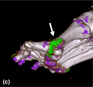
Yan, C.
Zhou, L.
Li, J.
Zhang, G.
Yang, C.
Gu, J.
Lu, X.
Zhang, L.
and
Zeng, M.
2024.
Improved small vessel visibility in diabetic foot arteriography using dual-energy CT.
Clinical Radiology,
Vol. 79,
Issue. 3,
p.
e424.