Hostname: page-component-76fb5796d-vvkck Total loading time: 0 Render date: 2024-04-27T02:53:42.275Z Has data issue: false hasContentIssue false

Minimal Resin Embedding of Multicellular Specimens for Targeted FIB-SEM Imaging

Published online by Cambridge University Press:  04 August 2017

Nicole Schieber
Affiliation:
Cell Biology and Biophysics Unit, European Molecular Biology Laboratory, Heidelberg, Germany
Pedro Machado
Affiliation:
Electron Microscopy Core Facility, European Molecular Biology Laboratory, Heidelberg, Germany.
Sebastian Markert
Affiliation:
University of Wurzburg, Imaging Core Facility, Biocenter, Germany.
Christian Stigloher
Affiliation:
University of Wurzburg, Imaging Core Facility, Biocenter, Germany.
Yannick Schwab
Affiliation:
Cell Biology and Biophysics Unit, European Molecular Biology Laboratory, Heidelberg, Germany Electron Microscopy Core Facility, European Molecular Biology Laboratory, Heidelberg, Germany.
Anna Steyer
Affiliation:
Cell Biology and Biophysics Unit, European Molecular Biology Laboratory, Heidelberg, Germany

Abstract

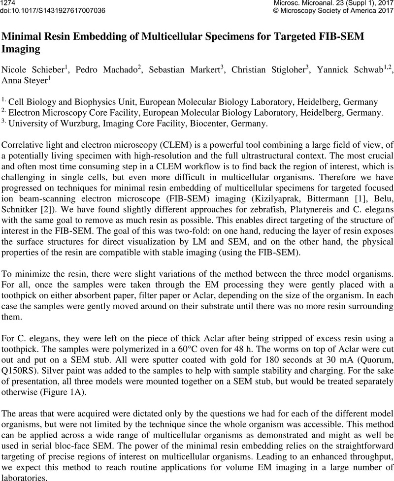
Image of the first page of this content. For PDF version, please use the ‘Save PDF’ preceeding this image.'
Type
Abstract
Copyright
© Microscopy Society of America 2017 

References

[1] Kizilyaprak, C., et al FIB-SEM tomography in biology. Methods Mol Biol 2014 1117, p. 541558.Google Scholar
[2] Belu, A., et al Ultra-thin resin embedding method for scanning electron microscopy of individual cells on high and low aspect ratio 3D nanostructures. J Microsc 2016 263(1p. 7886.Google Scholar
[3] We would like to thank Sevi Durdu (Gilmour lab, EMBL Heidelberg) for providing the zebrafish. Darren Gilmour for giving valuable feedback to the project. Hernando Martinez Vergara (Arendt lab; EMBL Heidelberg) for providing the Platynereis and Detlev Arendt for his support in the project. We would like to thank the Electron Microscopy Core Facility (EMCF, EMBL Heidelberg) for their support.Google Scholar