No CrossRef data available.
Article contents
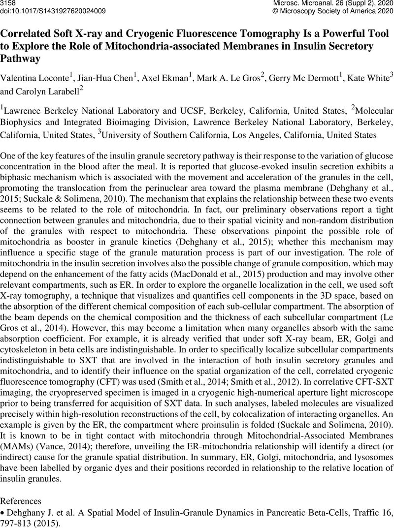
Correlated Soft X-ray and Cryogenic Fluorescence Tomography Is a Powerful Tool to Explore the Role of Mitochondria-associated Membranes in Insulin Secretory Pathway
Published online by Cambridge University Press: 30 July 2020
Abstract
An abstract is not available for this content so a preview has been provided. As you have access to this content, a full PDF is available via the ‘Save PDF’ action button.

- Type
- Biological Soft X-Ray Tomography
- Information
- Copyright
- Copyright © Microscopy Society of America 2020
References
Dehghany, J. et al. . A Spatial Model of Insulin-Granule Dynamics in Pancreatic Beta-Cells, Traffic 16, 797-813 (2015).10.1111/tra.12286CrossRefGoogle ScholarPubMed
Suckale, J. and Solimena, M. The insulin secretory granule as a signaling hub. Trends Endocrinol Metab 21, 599-609 (2010).10.1016/j.tem.2010.06.003CrossRefGoogle ScholarPubMed
MacDonald, MJ., et al. ., Characterization of Phospholipids in Insulin Secretory Granules and Mitochondria in Pancreatic Beta Cells and Their Changes with Glucose Stimulation. J Biol CHem 290(17): 11075–11092 (2015).10.1074/jbc.M114.628420CrossRefGoogle ScholarPubMed
Le Gros, M. A. et al. . Biological soft X-ray tomography on beamline 2.1 at the Advanced Light Source. Journal of synchrotron radiation 21, 1370-1377, doi:10.1107/S1600577514015033 (2014).CrossRefGoogle ScholarPubMed
McDermott, G. et al. . Visualizing cell architecture and molecular location using soft x-ray tomography and correlated cryo-light microscopy. Annu Rev Phys Chem 63, 225-239, doi:10.1146/annurevphyschem-032511-143818 (2012).CrossRefGoogle ScholarPubMed
Smith, E. A. et al. . Quantitatively imaging chromosomes by correlated cryo-fluorescence and soft xray tomographies. Biophys J 107, 1988-1996, doi:10.1016/j.bpj.2014.09.011 (2014).CrossRefGoogle Scholar
Vance, J. E., MAM(Mitochondria-Associated Membrans) in Mammalian cells: Lipids and Beyond. Biochim et Biophys Acta 1841, 595-609 (2014).10.1016/j.bbalip.2013.11.014CrossRefGoogle Scholar


