Hostname: page-component-8448b6f56d-wq2xx Total loading time: 0 Render date: 2024-04-18T01:10:05.326Z Has data issue: false hasContentIssue false

Inflammatory Biomarkers and Intracranial Hemorrhage after Endovascular Thrombectomy

Published online by Cambridge University Press:  20 August 2021

Jose Danilo Bengzon Diestro*
Affiliation:
Division of Diagnostic and Therapeutic Neuroradiology, Department of Medical Imaging, St. Michael’s Hospital, University of Toronto, Toronto, Canada
Carmen Parra-Farinas
Affiliation:
Division of Diagnostic and Therapeutic Neuroradiology, Department of Medical Imaging, St. Michael’s Hospital, University of Toronto, Toronto, Canada
Michael Balas
Affiliation:
Division of Neurosurgery, Department of Surgery, St. Michael’s Hospital, University of Toronto, Toronto, Canada
Zsolt Zador
Affiliation:
Division of Neurosurgery, Department of Surgery, St. Michael’s Hospital, University of Toronto, Toronto, Canada
Noora Almusalam
Affiliation:
Division of Neurology, Department of Medicine, St. Michael’s Hospital, University of Toronto, Toronto, Canada
Adam A. Dmytriw
Affiliation:
Department of Medical Imaging, St. Michael’s Hospital, University of Toronto, Toronto, Canada
Phavalan Rajendram
Affiliation:
Division of Neurology, Department of Medicine, St. Michael’s Hospital, University of Toronto, Toronto, Canada
Rebecca Phillips
Affiliation:
Cumming School of Medicine, University of Calgary, Calgary, Canada
Abdullah Alqabbani
Affiliation:
Department of Medical Imaging, St. Michael’s Hospital, University of Toronto, Toronto, Canada
Emmad Qazi
Affiliation:
Department of Medical Imaging, St. Michael’s Hospital, University of Toronto, Toronto, Canada
Yangmei Li
Affiliation:
Division of Neurosurgery, Department of Surgery, St. Michael’s Hospital, University of Toronto, Toronto, Canada
Walter Montanera
Affiliation:
Division of Diagnostic and Therapeutic Neuroradiology, Department of Medical Imaging, St. Michael’s Hospital, University of Toronto, Toronto, Canada Division of Neurosurgery, Department of Surgery, St. Michael’s Hospital, University of Toronto, Toronto, Canada
Dipanka Sarma
Affiliation:
Division of Diagnostic and Therapeutic Neuroradiology, Department of Medical Imaging, St. Michael’s Hospital, University of Toronto, Toronto, Canada Division of Neurosurgery, Department of Surgery, St. Michael’s Hospital, University of Toronto, Toronto, Canada
Thomas R. Marotta
Affiliation:
Division of Diagnostic and Therapeutic Neuroradiology, Department of Medical Imaging, St. Michael’s Hospital, University of Toronto, Toronto, Canada Division of Neurosurgery, Department of Surgery, St. Michael’s Hospital, University of Toronto, Toronto, Canada
Aditya Bharatha
Affiliation:
Division of Diagnostic and Therapeutic Neuroradiology, Department of Medical Imaging, St. Michael’s Hospital, University of Toronto, Toronto, Canada Division of Neurosurgery, Department of Surgery, St. Michael’s Hospital, University of Toronto, Toronto, Canada
Julian Spears
Affiliation:
Division of Diagnostic and Therapeutic Neuroradiology, Department of Medical Imaging, St. Michael’s Hospital, University of Toronto, Toronto, Canada Division of Neurosurgery, Department of Surgery, St. Michael’s Hospital, University of Toronto, Toronto, Canada
*
Correspondence to: Jose Danilo Bengzon Diestro, Division of Diagnostic and Therapeutic Neuroradiology, Department of Medical Imaging, St. Michael’s Hospital, University of Toronto, Toronto, Canada. Email: danni.diestro@gmail.com
Rights & Permissions [Opens in a new window]

Abstract:

Background:

Intracranial hemorrhage after endovascular thrombectomy is associated with poorer prognosis compared with those who do not develop the complication. Our study aims to determine predictors of post-EVT hemorrhage – more specifically, inflammatory biomarkers present in baseline serology.

Methods:

We performed a retrospective review of consecutive patients treated with EVT for acute large vessel ischemic stroke. The primary outcome of the study is the presence of ICH on the post-EVT scan. We used four definitions: the SITS-MOST criteria, the NINDS criteria, asymptomatic hemorrhage, and overall hemorrhage. We identified nonredundant predictors of outcome using backward elimination based on Akaike Information Criteria. We then assessed prediction accuracy using area under the receiver operating curve. Then we implemented variable importance ranking from logistic regression models using the drop in Naegelkerke R2 with the exclusion of each predictor.

Results:

Our study demonstrates a 6.3% SITS (16/252) and 10.0% NINDS (25/252) sICH rate, as well as a 19.4% asymptomatic (49/252) and 29.4% (74/252) overall hemorrhage rate. Serologic markers that demonstrated association with post-EVT hemorrhage were: low lymphocyte count (SITS), high neutrophil count (NINDS, overall hemorrhage), low platelet to lymphocyte ratio (NINDS), and low total WBC (NINDS, asymptomatic hemorrhage).

Conclusion:

Higher neutrophil counts, low WBC counts, low lymphocyte counts, and low platelet to lymphoycyte ratio were baseline serology biomarkers that were associated with post-EVT hemorrhage. Our findings, particularly the association of diabetes mellitus and high neutrophil, support experimental data on the role of thromboinflammation in hemorrhagic transformation of large vessel occlusions.

Résumé :

RÉSUMÉ :

Biomarqueurs inflammatoires et hémorragie intracrânienne à la suite d’une thrombectomie endovasculaire.

Contexte :

Le fait d’être victime d’une hémorragie intracrânienne (HIC) à la suite d’une thrombectomie endovasculaire (TEV) est associé à un pronostic plus défavorable par rapport à des patients qui ne développent pas cette complication. Notre étude vise ainsi à déterminer les prédicteurs d’une hémorragie post-TEV, plus particulièrement les biomarqueurs inflammatoires présents dans la sérologie de base.

Méthodes :

Nous avons réalisé une étude rétrospective de patients vus consécutivement qui ont été traités au moyen d’une TEV dans le cas d’AVC ischémiques aigus affectant de larges vaisseaux sanguins. Le principal aspect mesuré dans cette étude a été la présence d’une HIC détectée à l’occasion d’un examen de tomodensitométrie post-TEV. Nous avons aussi fait appel à quatre définitions : SITS-MOST (safe implementation of thrombolysis in stroke-monitoring study), NINDS (National Institute of Neurological Disorders and Stroke), hémorragie asymptomatique et hémorragie globale. De plus, nous avons identifié des prédicteurs non-redondants en utilisant une rétro-élimination basée sur le critère d’information d’Akaike. Nous avons ensuite évalué la précision de ces prédicteurs en utilisant la fonction d’efficacité du récepteur. Finalement, nous avons mis en œuvre le classement par importance des variables à partir de modèles de régression logistique en utilisant la diminution du R2 de Naegelkerke avec l’exclusion de chaque prédicteur.

Résultats :

En fonction des définitions SITS-MOST et NINDS, notre étude a montré respectivement un taux d’HIC de 6,3 % (16/252) et de 10,0 % (25/252). Les taux d’hémorragie asymptomatique et d’hémorragie globale ont été respectivement de 19,4 % (49/252) et de 29,4 % (74/252). Les marqueurs sérologiques qui ont montré une association avec des cas d’hémorragie post-EVT ont été les suivants : faible nombre de lymphocytes (SITS), nombre élevé de neutrophiles (NINDS, hémorragie globale), faible rapport plaquettes/lymphocytes (NINDS) et faible total de globules blancs (NINDS, hémorragie asymptomatique).

Conclusion :

Un nombre élevé de neutrophiles, un faible total de globules blancs, un faible nombre de lymphocytes ainsi qu’un faible rapport plaquettes/lymphocytes sont les biomarqueurs sérologiques de base qui ont été associés à des cas d’hémorragie post-TEV. Nos résultats, en particulier l’association entre le diabète sucré et un taux élevé de neutrophiles, confirment les données expérimentales quant au rôle de la thrombo-inflammation dans les transformations hémorragiques consécutives à l’occlusions de larges vaisseaux sanguins.

Type
Original Article
Copyright
© The Author(s), 2021. Published by Cambridge University Press on behalf of Canadian Neurological Sciences Federation

Background

Endovascular thrombectomy (EVT) improves 3-month functional outcomes in patients with large vessel acute ischemic stroke. Reference Goyal, Menon and van Zwam1 However, 4.4% of patients undergoing EVT still develop symptomatic intracranial hemorrhage (sICH). Reference Goyal, Tsivgoulis and Malhotra2 Real-world studies show even higher sICH rate at 9–16%. Reference Zaidat, Castonguay and Gupta3,Reference Hao, Yang and Wang4 Post-EVT patients who develop sICH have significantly higher morbidity and mortality. Reference Berger, Fiorelli and Steiner5 Even asymptomatic intracranial hemorrhage (ICH) in patients undergoing EVT, has been found to decrease the likelihood of attaining an excellent (MRS 0-1) functional outcome. Reference Hao, Liu and Wang6

Given the prognostic implications of post-EVT ICH, identifying factors associated with its occurrence has significant clinical relevance. In particular, we sought to determine if serum biomarkers of neuroinflammation such as neutrophil count, neutrophil-to-lymphocyte ratio, and platelet-to-lymphocyte ratio had any predictive value for ICH after EVT. The goal of this study was to determine factors associated with post-EVT hemorrhage in patients undergoing EVT for large vessel acute ischemic stroke.

Methods

Patient Selection and Study Site

We performed a retrospective review of consecutive patients treated with EVT for acute large vessel ischemic stroke at our institution, a regional stroke center, from March 2011 to April 2020. The inclusion criteria were as follows: age >18 years, diagnosis of acute ischemic stroke with large vessel occlusion (LVO) of the middle cerebral artery (MCA), and/or internal carotid artery (ICA) on computed tomography angiography (CTA), treatment with standard-of-care EVT (stent retriever and/or large bore catheter aspiration), availability of post-EVT head computed tomography (CT), and pre-EVT complete blood counts. Patients with hemorrhage due to vessel perforation were excluded from the study.

The study was approved by the institutional review board. A complete blood count is done for all new acute stroke patients. The hospital uses the Sysmex XN-9000 (Kobe, Japan), a quantitative, multiparameter automated hematology analyzer. The machine uses flow cytometry for cell analysis and sheath flow direct current detection method for counting red blood cells and platelets. Image analysis is also utilized for the differential count

A CTA follows to identify an LVO. Then, the neurovascular team assesses EVT candidacy. Twenty-four hours after EVT, a plain head CT is done to screen for hemorrhage. In cases where it is difficult to delineate HT from contrast staining, a repeat CT is done after another 24 hours to establish a final radiologic diagnosis.

Outcome Measures

The primary outcome of the study is the presence of ICH on the post-EVT scan. We used four definitions Reference Yaghi, Willey and Cucchiara7 : the SITS-MOST criteria (parenchymal hemorrhage >30% of the territory of the infarcted region, with an associated NIHSS worsening of >4 points within 36 hours of stroke onset), the NINDS criteria (any clinical deterioration with any hemorrhage), asymptomatic hemorrhage (no symptoms with any hemorrhage), and overall hemorrhage (includes all types of hemorrhage).

Data Collection

Patient medical records were reviewed by study personnel. We obtained patient demographics, treatment details, and both radiologic and clinical treatment outcomes. All post-EVT cranial scans were again reviewed for the presence of ICH by interventional neuroradiologists.

Statistical Analysis

Methods Overview

Briefly to overview, we identified nonredundant predictors of outcome using backward elimination based on Akaike Information Criteria. We then assessed prediction accuracy using area under the receiver operating curve. To further assess the influence of our predictors, we implemented variable importance ranking from logistic regression models using the drop in Naegelkerke R2 with the exclusion of each predictor.

Predictive Models

The following variables were considered for the models: age, sex, comorbid conditions (atrial fibrillation, congestive heart failure, hypertension, diabetes, coronary artery disease, hyperlipidemia) NIHSS score at presentation, prior anticoagulation, prior antiplatelet use, prior ischemic stroke, prior hemorrhagic stroke, treatment with intravenous or intraarterial thrombolysis, time from symptom onset to reperfusion, degree of recanalization, use of general anesthesia, ASPECTS score on admission, exact location of clot, total number of thrombectomy passes, and serum biomarkers (WBC count, neutrophil count, lymphocyte count, neutrophil-to-lymphocyte ratio, and platelet-to-lymphocyte ratio). We applied logistic regression models for predictions and to assess variable correlation with clinical outcome. For model selection, we will use Akaike information criterion with backward elimination to optimize the balance of model complexity against goodness of fit. Reference Akaike, Atkinson and Fienberg8 Predictions will be undertaken with threefold cross-validation to avoid overfitting. Reference Steyerberg, Mushkudiani and Perel9 This method has been found superior in terms of discriminatory ability, calibration, and overall accuracy to the split-sample method by the comparative study of Steyerberg et al. Reference Steyerberg, Harrell, Borsboom, Eijkemans, Vergouwe and Habbema10 Predictive performance of the different models described below will be assessed by computing the AUC and compared using DeLong’s test. Reference DeLong, DeLong and Clarke-Pearson11

Variable Importance Ranking

We used the Nagelkerke R2 value, Reference Nagelkerke12 a measure for goodness of fit, to rank variable importance. Nagelkerke R2 numerically expresses the percentage of variability attributed to a predictor. The ranking of variables was extracted from the drop in the Nagelkerke R2 value that occurs in response to excluding variables of interest from the model. We used this ranking to identify the most influential variables and include them to create a more simplistic model with fewer variables. There are recognized limitations for “pseudo” R2 methods: (1) they can be argued to give artificially high R2 scores that may suggest the model fits better than it really does, and (2) there are a variety of “pseudo” R2 measures to choose from, each of which interprets the model differently and therefore gives different results. In our study, we used the same modality of R2 value to assess the change in model fit rather than focus on the numeric value, a technique that has been applied in past papers. Reference Murray, Butcher and McHugh13,Reference Zador, Sperrin and King14

Statistical Software

Statistical analysis and modeling were carried out in R, 15 an open-source software environment for statistical programming and graphics (https://www.r-project.org/). Receiver operating curve analysis was done using the “pROC” package. Reference Robin, Turck and Hainard16 Threshold optimization was performed using packages “pROC” and “SDMTools” Reference VanDerWal, Falconi, Januchowski, Shoo and Storlie17 AUCs were compared using DeLong’s test. Reference DeLong, DeLong and Clarke-Pearson11 Nagelkerke R2 was implemented using the “fmsb” package. Reference Nakazawa18

Ethics Approval

The study was approved by our institutional research ethics board (18-397). The retrospective nature of the study precluded informed consent from the patients.

Results

Patient Characteristics

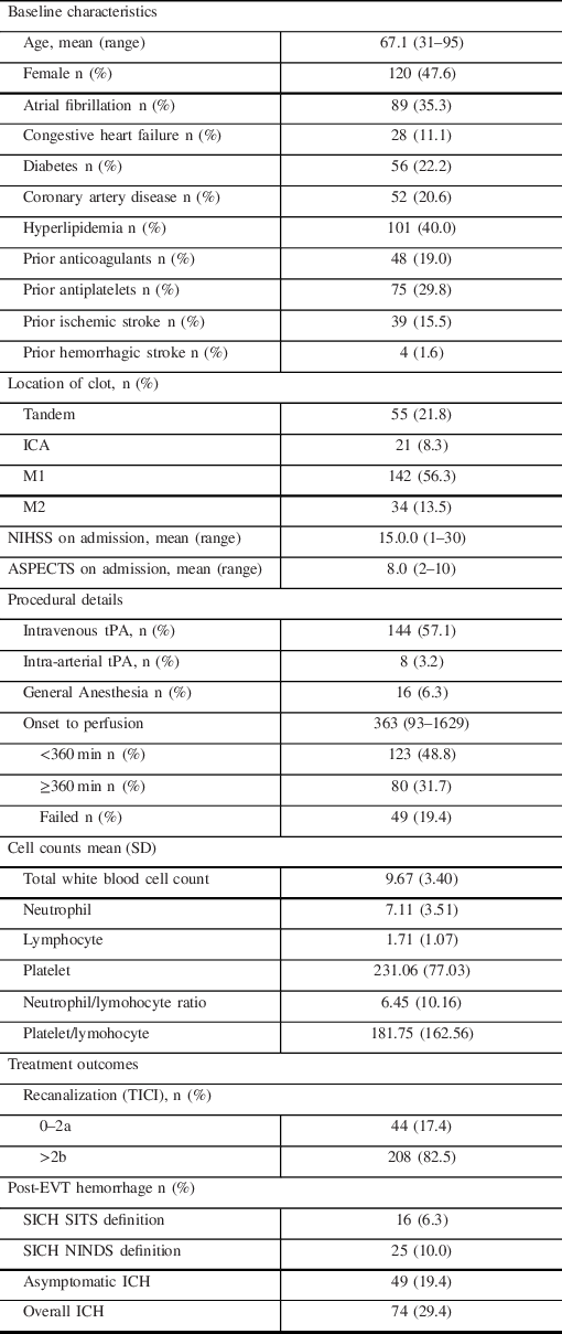
A total of 335 acute stroke patients underwent EVT, of which 252 patients met the inclusion criteria (See Online Supplement Figure 1). The baseline characteristics of the patients are summarized in Table 1.

Table 1: Baseline characteristics and treatment outcomes

Predictive Models

On logistic regression modeling, we yielded 5, 6, 7, and 6 variables significantly associated with occurrence of post-EVT intracerebral hemorrhage as described by SITS, NINDS, asymptomatic, and overall ICH definitions (Table 2) respectively. Consistent predictors across the four models were the lower white blood cell (WBC) or lymphocyte count, and number of passes. Significance values and odds ratios are summarized in Table 2.

Table 2: Table summary of confidence intervals, odds ratios, and significance levels for the post EVT intracerebral hemorrhage definitions

Predictive Performance

AUCs were 0.69 (95% CI: 0.55–0.83), 0.78 (95% CI: 0.69–0.86), 0.69 (95% CI: 0.61–0.77), and 0.72 (95% CI: 0.65– 0.79) for the SITS, NINDS, asymptomatic, and overall hemorrhages, respectively. These values reflect only fair diagnostic accuracy of the models. Predictions were carried out using threefold cross-validation approach to avoid model overfitting. Using DeLong’s test, there were no significant differences between all combinations of ROC curves.

Importance Ranking of Outcome Predictors

Based on the drop in Nagelkerke R2 values, a pseudo-R2 value used to describe goodness of model fit, we ranked the influence of each variable on the outcome of interest. While there was some overlap in significant predictors between models, the highest-ranking variables were distinct for each model as summarized in Table 3.

Table 3: Variable importance ranking

Discussion

Our study demonstrates a 6.3% (SITS) and 10.0% (NINDS) sICH rate, as well as a 19.4% asymptomatic and 29.4% overall hemorrhage rate. Serologic markers that demonstrated association with post-EVT hemorrhage were: low lymphocyte count (SITS), high neutrophil count (NINDS, overall hemorrhage), low platelet-to-lymphocyte ratio (NINDS), and low total WBC (NINDS, asymptomatic hemorrhage). Other factors that showed previous association with post EVT hemorrhage were also seen in our study: diabetes mellitus, Reference Enomoto, Shigeta and Ota19 higher number of passes, Reference Hao, Yang and Wang4,Reference Enomoto, Shigeta and Ota19 onset to perfusion time, Reference Hao, Yang and Wang4,Reference Bin, Yoon and Lee20 lower ASPECTS, Reference Hao, Liu and Wang6,Reference Boisseau, Fahed and Lapergue21 and intravenous thrombolysis. Reference Enomoto, Shigeta and Ota19,Reference Nogueira, Gupta and Jovin22 The neutrophil-to-lymphocyte ratio has previously been associated with early neurological deterioration, Reference Nam, Kim and Lee23 post-thrombolysis hemorrhagic transformation, Reference Liu, Lu and Yin24 and poor outcomes in acute ischemic stroke patients. Reference Song, Zhao and Rajah25 However, it was not associated with post-EVT hemorrhage in our study using any of the definitions.

Blood–brain barrier disruption induced by the inflammatory response to acute ischemic stroke may play a role in post-EVT hemorrhage. Neutrophils are a source of matrix metalloproteinase (MMP)-9, which play a direct role in degrading tight junction proteins. Conceivably, higher neutrophil counts may lead to higher levels of MMP-9, potentially translating to a higher risk of HT. Reference Kanazawa, Takahashi, Nishizawa and Shimohata26 The role of neutrophils is emphasized further by decrease in ischemic volume in rats undergoing middle cerebral artery occlusion after the administration of anti-neutrophil antibodies compared with those with a normal complement of neutrophils. Reference Dawson, Ruetzler, Carlos, Kochanek and Hallenbeck27 These pathomechanisms may underlie a recent finding that high neutrophil count predicts poor clinical outcomes despite recanalization in patients with large vessel occlusion. Reference Boisseau, Desilles and Fahed28 Large cohorts of patients undergoing intravenous thrombolysis Reference Maestrini, Strbian and Gautier29 and endovascular thrombectomy Reference Hao, Yang and Wang4 also demonstrated an increased risk of hemorrhagic transformation in patients with higher neutrophil counts and ratios. Circulating neutrophil transcriptomes have also been demonstrated to correlate with the presence of unruptured intracranial aneurysms. Reference Tutino, Poppenberg and Li30Reference Poppenberg, Tutino and Li32

Diabetes has been associated with post EVT hemorrhage in past studies. Reference Enomoto, Shigeta and Ota19,Reference Nogueira, Gupta and Jovin22 Chronic hyperglycemia in diabetic mice undergoing MCA occlusion has been demonstrated to aggravate HT by inciting mitochondrial dysfunction leading to endothelial cell death. Reference Mishiro, Imai and Sugitani33 Classically, HT had been attributed to free radical damage from reperfusion injury. However, similar to our findings, a large multicenter study demonstrated that reperfusion status did not correlate with HT after EVT. Reference Boisseau, Fahed and Lapergue21 More recent basic science research has focused on downstream microvascular thromboinflammation (DMT), a phenomenon characterized by leukocyte margination (mostly neutrophils) and thrombosis in the venules after proximal large vessel occlusion. DMT by inducing vascular leakage may account for microhemorrhages and incomplete reperfusion despite large vessel recanalization. Reference Desilles, Syvannarath and Di Meglio34

Hyperglycemia has been demonstrated to precipitate infarct growth and HT by exacerbating DMT through preactivating neutrophils in rats undergoing experimental large vessel occlusion. Reference Desilles, Syvannarath and Ollivier35 Earlier studies noted that diabetic rats had a more robust neutrophil–endothelial cell interaction compared with nondiabetic ones in the setting of large vessel ischemia reperfusion injury models. Reference Ritter, Davidson and Henry36 This interplay between diabetes and neutrophils may be the pathomechanism behind the association of both factors with post-EVT ICH in our study.

Higher WBC count has already been associated with less hematoma expansion in spontaneous ICH. Reference Morotti, Phuah and Anderson37 However, its association with less post-EVT hemorrhage has not yet been described elsewhere. A higher platelet lymphocyte ratio has been associated with poorer clinical outcomes in patients undergoing thrombolysis and endovascular therapy Reference Altintas, Altintas, Tasal, Kucukdagli and Asil38,Reference Xu, He and Li39 ; counterintuitively, our study demonstrates that a lower ratio is associated with sICH (NINDS). Similarly, the association of lower lymphocyte to our outcome of interest (SITS) is also a new finding.

Limitations of our study include its retrospective nature, lack of a core laboratory, and low sample size. Next, perfusion imaging and MRI data were not included in the analysis. The model also needs validation using a different large independent cohort. Furthermore, the models obtained by the study only have moderate accuracy. Lastly, the paper is exploratory in nature. It is a proof of concept of the association of serology markers to post-EVT hemorrhage.

Conclusion

Higher neutrophil counts, low WBC counts, low lymphocyte counts, and low platelet-to-lymphoycyte ratio were baseline serology biomarkers that were associated with post-EVT hemorrhage. Our findings, particularly the association of diabetes mellitus and high neutrophil count with post-EVT hemorrhage, support experimental data on the role of thromboinflammation in hemorrhagic transformation of large vessel occlusions. Larger stroke registries with data on inflammatory biomarkers are necessary to confirm our findings.

Conflict of Interest

The authors have no conflicts of interest to declare.

Statement of Authorship

All authors fulfilled the following ICMJE criteria for authorship; substantial contributions to the conception or design of the work; or the acquisition, analysis, or interpretation of data for the work; AND Drafting the work or revising it critically for important intellectual content; AND Final approval of the version to be published; AND Agreement to be accountable for all aspects of the work in ensuring that questions related to the accuracy or integrity of any part of the work are appropriately investigated and resolved.

Data Statement

The full dataset of the study is available upon reasonable request.

Supplementary Material

To view supplementary material for this article, please visit https://doi.org/10.1017/cjn.2021.197.

References

Goyal, M, Menon, BK, van Zwam, WH, et al. Endovascular thrombectomy after large-vessel ischaemic stroke: a meta-analysis of individual patient data from five randomised trials. Lancet. 2016;387:1723–31. DOI 10.1016/S0140-6736(16)00163-X.CrossRefGoogle ScholarPubMed
Goyal, N, Tsivgoulis, G, Malhotra, K, et al. Medical management vs mechanical thrombectomy for mild strokes. JAMA Neurol. 2019;77:1624. DOI 10.1001/jamaneurol.2019.3112.CrossRefGoogle Scholar
Zaidat, OO, Castonguay, AC, Gupta, R, et al. North American Solitaire Stent Retriever Acute Stroke registry: post-marketing revascularization and clinical outcome results. J Neurointerv Surg. 2014;6:584–8. DOI 10.1136/neurintsurg-2013-010895.CrossRefGoogle ScholarPubMed
Hao, Y, Yang, D, Wang, H, et al. Predictors for symptomatic intracranial hemorrhage after endovascular treatment of acute ischemic stroke. Stroke. 2017;48:1203–9. DOI 10.1161/STROKEAHA.116.016368.CrossRefGoogle ScholarPubMed
Berger, C, Fiorelli, M, Steiner, T, et al. Hemorrhagic transformation of ischemic brain tissue. Stroke. 2001;32:1330–5. DOI 10.1161/01.STR.32.6.1330.CrossRefGoogle ScholarPubMed
Hao, Y, Liu, W, Wang, H, et al. Prognosis of asymptomatic intracranial hemorrhage after endovascular treatment. J Neurointerv Surg. 2019;11:123–6. DOI 10.1136/neurintsurg-2018-013848.CrossRefGoogle ScholarPubMed
Yaghi, S, Willey, JZ, Cucchiara, B, et al. Treatment and outcome of hemorrhagic transformation after intravenous alteplase in acute ischemic stroke: a scientific statement for healthcare professionals from the American Heart Association/American Stroke Association. Stroke. 2017;48:1050. DOI 10.1161/STR.0000000000000152.CrossRefGoogle ScholarPubMed
Akaike, H. Prediction and entropy. In: Atkinson, AC, Fienberg, SE, editors. A celebration of statistics. New York, NY: Springer; 1985.Google Scholar
Steyerberg, EW, Mushkudiani, N, Perel, P, et al. Predicting outcome after traumatic brain injury: development and international validation of prognostic scores based on admission characteristics. PLoS Med. 2008;5:1251–61. DOI 10.1371/journal.pmed.0050165.CrossRefGoogle ScholarPubMed
Steyerberg, EW, Harrell, FE, Borsboom, GJJM, Eijkemans, MJC, Vergouwe, Y, Habbema, JDF. Internal validation of predictive models: efficiency of some procedures for logistic regression analysis. J Clin Epidemiol. 2001;54:774–81. DOI 10.1016/S0895-4356(01)00341-9.CrossRefGoogle ScholarPubMed
DeLong, ER, DeLong, DM, Clarke-Pearson, DL. Comparing the areas under two or more correlated receiver operating characteristic curves: a nonparametric approach. Biometrics. 1988;44:837. DOI 10.2307/2531595.CrossRefGoogle ScholarPubMed
Nagelkerke, NJD. A note on a general definition of the coefficient of determination. Biometrika. 1991;78:691–2. DOI 10.1093/biomet/78.3.691.CrossRefGoogle Scholar
Murray, GD, Butcher, I, McHugh, GS, et al. Multivariable prognostic analysis in traumatic brain injury: results from the IMPACT study. J Neurotrauma. 2007;24:329–37. DOI 10.1089/neu.2006.0035.CrossRefGoogle ScholarPubMed
Zador, Z, Sperrin, M, King, AT. Predictors of outcome in traumatic brain injury: new insight using receiver operating curve indices and Bayesian network analysis. PLoS One. 2016;11:118. DOI 10.1371/journal.pone.0158762.CrossRefGoogle ScholarPubMed
R Development Core Team. R: a language and environment for statistical computing. Vienna: R Foundation for Statistical Computing; 2016, ISBN: 3-900051-07-0, 10.1038/sj.hdy.6800737,Google Scholar
Robin, X, Turck, N, Hainard, A, et al. pROC: an open-source package for R and S+ to analyze and compare ROC curves. BMC Bioinformatics. 2011;12:77. DOI 10.1186/1471-2105-12-77.Google Scholar
VanDerWal, J, Falconi, L, Januchowski, S, Shoo, L, Storlie, C. SDMTools: Species Distribution Modelling Tools: Tools for processing data associated with species distribution modelling exercises. Version 1.1-221. 2014. Available at: https://www.rforge.net/SDMTools/.Google Scholar
Nakazawa, M. fmsb: Functions for medical statistics book with some demographic data. CRAN.R-project. 2015.Google Scholar
Enomoto, M, Shigeta, K, Ota, T, et al. Predictors of intracranial hemorrhage in acute ischemic stroke after endovascular thrombectomy. Interv Neuroradiol. 2020;26:368–75. DOI 10.1177/1591019920926335.CrossRefGoogle ScholarPubMed
Bin, Lee Y, Yoon, W, Lee, YY, et al. Predictors and impact of hemorrhagic transformations after endovascular thrombectomy in patients with acute large vessel occlusions. J Neurointerv Surg. 2019;11:469–73. DOI 10.1136/neurintsurg-2018-014080.Google Scholar
Boisseau, W, Fahed, R, Lapergue, B, et al. Predictors of parenchymal hematoma after mechanical thrombectomy. Stroke. 2019;50:2364–70. DOI 10.1161/STROKEAHA.118.024512.CrossRefGoogle ScholarPubMed
Nogueira, RG, Gupta, R, Jovin, TG, et al. Predictors and clinical relevance of hemorrhagic transformation after endovascular therapy for anterior circulation large vessel occlusion strokes: a multicenter retrospective analysis of 1122 patients. J Neurointerv Surg. 2015;7:1621. DOI 10.1136/neurintsurg-2013-010743.CrossRefGoogle ScholarPubMed
Nam, K-W, Kim, TJ, Lee, JS, et al. Neutrophil-to-lymphocyte ratio predicts early worsening in stroke due to large vessel disease. PLoS One. 2019;14:e0221597. DOI 10.1371/journal.pone.0221597.Google ScholarPubMed
Liu, Y-L, Lu, J-K, Yin, H-P, et al. High neutrophil-to-lymphocyte ratio predicts hemorrhagic transformation in acute ischemic stroke patients treated with intravenous thrombolysis. Int J Hypertens. 2020;2020:5980261. DOI 10.1155/2020/5980261.CrossRefGoogle ScholarPubMed
Song, S-Y, Zhao, X-X, Rajah, G, et al. Clinical significance of baseline neutrophil-to-lymphocyte ratio in patients with ischemic stroke or hemorrhagic stroke: an updated meta-analysis. Front Neurol. 2019;10:517. DOI 10.3389/fneur.2019.01032.CrossRefGoogle ScholarPubMed
Kanazawa, M, Takahashi, T, Nishizawa, M, Shimohata, T. Therapeutic strategies to attenuate hemorrhagic transformation after tissue plasminogen activator treatment for acute ischemic stroke. J Atheroscler Thromb. 2017;24:240–53. DOI 10.5551/jat.RV16006.CrossRefGoogle ScholarPubMed
Dawson, DA, Ruetzler, CA, Carlos, TM, Kochanek, PM, Hallenbeck, JM. Polymorphonuclear leukocytes and microcirculatory perfusion in acute stroke in the SHR. Keio J Med. 1996;45:248–53. DOI 10.2302/kjm.45.248.CrossRefGoogle ScholarPubMed
Boisseau, W, Desilles, J-P, Fahed, R, et al. Neutrophil count predicts poor outcome despite recanalization after endovascular therapy. Neurology. 2019;93:e467–75. DOI 10.1212/WNL.0000000000007859.CrossRefGoogle ScholarPubMed
Maestrini, I, Strbian, D, Gautier, S, et al. Higher neutrophil counts before thrombolysis for cerebral ischemia predict worse outcomes. Neurology. 2015;85:1408–16. DOI 10.1212/WNL.0000000000002029.CrossRefGoogle ScholarPubMed
Tutino, VM, Poppenberg, KE, Li, L, et al. Biomarkers from circulating neutrophil transcriptomes have potential to detect unruptured intracranial aneurysms. J Transl Med. 2018;16:373. DOI 10.1186/s12967-018-1749-3.CrossRefGoogle ScholarPubMed
Tutino, VM, Poppenberg, KE, Jiang, K, et al. Circulating neutrophil transcriptome may reveal intracranial aneurysm signature. PLoS One. 2018;13:e0191407. DOI 10.1371/journal.pone.0191407.CrossRefGoogle ScholarPubMed
Poppenberg, KE, Tutino, VM, Li, L, et al. Classification models using circulating neutrophil transcripts can detect unruptured intracranial aneurysm. J Transl Med. 2020;18:392. DOI 10.1186/s12967-020-02550-2.CrossRefGoogle ScholarPubMed
Mishiro, K, Imai, T, Sugitani, S, et al. Diabetes mellitus aggravates hemorrhagic transformation after ischemic stroke via mitochondrial defects leading to endothelial apoptosis. PLoS One. 2014;9:e103818. DOI 10.1371/journal.pone.0103818.CrossRefGoogle ScholarPubMed
Desilles, J, Syvannarath, V, Di Meglio, L, et al. Downstream microvascular thrombosis in cortical venules is an early response to proximal cerebral arterial occlusion. J Am Heart Assoc. 2018;7:188. DOI 10.1161/JAHA.117.007804.CrossRefGoogle ScholarPubMed
Desilles, J-P, Syvannarath, V, Ollivier, V, et al. Exacerbation of thromboinflammation by hyperglycemia precipitates cerebral infarct growth and hemorrhagic transformation. Stroke. 2017;48:1932–40. DOI 10.1161/STROKEAHA.117.017080.CrossRefGoogle ScholarPubMed
Ritter, L, Davidson, L, Henry, M, et al. Exaggerated neutrophil-mediated reperfusion injury after ischemic stroke in a rodent model of type 2 diabetes. Microcirculation. 2011;18:552–61. DOI 10.1111/j.1549-8719.2011.00115.x.CrossRefGoogle Scholar
Morotti, A, Phuah, C-L, Anderson, CD, et al. Leukocyte count and intracerebral hemorrhage expansion. Stroke. 2016;47:1473–8. DOI 10.1161/STROKEAHA.116.013176.CrossRefGoogle ScholarPubMed
Altintas, O, Altintas, MO, Tasal, A, Kucukdagli, OT, Asil, T. The relationship of platelet-to-lymphocyte ratio with clinical outcome and final infarct core in acute ischemic stroke patients who have undergone endovascular therapy. Neurol Res. 2016;38:759–65. DOI 10.1080/01616412.2016.1215030.CrossRefGoogle ScholarPubMed
Xu, J-H, He, X-W, Li, Q, et al. Higher platelet-to-lymphocyte ratio is associated with worse outcomes after intravenous thrombolysis in acute ischaemic stroke. Front Neurol. 2019;10: 439. DOI 10.3389/fneur.2019.01192.CrossRefGoogle ScholarPubMed
Figure 0

Table 1: Baseline characteristics and treatment outcomes

Figure 1

Table 2: Table summary of confidence intervals, odds ratios, and significance levels for the post EVT intracerebral hemorrhage definitions

Figure 2

Table 3: Variable importance ranking

Supplementary material: PDF

Diestro et al. supplementary material

Figure S1

Download Diestro et al. supplementary material(PDF)
PDF 72.4 KB