


Introduction
Active microbial communities play an important role in biogeochemical cycling in glacial environments and thus exert a strong influence on the nature of dissolved material exported to downstream aquatic ecosystems (Reference Skidmore, Anderson, Sharp, Foght and LanoilSkidmore and others, 2005). Dissolved organic matter (DOM) is the primary form of organic matter (OM) in glacial (Reference Hagler, Bergin, Smith, Dibb, Anderson and SteigHagler and others, 2007) and other aquatic ecosystems (Reference Findlay and SinsabaughFindlay and Sinsabaugh, 2003). It is an important component of aquatic carbon and nutrient budgets and is a metabolic substrate for heterotrophic bacteria and other organisms at the base of aquatic food chains (Reference AzamAzam, 1998; Reference JonesJones, 1992; Reference TranvikTranvik, 1992). DOM quantity and characteristics are important factors in aquatic ecosystems because DOM abundance influences OM availability to heterotrophic organisms while the molecular and chemical characteristics determine whether a given heterotrophic organism has the capability to metabolize it. While there have been numerous studies of fluxes of inorganic solutes in glacier meltwater (e.g. Reference Tranter, Sharp, Lamb, Brown, Hubbard and WillisTranter and others, 2002, and references therein) the difficulty involved in characterizing OM (e.g. Reference Fimmen, Cory, Chin, Trouts and McKnightFimmen and others, 2007) and the low concentrations of DOM in glacial meltwaters have limited the study of OM fluxes from glacier environments (e.g. Reference Lafreniere and SharpLafrenie`re and Sharp, 2004; Reference Barker, Sharp, Fitzsimons and TurnerBarker and others, 2006, Reference Barker, Sharp and Turner2009; Reference Bhatia, Das, Longnecker, Charette and KujawinskiBhatia and others, 2010).
Fluorescence spectroscopy is an analytical technique capable of describing DOM characteristics in dilute aqueous samples (Reference CobleCoble, 1996; Reference Parlanti, Wortz, Geoffroy and LamotteParlanti and others, 2000; Reference McKnight, Boyer, Westerhoff, Doran, Kulbe and AndersenMcKnight and others, 2001; Reference Stedmon, Markager and BroStedmon and others, 2003; Reference Barker, Sharp and TurnerBarker and others, 2009). While it is limited to identifying only fluorescing organic compounds, and thus less descriptively robust than techniques such as carbon nuclear magnetic resonance (13C-NMR) spectroscopy or electrospray ionization Fourier transform ion cyclotron resonance mass spectroscopy (ESI-FT-ICR MS), it is sensitive and requires less sample preparation. This is an important advantage because sample preparation (e.g. DOM isolation) may selectively isolate a portion of the bulk DOM, resulting in a biased characterization of the bulk DOM pool (Reference CobleCoble, 2007).
Most published fluorescence spectroscopic analyses refer to DOM from riverine, estuarine and marine aquatic environments (e.g. Reference CobleCoble, 1996; Reference Stedmon and MarkagerStedmon and Markager, 2005; Reference HoodHood and others, 2009). Five classes of fluorescing components (fluorophores) have been identified and associated with specific compounds in DOM (Table 1). Of these five classes, two ‘protein-like’ fluorophores are particularly interesting in the context of glacier biogeochemistry because they represent environmentally labile compounds and have been taken as indicators of recent microbial activity (Reference CobleCoble, 2007). Compounds such as proteins and carbohydrates are typically the most labile sources of carbon and nutrients in aquatic ecosystems, and the export of these compounds in glacier meltwater is potentially important for meltwater-fed ecosystems.
Table 1. Fluorophore identification and associated compound. Exmax is the excitation wavelength that promotes the highest fluorescence intensity while Emmax is the wavelength at which the fluorescence maximum occurs. After Reference CobleCoble (2007)

To date, no studies have linked the distribution of specific fluorophores to the distribution of microbes in glacial environments. Previous investigations have interpreted fluorophores found in glacial environments on the basis of studies from riverine, estuarine and marine waters (e.g. Reference Barker, Sharp, Fitzsimons and TurnerBarker and others, 2006, Reference Barker, Sharp and Turner2009). However, little is known about the types of compounds produced by microbes in glacial environments. Here we seek to determine whether the occurrence in glacier ice of fluorophores indicative of recent microbial activity is linked to the presence of culturable microbial communities containing organisms that could have produced them. The samples analyzed were obtained from the basal ice of Victoria Upper Glacier (VUG) in the McMurdo Dry Valleys of Antarctica.
Field Site
Basal ice samples were collected from a ∼ 3 m section of the terminal ice cliff of VUG (77817’ S, 161°32’ E). The transect sampled extended downwards from the contact between glacier ice and underlying basal ice. The sampling site was located within 3 m of the site sampled for analysis of DOM properties by Reference Barker, Sharp, Fitzsimons and TurnerBarker and others (2006).
VUG is a cold-based glacier (basal temperature –23°C) in the polar desert environment of the McMurdo Dry Valleys. It terminates in a ∼40m high ice cliff, the lower ∼15m of which is composed of basal ice (Reference Fitzsimons, Webb, Mager, MacDonell, Lorrain and SamynFitzsimons and others, 2008). The basal ice is easily discernible from the overlying glacier ice by its darker hue, which is due to the presence of dispersed fine sediment throughout, and by the presence of fine laminations consisting of sand-sized debris which extend horizontally across the ice cliff (Fig. 1b).

Fig. 1. (a) DOC concentration in trench 1 basal ice; (b) glacier ice/basal ice contact in trench 1 .
The VUG terminus rests against a frontal apron (Reference Fitzsimons, Webb, Mager, MacDonell, Lorrain and SamynFitzsimons and others, 2008) which abuts, and in some locations spills into, an ice-covered proglacial lake (Lake Upper Victoria). Reference Hall, Denton, Overturf and HendyHall and others (2002) and Reference Kelly, Denton and HallKelly and others (2002) provide evidence for a much larger lake in Victoria Valley (Glacial Lake Victoria) during the Last Glacial Maximum, 45 000–8600 14CyearsBP. Reference Kelly, Denton and HallKelly and others (2002, and references therein) provide evidence for VUG having advanced into Glacial Lake Victoria between 45 000 and 11054 14C years BP. Given the close association now and in the past between VUG and proglacial lakes in the Victoria Valley, it seems likely that, during periods of glacial advance, VUG has overridden water-saturated lacustrine and/or deltaic sediments and that OM contained within these sediments (either as particulate OM or as DOM within sediment pore-waters) has been incorporated into the basal ice of VUG.
Several models have been proposed for basal ice formation beneath cold-based glaciers overlying water-saturated sediment (Reference O’Neill and MillerO’Neill and Miller, 1985; Reference Souchez, Samyn, Lorrain, Pattyn and FitzsimonsSouchez and others, 2004; Reference Christoffersen, Tulaczyk, Carsey and BeharChristoffersen and others, 2006; Reference RempelRempel, 2008). A common feature of these models is that they all suggest that debris-rich ice is accreted to the base of the overlying glacier when sediment pore-water migrates upwards towards the glacier sole and freezes to it. Kinetic energy and the chemical potential (in the case of dissolved solutes) associated with pore-water circulation will transport material towards the glacier sole where it will be incorporated into the basal ice sequence during refreezing (Reference Souchez, Samyn, Lorrain, Pattyn and FitzsimonsSouchez and others, 2004). According to such models, the OM content of basal ice sequences, such as that described from VUG, will be derived from local subglacial sources as either particulate OM found in overridden sediments or DOM in sediment pore-waters. The absence of folding or thrusting within the VUG basal ice (Reference Fitzsimons, Webb, Mager, MacDonell, Lorrain and SamynFitzsimons and others, 2008) suggests that the basal ice sequence is conformable, with the oldest basal ice located immediately below the contact with overlying glacier ice (Fig. 1b) and the basal ice becoming younger with increasing distance from the contact. This mechanism for basal ice formation requires the presence of water-saturated basal sediment, such as is found at the bottom of proglacial Lake Upper Victoria.
Methodology
Basal ice samples were collected as follows. A sampling trench (trench 1) was excavated by hand to 50 cm depth using an ethanol-bathed and flame-sterilized chisel. Basal ice was sampled from trench 1 at 10cm intervals along the glacier face to a distance of 310 cm below the basal-ice/ glacier-ice contact. Sterile sampling was performed using ethanol-bathed and flame-sterilized chisels and aluminum collection trays. Ice samples were transferred into sterile Whirl-Pak bags without handling. All samples were stored frozen (<–20°C) and in the dark during transport to, and storage at, the University of Alberta. There was no evidence of melting during transport.
Ice samples were melted in their Whirl-Pak bags in the dark at room temperature. To extract DOM, meltwater was filtered under vacuum through pre-combusted (450°C for 8 hours) glass fibre F grade (GF/F) filter papers (0.7 μm) using a sterile (bathed in trace-metal grade hydrochloric acid (HCl)) glass filtration apparatus that had been combusted at 450°C for 8 hours. Duplicate filtered samples were stored in the dark at 4°C in pre-combusted amber borosilicate glass vials until analysis (<7 days).
Dissolved organic carbon (DOC) was used as a proxy for DOM in the filtered samples (Reference McKnight, Hood, Klapper, Findlay and SinsabaughMcKnight and others, 2003). DOC concentration was determined by catalytic high-temperature combustion and non-dispersive infrared detection using a Shimadzu TOC-5000A Total Organic Carbon Analyzer equipped with a high-sensitivity platinum catalyst. Prior to analysis, each sample was acidified to pH 2 using trace-metal grade HCl and sparged for 5 min with TOC grade air to remove dissolved carbonate species. Each sample was analyzed in triplicate. Five replicates were analyzed if the coefficient of variation exceeded 2%. The detection limit was 0.05 ppm (Reference Miller and MillerMiller and Miller, 1988).
DOM fluorescence was characterized using total emission spectra of filtered samples collected with a SPEX Fluorolog-3 spectrofluorometer equipped with excitation and emission monochrometers and a xenon lamp source. Total fluorescence scans, known as excitation–emission matrices (EEMs), record fluorescent emission over a predetermined range of excitation and emission wavelengths. EEMs reveal much more information about the fluorophore composition of DOM than do other types of fluorescence measurements, such as single excitation wavelength emission and synchronous fluorescence, because EEMs collect data from multiple emission wavelengths at multiple excitation wavelengths.
Fluorescence scans were performed at 10 nm intervals from 250 to 450 nm excitation and monitored at 10nm intervals from 260 to 700 nm emission. All samples were scanned at room temperature using a quartz glass cuvette with a 10 mm path length. All scans were Raman-corrected by subtracting the spectrum for deionized water scanned under identical conditions. All scans were dark-corrected and normalized relative to the intensity of the Raman scatter band (Reference Huguet, Vacher, Relexans, Saubusse, Froidefond and ParlantiHuguet and others, 2009) of the deionized water blank to correct for lamp fluctuation (<1.6% over the course of the entire analysis). Fluorophores in the EEMs were identified visually and assigned identities based upon existing literature (Table 1).
We chose three basal ice samples from trench 1 for microbial incubation and analyses to determine whether specific fluorophores are associated with culturable bacteria. Basal ice samples were chosen without any a priori knowledge of DOC concentration or fluorescence characteristics. Microbial incubations and analyses were carried out as follows. Basal ice samples from the 10–20, 60–70 and 220–230 cm intervals below the glacier-ice/basal-ice interface were transferred in a UV- and Roccol-sterilized biosafety hood to doubly sterilized 4M HCl-washed beakers and melted at 4°C. 10mL of the meltwater was added to N2-flushed serum bottles containing 90 mL of 0.1% sodium pyrophosphate, shaken by hand for 5 min and serially diluted into similar bottles with inversion. 0.1 mL of this dilution series was plated in triplicate onto pre-chilled R2A plates (Difco) for heterotrophic plate counts, which were incubated for 6weeks at 4°C, 10°C and 20°C in the dark and counted weekly. Newly observed and morphologically distinct colonies were subcultured each week onto R2A agar and incubated at their isolation temperature. No new colonies were observed beyond 6weeks of incubation. Genomic DNA was extracted from all strains according to Reference FoghtFoght and others (2004) and the partial 16S rRNA gene (approximately 1400 base pairs) was amplified using the primers PB36F and PB38R according to Reference Cheng and FoghtCheng and Foght (2007). PCR products were screened by restriction fragment length polymorphism (RFLP) analysis using Cfol and Haelll (Roche). Restriction fragment sizes were calculated using Gel-Pro Analyzer version 4.5 image-analysis software (MediaCybernetics, Silver Spring, MD). PCR products exhibiting unique RFLP patterns were partially sequenced using the primer PB36F, compared with the US National Center for Biotechnology Information (NCBI) nucleotide database using BLAST (Reference Altschul, Gish, Miller, Myers and LipmanAltschul and others, 1990) and the nearly full-length 16S rRNA gene sequenced for representative members according to Reference Cheng and FoghtCheng and Foght (2007; GenBank accession numbers: EU155008–EU155017, GQ454797–GQ454806, GQ454841–GQ454859). The identification of the cultured microbes by phylogenetic relation serves two purposes here. Firstly, the presence of microbes that have not been identified previously in cold environments may serve as an indication of sample contamination. Secondly, the presence in basal ice of microorganisms that have been isolated previously from specific Antarctic environments may serve as an indication of the source of OM to basal ice. For example, the presence of microorganisms that are specific to Antarctic lacustrine environments would indicate that OM from VUG basal ice was derived from overridden lacustrine sediment. It is important to note that the incubation conditions used here discriminate preferentially for aerobic heterotrophic bacteria.
Results
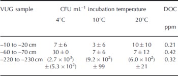
The DOC concentrations from each cultured interval did not vary with culturable bacterial abundance (Table 2), suggesting that the cultured bacterial biomass does not account for all of the DOM in VUG basal ice.
Table 2. Incubation results and DOC concentration in VUG basal ice samples. CFU: colony-forming units
DOC concentrations along trench 1 ranged from 0.13 to 6.34 ppm (mean = 0.5 ppm, mode = 0.2 ppm, SD = 1.1, n = 3 1 ; Fig 1a) and were lower than those reported by Reference Barker, Sharp, Fitzsimons and TurnerBarker and others (2006; Table 1) (1.78–46.66 ppm, mean = 5.89 ppm). Even ignoring the exceptionally high DOC concentration close to the glacier-ice/basal-ice contact reported by Reference Barker, Sharp, Fitzsimons and TurnerBarker and others (2006), a comparison of DOC concentrations between trench 1 basal ice (Fig. 1a) and concentrations reported by Reference Barker, Sharp, Fitzsimons and TurnerBarker and others (2006) from identical depth intervals indicates that DOC concentrations are significantly different (DOCtrench 1 = 0.16–0.57 ppm, DOCVUG= 2.19–3.77 ppm; P = 0.0001). Previous investigations have noted a heterogeneous distribution of OC in basal ice (Reference Skidmore, Foght and SharpSkidmore and others, 2000; Reference Barker, Sharp, Fitzsimons and TurnerBarker and others, 2006, Reference Barker, Sharp and Turner2009; Reference Bhatia, Sharp and FoghtBhatia and others, 2006, Reference Bhatia, Das, Longnecker, Charette and Kujawinski2010). While the reason for this variation is unclear, perhaps autotrophic microbial activity is occurring and creating OM in ice closer to the ice face where light is less attenuated, or perhaps ice sublimation at the ice face surface is concentrating OM in the ice closest to the ice face. These points may warrant further investigation because if an increase in OM concentrations at the ice surface is widespread then this phenomenon would need to be considered if attempting to forecast the contribution of glacially derived OM flux to the downstream environments.
The average EEM for all sampled intervals sampled in trench 1 shows that basal ice DOM contains several fluorophores (Fig. 2), while the individual EEMs show that the relative intensity of these fluorophores changes spatially between samples (Fig. 3; data not shown for non-cultured samples). The limited number of sample EEMs in the study precludes a more objective identification of fluorophores in the DOM, such as would be possible with statistically based approaches such as parallel factor analysis (PARAFAC; Reference Stedmon, Markager and BroStedmon and others, 2003; Reference Stedmon and BroStedmon and Bro, 2008). Nevertheless, visual identification of fluorophores in the EEMs from trench 1 does help to characterize the DOM from the VUG basal ice.

Fig. 2. The average EEM for basal ice samples from trench 1 (n = 31).

Fig. 3. The EEM from each of the basal ice samples that were incubated. Note that while the protein-like fluorophore is the most prominent fluorophore in all of the samples, the 220–230 cm sample displays the most well-defined marine humic-like material fluorophore.
The most prominent feature in the average EEM is the strong fluorescence peak at 270/320 nm (ex/em) (Fig. 2), which has been identified as a fluorophore corresponding to protein-like material (the amino acid tyrosine; Table 1) and possibly indicative of associated non-fluorescing total hydrolysable amino acids (Reference Yamashita and TanoueYamashita and Tanoue, 2008). This supports the finding of Reference Barker, Sharp, Fitzsimons and TurnerBarker and others (2006) that the protein-like fluorophore is the most prominent component of the fluorescence spectra of VUG basal ice. Other fluorophores detected in the VUG basal ice occurring at 290/395 and 345/440nm (ex/em) (Fig. 2) are indicative of marine humic-like material and humic-like material, respectively (Table 1). Humic substances are derived from the carbon of organic residues from non-mineralized OM and have undergone secondary synthesis reactions to produce a material that is generally resistant to microbial oxidation (Reference Sylvia, Fuhrmann, Hartel and ZubererSylvia and others, 1999). Humic material accumulates in environments where OM is not completely degraded and accumulates over time. Marine humic material is most common in the open ocean, away from rivers and other OM sources (Reference Yamashita and TanoueYamashita and Tanoue, 2008). Spatial and temporal trends in marine humic-like, humic-like and protein-like fluorescence are interpreted as indicators of changes in biogeochemical activity and OM source in estuarine and marine systems (Reference CobleCoble, 2007, and references therein). For example, increases in humic-like fluorescence in estuaries are often interpreted as indicating an increase in terrestrial riverine contribution to the OC pool (Reference Stedmon and MarkagerStedmon and Markager, 2005). It should be noted that the term ‘marine humic material’ does not imply a marine source of the humic material. It is so-named because it is most commonly detected in the open ocean (e.g. Reference Yamashita and TanoueYamashita and Tanoue, 2008).
EEMs for each of the samples used for microbial analyses are shown in Figure 3. The 10–20 cm sample contains a prominent fluorophore at 270/320 nm which is indicative of protein-like material. A secondary fluorophore that appears as a shoulder on the main peak at 282/370 nm is indicative of marine humic-like material. Trench 1 (60–70cm) also contains a prominent fluorophore indicative of protein-like material; however, the DOM from this interval exhibits a broadly shaped EEM without distinct peaks, suggestive of the existence of multiple fluorophores and potential peak overlap between fluorophores. Trench 1 (220–230cm) displays the protein-like fluorophore but also displays a well-resolved fluorophore at 300/410nm, which is indicative of marine humic-like material.
In total, 131 bacterial strains were isolated and identified from the VUG basal ice samples. Clustering of these strains using RFLP and 16S rRNA gene sequencing, which permits differentiation between species, revealed 29 unique sequence groups including those related to the genera Hymenobacter (58 strains), Knoellia (11 strains), Flavobacterium and Polarmonas (10 strains), Arthrobacter, Frigoribacterium and Janthinobacterium (7 strains each), Nocardiodes (6 strains), Variovorax (2 strains), Brevundimonas, Deinococcus and Sphingomonas (1 strain each) and several divergent α-Proteobacteria (10 strains). Hymenobacter strains represented 44% of the isolates from the 220–230 cm sample, many of which belong to novel species (Klassen and Foght, in press).
Microbes were isolated at 4°C, 10°C and 20°C from each of the three VUG basal ice samples (Table 2). Most microbes were recovered from the 220–230cm sample interval and very few microbes were recovered from the 10–20 and 60–70 cm sample intervals. The numbers of colony-forming units (CFU) per milliliter isolated from the 10–20 and 60–70cm samples are similar to those reported in other glaciers by Reference Christner, Mosley-Thompson, Thompson and ZagorodnovChristner and others (2000). More isolates were recovered from the 220–230cm sample incubated at 4°C than at 10°C and 20°C (Table 1), indicating their cold-adaptation or that cold temperatures permitted a higher rate of resuscitation.
Discussion
DOC is present at detectable but variable concentrations in the basal ice from trench 1. This is consistent with previous reports of high spatial variability in DOC concentrations in basal ice (Reference Skidmore, Foght and SharpSkidmore and others, 2000; Reference Barker, Sharp, Fitzsimons and TurnerBarker and others, 2006, Reference Barker, Sharp and Turner2009; Reference Bhatia, Sharp and FoghtBhatia and others, 2006, Reference Bhatia, Das, Longnecker, Charette and Kujawinski2010).
The similarity of the bacterial isolates from VUG basal ice samples to bacteria isolated from other polar environments suggests that the samples from trench 1 are uncontaminated. Several of the taxa identified (Sphingomonas-like α-Proteo-bacteria, Commomonas-like β-Proteobacteria, Arthrobacter, Flavobacterium, Frigoribacterium, Janthinobacterium, Kocuria, Microbacterium) have been isolated previously from other glaciers (Reference Christner, Mosley-Thompson, Thompson and ZagorodnovChristner and others, 2000; Reference FoghtFoght and others, 2004; Reference Miteva, Sheridan and BrenchleyMiteva and others, 2004; Reference Miteva and BrenchleyMiteva and Brenchley 2005; Reference Skidmore, Anderson, Sharp, Foght and LanoilSkidmore and others, 2005; Reference Xiang, Yao, An, Xu and WangXiang and others, 2005; Reference Yao, Xiang, Zhang, Wang and WangYao and others, 2006; Reference Cheng and FoghtCheng and Foght, 2007; Reference Mikucki and PriscuMikucki and Priscu, 2007; Reference Zhang, Yao, Tian, Xu and AnZhang and others, 2008) while others (e.g. Hymenobacter and Deinococcus) are well known for their recovery from Antarctic soil and cryptoendolithic environments (Reference Hirsch, Ludwig, Hethke, Sittig, Hoffman and GallikowskiHirsch and others, 1998; Reference AdamsAdams and others, 2006; Reference AislabieAislabie and others, 2006).
Hymenobacter-like bacteria have a cosmopolitan distribution but are most often found in environments characterized by oxidative and desiccation stress (e.g. Reference Hirsch, Ludwig, Hethke, Sittig, Hoffman and GallikowskiHirsch and others, 1998; Reference AislabieAislabie and others 2006; Reference Zhang, Yao, An, Tian and XuZhang and others, 2006). The genus Hymenobacter consists of obligatory aerobic red-pigmented Gram-negative chemoheterotrophic bacteria (Reference Oren, Dworkin, Falkow, Rosenberg, Schleifer and StackebrandtOren, 2006). Hymenobacter roseosalvarius has been isolated from Antarctic and high-altitude soils, sandstone and permafrost (Reference Hirsch, Ludwig, Hethke, Sittig, Hoffman and GallikowskiHirsch and others, 1998; Reference AislabieAislabie and others, 2006; Reference Zhang, Yao, Tian, Xu and AnZhang and others 2008). Basal ice formation models predict that pore-water from unfrozen subglacial sediments flows towards, and freezes onto, the base of an overriding cold-based glacier.
Bacteria were cultured from each of the basal ice samples, suggesting that culturable aerobic heterotrophic bacteria may be widespread in VUG basal ice. However, the presence of culturable bacteria does not imply biogeochemical activity in situ, because the conditions under which the bacteria were cultured differ from those experienced within basal ice (e.g. temperature, pressure). The results of the spectrofluorescence analysis may provide a better indication of the presence of potentially biogeochemically active microbes in basal ice.
Fluorescence spectroscopy indicates that the DOM in VUG basal ice includes both protein-like and humic-like material (Fig. 3), while the average EEM (Fig. 2) shows that the protein-like fluorophore is the most prominent fluorophore in VUG DOM, similar to DOM characterized from other glaciers (Reference Barker, Sharp and TurnerBarker and others 2009; Reference HoodHood and others, 2009; Reference Bhatia, Das, Longnecker, Charette and KujawinskiBhatia and others, 2010). The presence of protein-like fluorescence in DOM has been interpreted to be indicative of the production of amino acids in DOM during microbial metabolism, which are considered to be environmentally labile and rapidly metabolized by microbes (Reference Yamashita and TanoueYamashita and Tanoue, 2003). For example, Reference Ren, Zhao, Wang and WangRen and others (2008) found that incubated dinoflagellates produce protein-like fluorescent DOM during their exponential and decadency growth phase. This proteinaceous fluorescence is derived as free amino acids and peptides from the aromatic amino acids phenylalanine, tyrosine and tryptophan (Reference Yamashita and TanoueYamashita and Tanoue, 2003) or partially from amino acids bound in cell wall proteins (Reference Determann, Lobbes, Reuter and RullkötterDetermann and others, 1998). The presence of aromatic amino acids in natural waters has also been associated with the presence of non-fluorescing total hydrolysable amino acids in marine water (Reference Yamashita and TanoueYamashita and Tanoue, 2003). These findings, and the association between protein-like fluorescence and marine algal and phytoplankton blooms, have been interpreted as evidence for in situ microbial activity and/or freshly produced DOM. While we have observed protein-like fluorescence in DOM derived from ice in which culturable microbes also exist, more prominent marine humic-like fluorescence occurred in the 260–270 cm sample from which more CFUmL−1 were isolated.
Marine humic material is a complex mixture of dissolved and colloidal substances produced in situ in sea water which cannot be separated fully into individual compounds (Reference CobleCoble, 2007). It fluoresces at relatively long wavelengths indicative of a more aromatic molecular structure than that of amino acids, similar to that of terrestrial humic material (Table 1). The marine humic-like fluorophore is produced in situ by the biological oxidation of pre-existing OM as a by-product of microbial respiration in both natural waters and incubations (Reference Parlanti, Wortz, Geoffroy and LamotteParlanti and others, 2000; Reference Rochelle-Newall and FisherRochelle-Newall and Fisher, 2002). Reference Yamashita and TanoueYamashita and Tanoue (2008) found that marine humic material is bio-refractory over time periods of at least 900 years in the open ocean and is thus considered to be refractory and a sink of oceanic carbon. By analogy, OM degradation by heterotrophic microorganisms in basal ice could produce similar refractory material that would accumulate over time. In contrast, labile protein-like material produced as a by-product of microbiological activity would be mineralized by subsequent heterotrophy.
The presence of the humic-like fluorophore is indicative of recalcitrant humic material in DOM. Humic material results from abiotic condensation reactions involving non-mineralized OM. As non-mineralized OM undergoes humification, it becomes polymerized, resulting in more condensed, aromatic and complex humic material which is resistant to microbial decay (Reference Sylvia, Fuhrmann, Hartel and ZubererSylvia and others, 1999). Humic material may be formed in any environment where OM accumulates (e.g. sediment) and is not indicative of microbial activity.
The most obvious drawback to this study is that only a small number of intervals in trench 1 were sampled for microbial characterization and DOC analysis. A more comprehensive sampling for microbial analysis would permit us to test the relationship between psychrotolerant bacterial abundance and marine humic-like fluorescence much more comprehensively. Additionally, culturing bacteria under a wider range of conditions would also be useful. The protocol employed here selected for aerobic heterotrophs and it is no surprise that the obligatory aerobic Hymenobacter was the genus most represented in the cultures. Its ability to survive oxidatively stressed conditions would be advantageous in the englacial environment where dissolved oxygen and nutrients are limited to a thin film of water located at intergrain boundaries, potentially far from gas and nutrient sources (e.g. bubbles and sediment grains). However, given the potentially oxygen-limited or anaerobic conditions that might be encountered in an englacial setting (e.g. micro-sites around sediment grains), culturing samples under oxygen-limited or anoxic conditions might yield a more representative bacterial enumeration. A second drawback of this investigation is that only fluorescing compounds in the OM were detected. A more rigorous characterization by 13C-NMR or ESI-FT-ICR MS would doubtless have provided additional insight into the types of organic compounds present in VUG basal ice OM. Unfortunately this drawback was unavoidable because the low concentrations of DOC in the ice samples, and the small volume of ice sampled precluded such analyses. However, fluorescence spectroscopy would be of use for identifying areas of interest in trench 1 where a larger sample volume may be collected for a more rigorous DOC characterization.
Summary and Conclusions
The presence of psychrotolerant bacteria and associated marine humic-like fluorescence in VUG basal ice is suggestive of in situ microbial degradation of OM within the basal ice of VUG. While previous investigations have cited the presence of proteinaceous fluorescence as evidence of possible in situ microbial metabolism, the results here indicate that proteinaceous fluorescence alone may be indicative of the presence of microbes and proteinaceous material associated with microbial cell walls, whereas it is the marine humic-like fluorescence that may be indicative of actual metabolic activity.
Fluorescent amino acids may exist in, and/or be derived from, microbial cell walls and organelles, and free amino acids are considered to be highly labile and quickly mineralized (Reference CobleCoble, 2007). However, the presence of marine humic-like material is indicative of the production and accumulation of refractory by-products of OM biooxidation. The fact that greater marine humic-like fluorescence is associated with higher abundances of culturable psychrotolerant bacteria supports the argument that marine humic-like fluorescence in basal ice may be a better indication of in situ microbial metabolism than the presence of the protein-like fluorophore.
The dominant fluorophore in all of the basal ice samples from VUG in trench 1 , and in other glacier ice and meltwater samples reported by Reference Barker, Sharp, Fitzsimons and TurnerBarker and others (2006, Reference Barker, Sharp and Turner2009) and Reference HoodHood and others (2009), is indicative of the ubiquitous presence of protein-like material, likely tyrosine, and possibly of total hydrolysable amino acids (Reference Yamashita and TanoueYamashita and Tanoue, 2008). These compounds represent a potential labile source of carbon and nutrients that can be transported to downstream aquatic ecosystems in glacier meltwater. Marine humic-like material in some basal ice samples would represent the transport of a recalcitrant form of DOM in meltwater to downstream environments. The conversion of proteinaceous material to marine humic-like material by in situ OM mineralization represents the conversion of a labile form of OM to a recalcitrant form of OM. The production of more recalcitrant marine humic-like material in glacier environments, similar to the open ocean environment, may function as a carbon sink over millennial timescales.
Acknowledgements
This project would not have been possible without the logistical support of personnel with Antarctica New Zealand. Financial assistance was provided by the Natural Sciences and Engineering Research Council (NSERC) of Canada through a Discovery grant to M.J. Sharp, and by an NSERC Discovery grant to J. Foght (University of Alberta). The thoughtful comments of two anonymous reviewers were helpful in improving the manuscript.


