Skip to main content Accessibility help
×
  • Cited by 10
  • Edited by Stephen Davis, Royal Melbourne Hospital and University of Melbourne, Marc Fisher, National Institute of Mental Health, Bethesda, Maryland, Steven Warach, National Institutes of Health, Baltimore
Publisher:
Cambridge University Press
Online publication date:
August 2009
Print publication year:
2003
Online ISBN:
9780511544927

Book description

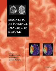
Magnetic resonance imaging provides non-invasive information about the brain's blood flow, water movement and biochemical abnormalities following stroke, and advances in magnetic resonance imaging (MRI) are transforming the investigation and treatment of cerebrovascular disease. Echoplanar techniques with diffusion and perfusion weighted imaging, together with developments in magnetic resonance spectroscopy and angiography, are replacing CT scanning as the diagnostic modality of choice. In this profusely illustrated book world leaders in these technologies review the scientific basis and clinical applications of MRI in stroke. It will appeal to a broad readership including stroke physicians, neurologists, neurosurgeons, rehabilitation specialists, and others with a clinical or research interest in cerebrovascular disease.

Reviews

‘This book provides a good overview of the use of MRI in stroke and is of educational benefit to both clinicians and radiologists involved in the care of patients with stroke and has started to take on some very important issues …’

Source: Neuroradiology

'This is an excellent intermediate level textbook that covers the topic of MR imaging in stroke and ischaemic stroke in particular. The book is well written throughout and easy to read. … The authorship reads like a Who's Who of the literature of stroke imaging and the quality of this book reflects it. … I would thoroughly recommend this book to all trainee physicians, neurovascular surgeons and neuroradiologists with an interest in ischaemic stroke …'

Source: Acta Neurochirugica

Refine List

Actions for selected content:

Select all | Deselect all
  • View selected items
  • Export citations
  • Download PDF (zip)
  • Save to Kindle
  • Save to Dropbox
  • Save to Google Drive

Save Search

You can save your searches here and later view and run them again in "My saved searches".

Please provide a title, maximum of 40 characters.
×

Contents

  • 1 - The importance of specific diagnosis in stroke patient management
    pp 1-14
  • View abstract

    Summary

    Stroke refers to any damage to the brain or spinal cord caused by a vascular abnormality. This chapter shows how specific diagnostic information available from non-invasive investigations can be applied to the management of individual patients. The complexity of managing stroke patients is increasing. Early stroke classifications relied on clinical information. Terms such as 'transient ischemic attack (TIA)', 'minor stroke', 'reversible ischemic neurologic deficit (RIND)', 'stroke in progress' and 'completed stroke' were used to distinguish stroke subtypes. The initial diagnostic step should be to determine if the event is due to stroke or a non-vascular stroke mimic. The next level of stroke diagnosis is primarily to distinguish hemorrhagic from ischemic stroke. A detailed diagnosis of stroke etiology is required to plan management strategies for secondary stroke prevention. Stroke severity is an important diagnostic consideration in determining stroke prognosis, which in turn influences management decisions.
  • 2 - Limitations of current brain imaging modalities in stroke
    pp 15-30
  • View abstract

    Summary

    The successful management of stroke patients requires the ability to confirm the diagnosis and determine underlying pathophysiology. Techniques such as computed tomography (CT), magnetic resonance imaging (MRI), positron emission tomography (PET) and single photon emission computed tomography (SPECT) provides a window onto the structural and functional changes that occur during stroke. This chapter focuses on the limitations of currently used brain imaging techniques. There has been a widespread belief that conventional MRI is less sensitive than CT in the detection of intracerebral hemorrhage. Ischemic changes may be seen in conventional MR studies within 1 to 2 hours of stroke onset. However, both CT and MRI have low sensitivities for acute ischemia in the hyper acute phase and neither imaging modality is able to identify hypoperfused but potentially salvageable ischemic tissue. SPECT and PET have added significantly to our knowledge of the pathophysiology of stroke.
  • 3 - Clinical efficacy of CT in acute cerebral ischemia
    pp 31-46
  • View abstract

    Summary

    Brain imaging is necessary to assess the exact diagnosis and the acute pathophysiological state of the brain. This chapter explores the questions of what unenhanced computed tomography (CT) is able to assess in patients with acute stroke, how accurate this information is, and whether imaging with CT has any impact on stroke diagnosis, stroke treatment and, finally, on the clinical outcome of patients. CT has the capacity to identify different pathophysiological states that results in the same clinical picture of an acute stroke syndrome: intracranial hemorrhage, ischemic edema and ischemia without ischemic edema. Surgery or autopsy regularly confirms the CT finding of intracranial hemorrhage. CT was the first imaging modality that could reliably differentiate between hemorrhagic and ischemic stroke. MRI was introduced into clinical practice as the second modality that can image brain parenchyma, and is now considered as an evolving standard of care in acute stroke.
  • 4 - Computerized tomographic-based evaluation of cerebral blood flow
    pp 47-54
  • View abstract

    Summary

    Computerized tomographic (CT)-based assessment of cerebral blood flow (CBF) offers many advantages in the care of patients with disorders of the central nervous system. The CT scanner detects small changes in attenuation due to the presence of Xe and the change in density is directly proportional to the concentration of Xe in the tissues. In patients with acute stroke, direct measurement of CBF is potentially helpful to maximize the benefit of acute stroke therapy and reduce the complications. The role of hemodynamic factors in the risk of recurrent stroke in patients with extra cranial carotid occlusive disease remains controversial. Xenon enhanced CT (XeCT) may provide valuable insights into the optimal blood pressure management of patients with Intracerebral hemorrhage (ICH). CT- based systems now offer the ability to provide exquisite tissue and vascular anatomy using conventional CT and CT angiography.
  • 5 - Technical introduction to MRI
    pp 55-68
  • View abstract

    Summary

    This chapter outlines the basic physical principles of magnetic resonance imaging (MRI), and overviews the MR techniques that have been developed for investigating stroke patients. Magnetic resonance angiography (MRA) is a major advance in application of MRI to imaging of patient blood flow in vessels. The signal intensity in MRI depends on proton density, T1, T2 and T2 relaxation process of any ensemble of the spins. Diffusion- and perfusion-weighted imaging combined with fast imaging capabilities on commercial systems has revolutionized our understanding of the pathophysiological mechanisms involved in the many clinical conditions especially cerebral ischemia and stroke. Another popular technique used for perfusion MR imaging is the arterial spin labelling (ASL). Magnetic resonance spectroscopy (MRS) of the brain is a noninvasive MR technique that gives the relative concentration of certain chemical compounds within 2 to 3 cm3 of tissue.
  • 6 - Clinical use of standard MRI
    pp 69-84
  • View abstract

    Summary

    The basic standard Magenetic Resonance Imaging (MRI) sequences which should be applied in screening for stroke are T1 and T2- weighted axial or sagittal scans through the whole brain. In hyperacute stroke MRA sequences provide valuable and accurate information in respect to the patency of the major intracranial vessels. The principal use for Magnetization Transfer Contrast (MTC) in the stroke context is in time of flight (TOF) MRA, where it suppresses background parenchymal signal, increasing the conspicuity of small vessels. Fluid Attenuated Inversion Recovery (FLAIR) is a routinely available technique which produces heavily T2-weighted images, at the same time nulling or completely subtracting the normally bright cerebrospinal fluid signal. Hyperacute parenchymal hemorrhage can be detected by MRI by utilizing one of the T2-weighted magnetic susceptibility sensitive sequences such as T2-weighted gradient echo (GE). Standard MRI sequences are extremely sensitive to the detection of acute, subacute and chronic infarcts.
  • 7 - MR angiography of the head and neck: basic principles and clinical applications
    pp 85-102
  • View abstract

    Summary

    This chapter opens with a discussion on the basic principles of Magnetic Resonance Angiogram (MRA), including time of flight and phase contrast techniques. It introduces the use of paramagnetic contrast agents for MRA. The advantages and pitfalls of MRA as compared with duplex sonography (DUS) and X-ray angiography (XRA) are discussed. MR uses a combination of magnetic fields and radiofrequency energy to produce images. MRI is highly accurate at measuring the carotid wall area and T2-weighted images can discriminate between the fibrous cap and lipid core of a plaque. The combination of MRI and MRA is an accurate non-invasive means for the detection of carotid and vertebral dissections. MRI depicts the nidus of a vascular malformation. MRA is reliable for detecting stenoses and occlusions involving the vertebrobasilar system. For the intracranial circulation, MRA is well accepted for imaging of the dural sinuses and larger intracranial veins.
  • 8 - Stroke MRI in intracranial hemorrhage
    pp 103-112
  • View abstract

    Summary

    Non-traumatic intracranial hemorrhage (ICH) accounts for 10-15% of all strokes, but up to 25% of more severe strokes. This chapter covers the magnetic resonance imaging (MRI) signatures of ICH in all stages, and focuses on the differential diagnosis in hyperacute stroke patients. It deals with subarachnoid hemorrhage (SAH), and discusses the future prospects such as the detection of perihemorrhagic pathological processes, which may contribute to the morbidity of ICH. The appearance of intracranial hemorrhage on MRI depends primarily on the age of the hematoma and the type of MR contrast. Computed tomography (CT) is also the imaging standard of care for the diagnosis of acute subarachnoid hemorrhage. Stroke MRI may be the diagnostic tool of choice not only for patients with subacute and chronic ICH and SAH but also in the initial assessment of patients with hyperacute ischemic or hemorrhagic stroke as well as hyperacute SAH.
  • 9 - Using diffusion-perfusion MRI in animal models for drug development
    pp 113-120
  • View abstract

    Summary

    Preclinical evaluation of purported acute stroke therapies plays a significant role in the drug development process. Animal stroke models and their use in evaluating treatment effects of potential acute stroke therapies are prone to many difficulties. Recently, it has been proposed that diffusion-perfusion MRI might help to bridge the gap between preclinical evaluation and advanced clinical trials. Several reports have appeared concerning the use of PWI and DWI to assess thrombolytic therapy in animal stroke models. Combining Perfusion MRI (PWI) and Diffusion MRI (DWI) in temporary occlusion models evaluating neuroprotective drugs may also be useful. The use of diffusion-perfusion MRI both in preclinical testing and in clinical development of acute stroke therapies will likely continue to expand, especially once the MRI modalities provide support for the approval of a new acute stroke therapy. Their utility will also increase as we move into the multitherapy era of acute stroke therapy.
  • 10 - Localization of stroke syndromes using diffusion-weighted MR imaging (DWI)
    pp 121-134
  • View abstract

    Summary

    Diffusion-weighted MR imaging (DWI) is a technique in which microscopic water motion is responsible for the contrast within the image. Diffusion of water molecules alters conventional T1- and T2-weighted MR imaging, because it induces a signal dephasing and a signal loss. Clinical practice uses different representations of the results of DWI data processing: diffusion weighted images, DWI trace and ADC maps, which are all equivalent. DWI is more accurate than CT in localizing ischemic lesions shortly after stroke onset. DWI can show small lesions adjacent to the cerebrospinal fluid. The NINDS and ECASS studies have demonstrated an increased risk of hemorrhagic transformation in stroke patients with a large area of hypodensity on admission CT when treated with thrombolytic therapy. DWI affords accurate localization of strokes. Finally, DWI can more frequently differentiate a small deep subcortical infarct from a cortical or a combined cortical/subcortical lesion than conventional MR imaging can.
  • 11 - MRI in transient ischemic attacks: clinical utility and insights into pathophysiology
    pp 135-146
  • View abstract

    Summary

    The brain responds dynamically to transient episodes of ischemic insult. Standard brain imaging techniques, computed tomography (CT) and conventional magnetic resonance imaging (MRI) are insensitive to dynamic and regionally varying neural parenchymal responses to tissue ischemia. MR spectroscopy is an interesting new application of MRI for the study of patients with transient ischemic attack (TIA). In an UCLA study, among TIA patients with early DWI abnormalities who had follow-up imaging, approximately one-half exhibited late CT or MRI evidence of established infarction. Both UCLA and Duke Series found a strong statistical correlation between duration of TIA symptoms and presence of a lesion on DWI. MR imaging studies have demonstrated the untenability of any definition of TIA based solely on clinical manifestations and an arbitrarily assigned time window, rather than tissue changes and physiologic processes. MRI has fundamentally altered our understanding of the pathophysiology of transient ischemic attack.
  • 12 - Perfusion-weighted MRI in stroke
    pp 147-160
  • View abstract

    Summary

    Perfusion-weighted magnetic resonance imaging (PWI) encompasses a set of techniques that create images depicting hemodynamics at the microvascular level. PWI offers the opportunity to study the pathophysiological events that lead most directly to ischemic damage. This chapter reviews the techniques employed in PWI with the role of contrast agents, and the MR pulse sequences that are usually chosen. Performing PWI with MRI rather than some other imaging modality offers the theoretical advantage that the contrast agent can be either an endogenous contrast agent that is naturally present in the blood or an exogenous one that is injected for the purpose of obtaining images. PWI techniques are usually designed to rely on gadolinium's susceptibility effect rather than its relaxivity effect, because these effects are exhibited over different ranges. PWI helps to identify tissue that is at risk of inclusion into growing infarcts.
  • 13 - Perfusion imaging with arterial spin labelling
    pp 161-174
  • View abstract

    Summary

    The spatially selective radiofrequency (RF) excitation and inversion capabilities of magnetic resonance make possible a totally non-invasive method for perfusion imaging, which is known as arterial spin labelling (ASL). The clearest distinction between labelling approaches is between continuous ASL and pulsed ASL. Continuous ASL usually employs a special RF labelling scheme known as flow driven adiabatic inversion. ASL perfusion measurement differs from dynamic susceptibility contrast (DSC) imaging in several important ways. Typically imaging of perfusion with ASL requires multiple averages to achieve an acceptable signal-to-noise ratio. ASL methods have been validated against existing methods for quantifying cerebral perfusion. CBF values obtained using continuous ASL has been validated against microspheres in a rat model of middle cerebral artery occlusion and against 15O-PET scanning in humans. Measurement of regional CBF is likely to have several other applications in cerebrovascular disease and stroke.
  • 14 - Clinical role of echoplanar MRI in stroke
    pp 175-190
  • View abstract

    Summary

    Stroke is a leading cause of death in Western countries, with a case mortality rate of 24% within the first month, higher than most forms of cancer. Echoplanar magnetic resonance imaging (EPI) enables rapid, non-invasive imaging and analysis of cerebral pathophysiology in acute stroke. Various functional imaging methods have been used in the evaluation of acute stroke. Positron emission tomography (PET) enables imaging and measurement of both perfusion and metabolism. At present, diffusion-weighted imaging (DWI) is the most used of the EPI parameters in clinical practice. DWI is both highly sensitive and specific for acute stroke. Magnetic resonance spectroscopy (MRS) allows the non-invasive assessment of metabolic changes during stroke. The addition of chemical shift imaging (CSI) to a multimodal acute stroke MRI examination may confer additional accuracy to the identification of at-risk ischemic tissue. EPI with DWI, PWI and MRS have now moved from the research arenainto routine clinical use.
  • 15 - The ischemic penumbra: the evolution of a concept
    pp 191-206
  • View abstract

    Summary

    The fundamental importance of the concept of the ischemic penumbra is the recognition that ischemic processes may be reversible. Although founded on the concept of critical changes of blood flow, the ischemic penumbra can also be described in molecular terms. A molecular delineation of the ischemic core employs analyses of appropriate proteins, many of which have a short half-life and hence rapid reductions in concentration. Multitracer PET imaging with 15O allows generation of quantitative brain maps for CBF, CMRO2, OEF, cerebral blood volume (CBV), and regional cerebral metabolic rate of glucose. Evolving MRI techniques are useful for assessment of penumbral tissue in acute stroke. DWI is increasingly available in the setting of acute stroke, and for rapid acquisition it is performed using echoplanar magnetic resonance imaging methods. The blood oxygen level dependent (BOLD) technique has been used to differentiate perfused and non-perfused tissues during experimental ischemia in cats.
  • 16 - New MR techniques to select patients for thrombolysis in acute stroke
    pp 207-222
  • View abstract

    Summary

    A landmark NINDS IV tPA trial showed that treatment with tPA within 3 hours of symptom onset improved neurologic impairment and functional outcome. Recently novel, semiquantitative PET measurements have been proposed to study the penumbra in humans that partially overcome the complexity of quantitative PET measurements. A perfusion-weighted imaging technique (PWI) is an evolving MR technology to study cerebral hemodynamics. The goal of hemodynamic imaging is to rapidly and accurately identify the area of hypoperfusion. Animal ischemia experiments have demonstrated that the lesion identified on diffusion-weighted imaging is larger than the area where ATP depletion has occurred; indicating that only part of the DWI lesion represents the ischemic core. The rationale of thrombolysis in acute stroke is to open occluded vessels and provide beneficial reperfusion. In animal stroke models, primary evidence of the efficacy of therapeutic intervention is established by a reduction in ischemic lesion volume compared to non-treated animals.
  • 17 - MRI as a tool in stroke drug development
    pp 223-232
  • View abstract

    Summary

    The principle of optimal patient selection by imaging confirmation of the relevant pathology was first demonstrated by the results of the intra arterial pro-urokinase stroke study, PROACT II. Four major factors are hypothesized to predict tissue response and clinical efficacy in stroke trials: time to treatment, the salvageable tissue-at-risk, the relevance of the patient sample to the treatment and the intrinsic effectiveness of the therapeutic strategy. In using MRI as a selection criterion, the goal would be a sample selection based upon a positive imaging diagnosis of a pathology rationally linked to the drug's mechanisms of action. MRI measurements of lesion volume acutely and chronically have proven to be a marker of clinical severity. MRI is increasingly used as a selection tool and an outcome measure in stroke trials, reflecting recognition that direct pathophysiological imaging may provide a more rational approach to stroke therapeutics.
  • 18 - Magnetic resonance spectroscopy in stroke
    pp 233-250
  • View abstract

    Summary

    Magnetic resonance spectroscopy (MRS) is a noninvasive method that allows the in vivo investigation of biochemical changes in both animals and humans. The application of MRS to the study of stroke has made possible dynamic studies of intracellular metabolism in cerebral ischemia. In vivo spectroscopy in humans is normally carried out at 1. 5 T and in animals can be carried out as high as 9. 4. Chemical shift imaging (CSI) allows assessment of the extent and distribution of in vivo biochemical changes. Histochemical and cell culture studies have shown that specific cell types or structures have metabolites that give rise to particular [1H]-MRS peaks. MRS has the potential to provide useful information to improve our understanding of the mechanisms underlying recovery and persistent disability. Further development of CSI and data processing techniques are required to move the use of spectroscopy further into the clinical domain.
  • 19 - Functional MRI and stroke
    pp 251-262
  • View abstract

    Summary

    The role of functional magnetic resonance imaging (FMRI) in patients with stroke is still in evolution. This chapter describes the history of FMRI development, and outlines recent contributions to the understanding of stroke-related brain dysfunction and ends with recommendations for future research in stroke using FMRI. The first report of the mapping of functional changes in regional human brain function using FMRI was done by Belliveau and his group. Imaging sequences are only one important aspect of FMRI. The majority of FMRI work to date has focused on academic studies clarifying brain-behaviour relationships. FMRI studies have significant advantages allowing non-invasive radioisotope-free serial studies of recovering association cortex. Little FMRI work has been done to date to examine recovery following damage to other brain areas or functions. Future integrative research correlating perfusion-diffusion characteristics and site of FMRI activation might shed light on factors predictive of recovery.

Metrics

Full text views

Total number of HTML views: 0
Total number of PDF views: 0 *
Loading metrics...

Book summary page views

Total views: 0 *
Loading metrics...

* Views captured on Cambridge Core between #date#. This data will be updated every 24 hours.

Usage data cannot currently be displayed.