Skip to main content Accessibility help
×
Hostname: page-component-8448b6f56d-m8qmq Total loading time: 0 Render date: 2024-04-18T00:07:28.625Z Has data issue: false hasContentIssue false

12 - Applications of Liquid Cell TEM in Corrosion Science

from Part II - Applications

Published online by Cambridge University Press:  22 December 2016

Frances M. Ross
Affiliation:
IBM T. J. Watson Research Center, New York
Get access

Summary

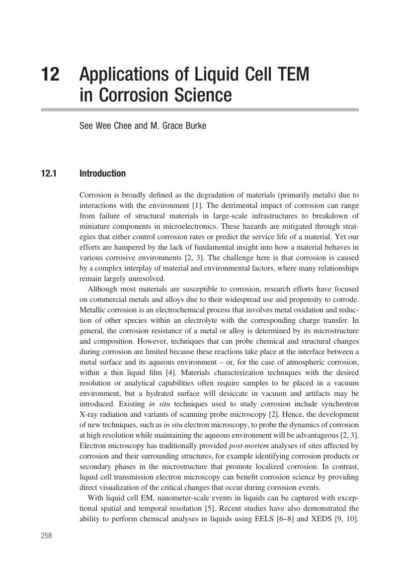
Image of the first page of this content. For PDF version, please use the ‘Save PDF’ preceeding this image.'
Type
Chapter
Information
Publisher: Cambridge University Press
Print publication year: 2016

Access options

Get access to the full version of this content by using one of the access options below. (Log in options will check for institutional or personal access. Content may require purchase if you do not have access.)

References

References and Notes

Shaw, B. and Kelly, R., What is corrosion? Interface, Electrochem. Soc., Spring (2006), 24–26.Google Scholar
Duquette, D. and Schafrik, R., Research Opportunities in Corrosion Science and Engineering (Washington, D.C.: The National Academies Press, 2011).Google Scholar
Song, G.-L., The grand challenges in electrochemical corrosion research. Front. Mater, 1 (2014), 2.Google Scholar
Frankel, G., Electrochemical techniques in corrosion: status, limitations, and needs. J. ASTM Int., 5 (2008), 127.Google Scholar
De Jonge, N. and Ross, F. M., Electron microscopy of specimens in liquid. Nat. Nanotechnol., 6 (2011), 695704.Google Scholar
Jungjohann, K. L., Evans, J. E., Aguiar, J. A., Arslan, I. and Browning, N. D., Atomic-scale imaging and spectroscopy for in situ liquid scanning transmission electron microscopy. Microsc. Microanal., 18 (2012), 621627.Google Scholar
Holtz, M. E., Yu, Y., Gao, J., Abruna, H. D. and Muller, D. A., In situ electron energy loss spectroscopy in liquids. Microsc. Microanal., 19 (2013), 10271035.CrossRefGoogle ScholarPubMed
Unocic, R. R., Baggetto, L., Unocic, K. et al., Coupling EELS/EFTEM imaging with environmental fluid cell microscopy. Microsc. Microanal., 18 (2012), 11041105.Google Scholar
Zaluzec, N. J., Burke, M. G., Haigh, S. J. and Kulzick, M. A., X-ray energy-dispersive spectrometry during in situ liquid cell studies using an analytical electron microscope. Microsc. Microanal., 20 (2014), 323329.Google Scholar
Schilling, S., Janssen, A., Zhong, Z. L., Zaluzec, N. J. and Burke, M. J., Liquid in situ analytical electron microscopy: examining SCC precursor events for Type 304 stainless steel in H2O. Microsc. Microanal., 21 (2015), 12911292.Google Scholar
McCafferty, E., Introduction to Corrosion Science (New York: Springer, 2010).Google Scholar
Frankel, G. and Sridhar, N., Understanding localized corrosion. Mater. Today, 11 (2008), 3844.Google Scholar
Frankel, G., Pitting corrosion of metals. J. Electrochem. Soc., 145 (1998), 21862198.Google Scholar
Soltis, J., Passivity breakdown, pit initiation and propagation of pits in metallic materials: review. Corros. Sci., 90 (2015), 522.Google Scholar
Accelerated dissolution of thin metal films had been observed during continuous imaging under the electron beam.Google Scholar
Unocic, R. R., Sacci, R. L., Brown, G. M. et al., Quantitative electrochemical measurements using in situ ec-S/TEM devices. Microsc. Microanal., 20 (2014), 452461.CrossRefGoogle ScholarPubMed
Frankel, G. and Rohwerder, M., Electrochemical techniques for corrosion. In Encyclopedia of Electrochemistry (Weinheim, Germany: Wiley-VCH, 2007).Google Scholar
Kelly, R. G., Scully, J. R., Shoesmith, D. and Buchheit, R., Electrochemical Techniques in Corrosion Science and Engineering (New York: Marcel Dekker, 2013).Google Scholar
Frankel, G., Techniques for Corrosion Quantification in the Characterization of Materials, 2nd edn. (Hoboken, NJ: John Wiley & Sons, 2012), pp. 850864.Google Scholar
Keddam, M., Application of advanced electrochemical techniques and concepts to corrosion phenomena. Corrosion, 62 (2006), 10561066.Google Scholar
Frankel, G., The growth of 2-D pits in thin film aluminum. Corros. Sci., 30 (1990), 1203.Google Scholar
Balazs, L. and Gouyet, J., Two-dimensional pitting corrosion of aluminium thin layers. Phys. A Stat. Mech. Appl., 217 (1995), 319338.Google Scholar
Frankel, G., Pit growth in thin metallic films. Mater. Sci. Forum, 247 (1997), 18.Google Scholar
Proost, J., Baklanov, M. and Verbeeck, R., Morphology of corrosion pits in aluminum thin film metallizations. J. Solid State Electrochem., 2 (1998), 150155.Google Scholar
Hernandez, S., Griffin, A. Jr., Brotzen, F. and Dunn, C., The effect of thickness on the corrosion susceptibility of Al thin film metallizations. J. Electrochem. Soc., 142 (1995), 12151220.Google Scholar
Zhao, Y.-P., Cheng, C.-F., Wang, G.-C. and Lu, T.-M., Characterization of pitting corrosion in aluminum films by light scattering. Appl. Phys. Lett., 73 (1998), 24322434.Google Scholar
Chee, S. W., Ross, F. M., Duquette, D. and Hull, R., Studies of corrosion of Al thin films using liquid cell transmission electron microscopy. MRS Proc., 1525 (2013), mrsf12-1525-ss11-03.Google Scholar
Chee, S. W., Duquette, D. J., Ross, F. M. and Hull, R., Metastable structures in Al thin films before the onset of corrosion pitting as observed using liquid cell transmission electron microscopy. Microsc. Microanal., 20 (2014), 462468.CrossRefGoogle ScholarPubMed
Chee, S. W., Pratt, S. H., Hattar, K. et al., Studying localized corrosion using liquid cell transmission electron microscopy. Chem. Commun., 51 (2015), 168171.Google Scholar
Chee, S. W., Hull, R. and Ross, F. M., Liquid cell TEM of the corrosion of metal films in aqueous solutions. Microsc. Microanal., 18 (2012), 11101111.Google Scholar
Liao, H.-G., Niu, K. and Zheng, H., Observation of growth of metal nanoparticles. Chem. Commun., 49 (2013), 1172011727.Google Scholar
Jiang, Y., Zhu, G., Lin, F., Zhang, H. and Jin, C., In situ study of oxidative etching of palladium nanocrystals by liquid cell electron microscopy. Nano Lett., 14 (2014), 37613765.CrossRefGoogle ScholarPubMed
Wu, J., Gao, W., Yang, H. and Zuo, J.-M., Imaging shape-dependent corrosion behavior of Pt nanoparticles over extended time using a liquid flow cell and TEM. Microsc. Microanal., 20 (2014), 15081509.Google Scholar
Sutter, E., Jungjohann, K., Bliznakov, S. et al., In situ liquid-cell electron microscopy of silver-palladium galvanic replacement reactions on silver nanoparticles. Nat. Commun., 5 (2014), 4946.Google Scholar
Chee, S. W., Park, J.-H., Pinkowitz, A. et al., Liquid cell TEM of Al thin film corrosion under potentiostatic polarization. Microsc. Microanal., 21 (2015), 973974.CrossRefGoogle Scholar
Park, J. H., Chee, S. W., Kodambaka, S. and Ross, F. M., In situ LC-TEM studies of corrosion of metal thin films in aqueous solutions. Microsc. Microanal., 21 (2015), 12911292.Google Scholar
Noh, K. W, Tai, K., Mao, S. and Dillon, S. J., Grain boundary parting limit during dealloying. Adv. Eng. Mater., 17 (2015), 157161.Google Scholar
Mayer, J., Giannuzzi, L. A., Kamino, T. and Michael, J., TEM sample preparation and FIB-induced damage. MRS Bull., 32 (2007), 400407.CrossRefGoogle Scholar
Unocic, R., Adamczyk, L., Dudney, N. et al., In-situ TEM characterization of electrochemical processes in energy storage systems. Microsc. Microanal., 17 (2011), 15641565.CrossRefGoogle Scholar
Zhong, X., Burke, M. G., Schilling, S., Haigh, S. J. and Zaluzec, N. J., Novel hybrid sample preparation method for in situ liquid cell TEM analysis. Microsc. Microanal., 20 (2014), 15141515.CrossRefGoogle Scholar
Woehl, T. J., Jungjohann, K. L., Evans, J. E. et al., Experimental procedures to mitigate electron beam induced artifacts during in situ fluid imaging of nanomaterials. Ultramicroscopy, 127 (2013), 5363.Google Scholar
Ring, E. A. and de Jonge, N., Microfluidic system for transmission electron microscopy. Microsc. Microanal., 16 (2010), 622629.Google Scholar
Hoppe, S. M., Sasaki, D. Y., Kinghorn, A. N. and Hattar, K., In-situ transmission electron microscopy of liposomes in an aqueous environment. Langmuir, 29 (2013), 99589961.Google Scholar
Abellan, P., Woehl, T. J., Parent, L. R. et al., Factors influencing quantitative liquid (scanning) transmission electron microscopy. Chem. Commun., 50 (2014), 48734880.CrossRefGoogle ScholarPubMed
Klein, K. L., Anderson, I. M. and de Jonge, N., Transmission electron microscopy with a liquid flow cell. J. Microsc., 242 (2011), 117123.Google Scholar
The liquid layer thicknesses quoted in atmospheric corrosion studies are normally in the tens of micrometers.Google Scholar
Sacci, R. L., Dudney, N. J., More, K. L. and Unocic, R. R., In operando transmission electron microscopy imaging of SEI formation and structure in Li-ion and Li-metal batteries. Microsc. Microanal., 20 (2014), 15981599.CrossRefGoogle Scholar
Schneider, N. M., Norton, M. M., Mendel, B. J. et al., Electron–water interactions and implications for liquid cell electron microscopy. J. Phys. Chem. C., 118 (2014), 2237322382.CrossRefGoogle Scholar
Grogan, J. M., Schneider, N. M., Ross, F. M. and Bau, H. H., Bubble and pattern formation in liquid induced by an electron beam. Nano Lett., 14 (2013), 359364.CrossRefGoogle ScholarPubMed
Kelm, M., Bohnert, E. and Pashalidis, I., Products formed from alpha radiolysis of chloride brines. Res. Chem. Intermed., 27 (2001), 503507.Google Scholar
Holtz, M. E., Yu, Y., Gunceler, D. et al., Nanoscale imaging of lithium ion distribution during in situ operation of battery electrode and electrolyte. Nano Lett., 14 (2014), 14531459.Google Scholar
Schilling, S., Janssen, A., Burke, M. G. et al., In situ analytical electron microscopy: imaging and analysis of steel in liquid water. 18th International Microscopy Congress (2014), www.microscopy.cz/proceedings/all.html#abstract-2947.Google Scholar
Bi-metallic exposure in the electrolyte frequently leads to galvanic corrosion but the effects of coupled metals are not so straightforward. Depending on the metals that are connected, it is possible that the more active metal becomes more resistant to corrosion. The reader is referred to general texts on corrosion for clarification.Google Scholar
Park, J.-H., Reuter, M. C., Kodambaka, S. and Ross, F. M., Electric field induced Au nanocrystal formation in aqueous solutions. Microsc. Microanal., 20 (2014), 15981599.CrossRefGoogle Scholar
Hoppe, S. M., Hernandez-Sanchez, B. A., Hattar, K. and Sasaki, D. Y., Progress towards in situ TEM of biofouling. Microsc. Microanal., 20 (2012), 11321133.Google Scholar

Save book to Kindle

To save this book to your Kindle, first ensure coreplatform@cambridge.org is added to your Approved Personal Document E-mail List under your Personal Document Settings on the Manage Your Content and Devices page of your Amazon account. Then enter the ‘name’ part of your Kindle email address below. Find out more about saving to your Kindle.

Note you can select to save to either the @free.kindle.com or @kindle.com variations. ‘@free.kindle.com’ emails are free but can only be saved to your device when it is connected to wi-fi. ‘@kindle.com’ emails can be delivered even when you are not connected to wi-fi, but note that service fees apply.

Find out more about the Kindle Personal Document Service.

Available formats
×

Save book to Dropbox

To save content items to your account, please confirm that you agree to abide by our usage policies. If this is the first time you use this feature, you will be asked to authorise Cambridge Core to connect with your account. Find out more about saving content to Dropbox.

Available formats
×

Save book to Google Drive

To save content items to your account, please confirm that you agree to abide by our usage policies. If this is the first time you use this feature, you will be asked to authorise Cambridge Core to connect with your account. Find out more about saving content to Google Drive.

Available formats
×