
Book contents
- Core Topics in Foot and Ankle Surgery
- Core Topics in Foot and Ankle Surgery
- Copyright page
- Contents
- Contributors
- Preface
- Abbreviations
- Chapter 1 Clinical Examination of the Foot and Ankle
- Chapter 2 Biomechanics of the Foot and Ankle
- Chapter 3 Radiology of the Foot and Ankle
- Chapter 4 Orthoses for the Foot and Ankle
- Chapter 5 Amputations, Prostheses, and Rehabilitation of the Foot and Ankle
- Chapter 6 Forefoot Pathology
- Chapter 7 Midfoot and Forefoot Arthritis
- Chapter 8 Ankle and Hindfoot Arthritis
- Chapter 9 The Cavus Foot
- Chapter 10 Adult Acquired Flat Foot Deformity
- Chapter 11 Sports Injuries of the Foot and Ankle
- Chapter 12 Ankle Arthroscopy
- Chapter 13 Foot Arthroscopy and Tendonoscopy
- Chapter 14 Heel Pain
- Chapter 15 Disorders of the Tendo Achillis
- Chapter 16 Rupture of the Tendo Achillis
- Chapter 17 Benign Tumors of the Foot and Ankle
- Chapter 18 Malignant Tumors of the Foot and Ankle
- Chapter 19 The Diabetic Foot
- Chapter 20 The Pediatric Foot
- Chapter 21 Fractures and Dislocations of the Ankle
- Chapter 22 Fractures and Dislocations of the Hindfoot and Midfoot
- Chapter 23 Fractures and Dislocations of the Forefoot
- Index
- References
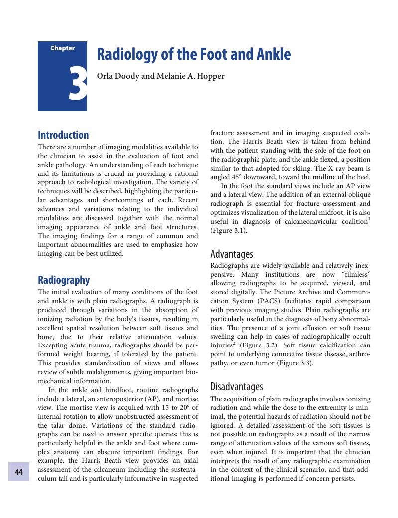
Chapter 3 - Radiology of the Foot and Ankle
Published online by Cambridge University Press: 29 March 2018

Book contents
- Core Topics in Foot and Ankle Surgery
- Core Topics in Foot and Ankle Surgery
- Copyright page
- Contents
- Contributors
- Preface
- Abbreviations
- Chapter 1 Clinical Examination of the Foot and Ankle
- Chapter 2 Biomechanics of the Foot and Ankle
- Chapter 3 Radiology of the Foot and Ankle
- Chapter 4 Orthoses for the Foot and Ankle
- Chapter 5 Amputations, Prostheses, and Rehabilitation of the Foot and Ankle
- Chapter 6 Forefoot Pathology
- Chapter 7 Midfoot and Forefoot Arthritis
- Chapter 8 Ankle and Hindfoot Arthritis
- Chapter 9 The Cavus Foot
- Chapter 10 Adult Acquired Flat Foot Deformity
- Chapter 11 Sports Injuries of the Foot and Ankle
- Chapter 12 Ankle Arthroscopy
- Chapter 13 Foot Arthroscopy and Tendonoscopy
- Chapter 14 Heel Pain
- Chapter 15 Disorders of the Tendo Achillis
- Chapter 16 Rupture of the Tendo Achillis
- Chapter 17 Benign Tumors of the Foot and Ankle
- Chapter 18 Malignant Tumors of the Foot and Ankle
- Chapter 19 The Diabetic Foot
- Chapter 20 The Pediatric Foot
- Chapter 21 Fractures and Dislocations of the Ankle
- Chapter 22 Fractures and Dislocations of the Hindfoot and Midfoot
- Chapter 23 Fractures and Dislocations of the Forefoot
- Index
- References
Summary
A summary is not available for this content so a preview has been provided. Please use the Get access link above for information on how to access this content.
- Type
- Chapter
- Information
- Core Topics in Foot and Ankle Surgery , pp. 44 - 62Publisher: Cambridge University PressPrint publication year: 2018
References
Crim, J. Imaging of tarsal coalition. Radiologic Clinics of North America. 2008; 46:6, 1017–26.Google Scholar
Clark, TW, Janzen, DL, Ho, K, Grunfeld, A, Connell, DG. Detection of radiographically occult ankle fractures following acute trauma: positive predictive value of an ankle effusion. AJR. American Journal of Roentgenology, 1995; 164:5, 1185–9.Google Scholar
Chandnani, VP, Harper, MT, Ficke, JR, et al. Chronic ankle instability: evaluation with MR arthrography, MR imaging, and stress radiography. Radiology. 1994; 192:1, 189–94.CrossRefGoogle ScholarPubMed
Dobbins, JT 3rd. Tomosynthesis imaging: at a translational crossroads. Medical Physics. 2009; 36:6, 1956–67.Google Scholar
Ottenin, MA, Jacquot, A, Grospretre, O, et al. Evaluation of the diagnostic performance of tomosynthesis in fractures of the wrist. AJR. American Journal of Roentgenology. 2012; 198:1, 180–6.Google Scholar
Crass, JR, van de Vegte, GL, Harkavy, LA. Tendon echogenicity: ex vivo study. Radiology. 1988; 167:2, 499–501.CrossRefGoogle ScholarPubMed
McNally, EG. The development and clinical applications of musculoskeletal ultrasound. Skeletal Radiology. 2011; 40:9, 1223–31.CrossRefGoogle ScholarPubMed
Drakonaki, EE, Allen, GM, Wilson, DJ. Real-time ultrasound elastography of the normal Achilles tendon: reproducibility and pattern description. Clinical Radiology. 2009; 64:12, 1196–202.Google Scholar
Nicolaou, S, Yong-Hing, CJ, Galea-Soler, S, et al. Dual-energy CT as a potential new diagnostic tool in the management of gout in the acute setting. AJR. American Journal of Roentgenology. 2010; 194:4, 1072–8.Google Scholar
Guggenberger, R, Gnannt, R, Hodler, J, et al. Diagnostic performance of dual-energy CT for the detection of traumatic bone marrow lesions in the ankle: comparison with MR imaging. Radiology. 2012; 264:1, 164–73.Google Scholar
Pache, G, Bulla, S, Baumann, T, et al. Dose reduction does not affect detection of bone marrow lesions with dual-energy CT virtual noncalcium technique. Academic Radiology. 2012; 19:12, 1539–45.CrossRefGoogle Scholar
Hunt, CH, Wood, CP, Lane, JI, Bolster, BD, Bernstein, MA, Witte, RJ. Wide, short bore magnetic resonance at 1.5 T: reducing the failure rate in claustrophobic patients. Clinical Neuroradiology. 2011; 21:3, 141–4.Google Scholar
Olsen, RV, Munk, PL, Lee, MJ, et al. Metal artifact reduction sequence: early clinical applications. Radiographics: A Review Publication of the Radiological Society of North America, Inc. 2000; 20:3, 699–712.CrossRefGoogle ScholarPubMed
Cerezal, L, Llopis, E, Canga, A, et al. MR arthrography of the ankle: indications and technique. Radiologic Clinics of North America. 2008; 46:6, 973–94.Google Scholar
Roemer, FW, Crema, MD, Trattnig, S, Guermazi, A. Advances in imaging of osteoarthritis and cartilage. Radiology. 2011; 260:2, 332–54.Google Scholar
Gold, GE, Chen, CA, Koo, S, Hargreaves, BA, Bangerter, NK. Recent advances in MRI of articular cartilage. AJR. American Journal of Roentgenology. 2009; 193:3, 628–38.CrossRefGoogle ScholarPubMed
Even-Sapir, E, Flusser, G, Lerman, H, Lievshitz, G, Metser, U. SPECT/multislice low-dose CT: a clinically relevant constituent in the imaging algorithm of nononcologic patients referred for bone scintigraphy. Journal of Nuclear Medicine: Official Publication, Society of Nuclear Medicine. 2007: 48:2, 319–24.Google ScholarPubMed
Flick, AB, Gould, N. Osteochondritis dissecans of the talus (transchondral fractures of the talus): review of the literature and new surgical approach for medial dome lesions. Foot Ankle. 1985; 5:4, 165–85.CrossRefGoogle ScholarPubMed
De Smet, AA, Fisher, DR, Burnstein, MI, Graf, BK, Lange, RH. Value of MR imaging in staging osteochondral lesions of the talus (osteochondritis dissecans): results in 14 patients. AJR. American Journal of Roentgenology. 1990; 154:3, 555–8.CrossRefGoogle ScholarPubMed
Rankine, JJ, Nicholas, CM, Wells, G, Barron, DA. The diagnostic accuracy of radiographs in Lisfranc injury and the potential value of a craniocaudal projection. AJR. American Journal of Roentgenology. 2012; 198:4, W365–9.CrossRefGoogle ScholarPubMed
Rosenberg, ZS, Beltran, J, Bencardino, JT. From the RSNA Refresher Courses. Radiological Society of North America. MR imaging of the ankle and foot. Radiographics. 2000; 20, S153–79.Google Scholar
Neustadter, J, Raikin, SM, Nazarian, LN. Dynamic sonographic evaluation of peroneal tendon subluxation. AJR. American Journal of Roentgenology. 2004; 183:4, 985–8.Google Scholar
Perrich, KD Goodwin, DW, Hecht, PJ, Cheung, Y. Ankle ligaments on MRI: appearance of normal and injured ligaments. AJR. American Journal of Roentgenology. 2009; 193:3, 687–95.Google Scholar
Klein, MA. MR imaging of the ankle: normal and abnormal findings in the medial collateral ligament. AJR. American Journal of Roentgenology. 1994; 162:2, 377–83.Google Scholar
Lipsky, BA, Pecoraro, RE, Wheat, LJ. The diabetic foot. Soft tissue and bone infection. Infectious Disease Clinics of North America. 1990; 4:3, 409–32.Google Scholar
Lipsky, BA, Aragón-Sánchez, J, Diggle, M, et al. IWGDF guidance on the diagnosis and management of foot infections in persons with diabetes. Diabetes Metab Res Rev. 2016; 32:Suppl 1, 45–74.CrossRefGoogle ScholarPubMed
Berkowitz, JF, Kier, R, Rudicel, S. Plantar fasciitis: MR imaging. Radiology. 1991; 179:3, 665–7.CrossRefGoogle ScholarPubMed
Morrison, WB, Schweitzer, ME, Wapner, KL, Lackman, RD. Plantar fibromatosis: a benign aggressive neoplasm with a characteristic appearance on MR images. Radiology. 1994; 193:3, 841–5.CrossRefGoogle ScholarPubMed
Hopper, MA, Robinson, P. Ankle impingement syndromes. Radiologic Clinics of North America. 2008; 46:6, 957–71.Google Scholar