
Book contents
- Pediatric Head and Neck Pathology
- Pediatric Head and Neck Pathology
- Copyright page
- Contents
- Contributors
- Preface
- Acknowledgments
- Chapter 1 Diagnostic Methods and Specimen Handling Techniques in Pediatric Surgical Pathology
- Chapter 2 Soft Tissue Tumors and Reactive and Inflammatory Lesions of the Oral Cavity and Head and Neck
- Chapter 3 Cutaneous Tumors and Pseudotumors of the Head and Neck
- Chapter 4 Mucocutaneous Pigmented Lesions, Nevi and Melanoma
- Chapter 5 Vesiculo-Erosive and Ulcerative Lesions of Oral Cavity and Skin
- Chapter 6 Oral Epithelial Neoplasms
- Chapter 7 Odontogenic and Non-Odontogenic Cysts
- Chapter 8 Odontogenic Tumors
- Chapter 9 Non-Neoplastic Diseases of Salivary Glands
- Chapter 10 Benign Neoplasms of Salivary Glands
- Chapter 11 Malignant Neoplasm of Salivary Glands
- Chapter 12 Non-Neoplastic Disorders of the Nasal Cavity, Paranasal Sinuses and Nasopharynx
- Chapter 13 Benign Neoplasms of the Nasal Cavity, Paranasal Sinuses and Nasopharynx
- Chapter 14 Malignant Neoplasms of the Nasal Cavity, Paranasal Sinuses and Nasopharynx
- Chapter 15 Disorders of the Larynx and Hypopharynx
- Chapter 16 Neoplasms of Bone and Fibro-Osseous Lesions of the Craniofacial Skeleton
- Chapter 17 Disorders of the Ear
- Chapter 18 Disorders of the Thyroid Gland and Parathyroid Glands
- Chapter 19 Reactive and Malignant Diseases of Hematopoietic and Lymphoid Tissues
- Chapter 20 Developmental and Syndromic Disturbances of the Craniofacial Region
- Index
- References
Chapter 17 - Disorders of the Ear
Published online by Cambridge University Press: 26 June 2017

Book contents
- Pediatric Head and Neck Pathology
- Pediatric Head and Neck Pathology
- Copyright page
- Contents
- Contributors
- Preface
- Acknowledgments
- Chapter 1 Diagnostic Methods and Specimen Handling Techniques in Pediatric Surgical Pathology
- Chapter 2 Soft Tissue Tumors and Reactive and Inflammatory Lesions of the Oral Cavity and Head and Neck
- Chapter 3 Cutaneous Tumors and Pseudotumors of the Head and Neck
- Chapter 4 Mucocutaneous Pigmented Lesions, Nevi and Melanoma
- Chapter 5 Vesiculo-Erosive and Ulcerative Lesions of Oral Cavity and Skin
- Chapter 6 Oral Epithelial Neoplasms
- Chapter 7 Odontogenic and Non-Odontogenic Cysts
- Chapter 8 Odontogenic Tumors
- Chapter 9 Non-Neoplastic Diseases of Salivary Glands
- Chapter 10 Benign Neoplasms of Salivary Glands
- Chapter 11 Malignant Neoplasm of Salivary Glands
- Chapter 12 Non-Neoplastic Disorders of the Nasal Cavity, Paranasal Sinuses and Nasopharynx
- Chapter 13 Benign Neoplasms of the Nasal Cavity, Paranasal Sinuses and Nasopharynx
- Chapter 14 Malignant Neoplasms of the Nasal Cavity, Paranasal Sinuses and Nasopharynx
- Chapter 15 Disorders of the Larynx and Hypopharynx
- Chapter 16 Neoplasms of Bone and Fibro-Osseous Lesions of the Craniofacial Skeleton
- Chapter 17 Disorders of the Ear
- Chapter 18 Disorders of the Thyroid Gland and Parathyroid Glands
- Chapter 19 Reactive and Malignant Diseases of Hematopoietic and Lymphoid Tissues
- Chapter 20 Developmental and Syndromic Disturbances of the Craniofacial Region
- Index
- References
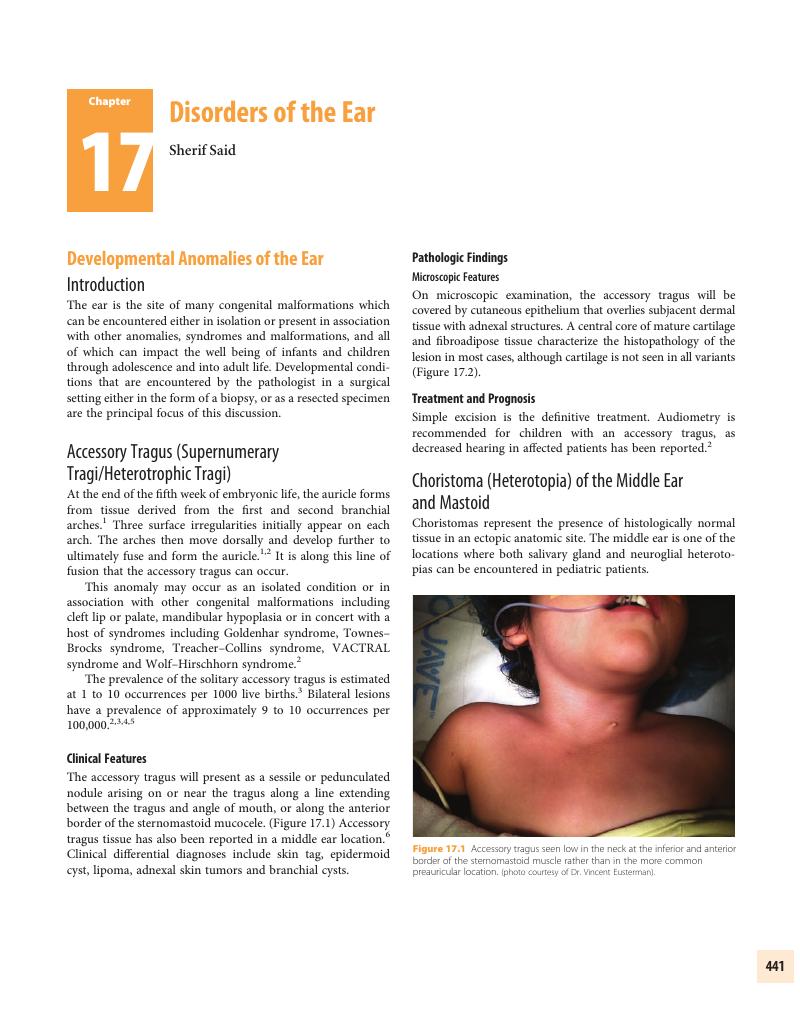
Summary
A summary is not available for this content so a preview has been provided. Please use the Get access link above for information on how to access this content.
- Type
- Chapter
- Information
- Pediatric Head and Neck Pathology , pp. 441 - 470Publisher: Cambridge University PressPrint publication year: 2016
References
Primary Sources
Wood-Jones, F, Chuan, IW. The development of the external ear. J Anat 1933–1934; 68:525–533.Google Scholar
Jansen, T, Romiti, R, Altmeyer, P. Accessory Tragus: report of two cases and review of the literature. Pediatr Dermatol 2000; 17:391–394.CrossRefGoogle ScholarPubMed
Melnick, M, Myrianthopolos, NC. External ear malformations: epidemiology, genetics, and natural history. Birth defects 1979; 15:1–138.Google Scholar
Weichmann, A. Briecht uber die in den jahren bis 1893 bis juli 1912 in der koniglichen universitats-Poliklinik ohern-, nasen- und halskrankheiten in Gottingen beobachtenen angeborenen missidungen des ohers (dissertation). Gottengen 1912.Google Scholar
Guszman, J. Beitrage Zur Lehre der branchiogenen ohr-und halsanhange. Z Ges Anat 1926; 81: 554–562.Google Scholar
Chintalapati, K, Gunasekaran, S, Frewer, J. Accessory tragus in the middle ear: A rare congenital anomaly. Int. J Pediatr Otorhinolaryngol 2010; 74:1338–1339.CrossRefGoogle ScholarPubMed
Secondary Sources
Taylor, GD, Martin, HF. Salivary gland tissue in the middle ear. A rare Tumor. Arch Otolaryngol. 1961; 73:651–657.Google Scholar
Ha, SL, Shin, JE, Yoon, TH. Salivary gland choristoma of the middle ear: a case report and review of the literature. Laryngoscope 1984; 94: 228–230.Google Scholar
Farneti, P, Balbi, M, Foschini, MP. Neurologlial choristoma of the middle ear. Acta Otorhinolaryngologica Italica 2007; 27:94–97.Google Scholar
Kartush, JM, Graham, MD, Arbor, A. Salivary gland choristoma of the middle ear: A case report and review of the literature. Laryngoscope 1984; 94:228–230.Google Scholar
Morimotot, N, Ogawa, K, Kanzaki. Salivary gland choristoma in the middle ear. A case report. Am J Otolaryngol 1999; 20: 232–235.Google Scholar
Enoz, M, Suoglu, Y. Salivary gland choristoma of the middle ear. Laryngoscope 2006; 116:1033–1034.Google Scholar
Gyure, AJ, Glasscock, ME, Pensak, ML. A clinicopathologic study of 15 patients with neuroglial heterotopias and encephaloceles of the middle ear and mastoid region. Laryngsocope 2000; 110:1731–1735.Google Scholar
Plontke, SK, Preyer, S, Pressler, H, Mundiger, PM, Plinkert, PK. Glial lesion of the infratemporal fossa presenting as a soft tissue middle ear mass – rudimentary encephalocele or neural crest remnant? Int. J Pediatr Otorhinolaryngol 2000; 56:141–147.Google Scholar
Nelson, EG, Kratz, RC. Sebaceous choristoma of the middle ear. Otolaryngol Head Neck Surg 1993; 108:372–373.Google Scholar
Lee, DK, Kim, JH, Cho, YS, et al. Salivary gland choristoma of the middle ear in an infant: a case report. Int J Pediatr Otolaryngol 2006; 70:167–170.Google Scholar
Saeed, YM, Bassis, ML. Mixed tumor of the middle ear. A case report. Arch Otolaryngol 1971; 93:433–434.CrossRefGoogle ScholarPubMed
Peters, BR, Maddox, HE III, Batsakis, JG. Pleomorphic adenoma of the middle ear and mastoid with posterior fossa extension. Arch Otolaryngol Head Neck Surg 1988, 114:676–678.Google Scholar
Triglia, JM, Nicollas, R, Ducroz, V, et al. First branchial cleft anomalies: a study of 39 cases and review of the literature. Arch Otolaryngol Head Neck Surg 1998; 124:291–295.Google Scholar
Olsen, KD, Maragos, NE, Weiland, LH. First branchial cleft anomalies. Laryngoscope 1980; 90:423–435.Google Scholar
Work, WP. Newer concepts of first branchial cleft defects. Laryngoscope 1972; 9:1581–1593.Google Scholar
Aronsohn, RS, Batsakis, JG, Rice, DH, Work, WP. Anomalies if the first brachial arch cleft. Arch Otolaryngol 1976; 102:737–740.Google Scholar
Lim, LH, Hartnick, CJ, Williging, JP. A rare case of combined Type I and Type II first branchial sinus. J Pediatr Surg 2003; 38:E12–E13.CrossRefGoogle ScholarPubMed
Greenway, RE, Hurst, L, Fenton, NA. An unusual first branchial cleft cyst. J Otolaryngol 1981; 10:219–225.Google Scholar
House, J, Wilkinson, E. External auditory exostoses: evaluation and treatment. Otolaryngol Head Neck Surg 2008; 138:672–678.Google Scholar
Harrison, DF. Exostoses of the external auditory meatus. J Laryngol Otol 1951; 65:704–714.Google Scholar
Timofeev, I, Notkina, N, Smith, I. Exostoses of the external auditory canal: a long term followup study of surgical treatment. Clin Otolaryngol 2004; 29:588–594.Google Scholar
Van Gilse, P. Des observations ulterieures sur la genese exostoses du conduitexterne par l’iratio d’eau froid. Acta Otolaryngol 1938; 26:343–352.Google Scholar
Fowler, E, Osmun, P. New bone growth due to cold water in the ears. Arch Otolaryngol 1942; 36:455–466.Google Scholar
Whitaker, SR, Cordier, A, Kosjakov, S, Charbonneau, R. Treatment of the external auditory canal exostoses. Laryngoscope 1998; 108: 195–199.Google Scholar
Schuknecht, HF. Exostoses of the external auditory canal. In: Pathology of the Ear. Philadelphia: Lea & Febiger 1993, 398–399.Google Scholar
Fisher, EW, McManus, TC. Surgery for external auditory canal exostoses and osteomata, J Laryngol Otol 1994; 108:106–110.Google Scholar
Alster, T, Tanzi, E. Hypertrophic scars and keloids. Etiology and management. Am J Clin Dermatol 2003; 4:235–243.Google Scholar
Urioste, SS, Arndt, KA, Dover, JS. Keloidal scars and hypertrophic scars: review and treatment strategies. Semin Cutan Med Surg 1999; 18:159–171.Google Scholar
Muir, I. On the nature of keloidal scars and hypertrophic scars. Br J Plast Surg 1990; 43: 61–69.Google Scholar
Niessen, FB, Spauwen, PH, Schalkwijk, J, et al. On the nature of keloidal scars and hypertrophic scars: a review. Plast Reconstr Surg 1999; 105: 1435–1458.Google Scholar
Marneros, AG, Norris, JE, Olsen, BR, et al. Clinical genetics of familial keloids. Arch Dermatol 2001; 137:1429–1434.Google Scholar
English, RS, Shenefelt, PD. Keloids and hypertrophic scars. Dermatol Surg 1999; 25:631–638.Google Scholar
Fong, EP, Chye, LT, Tan, WL. Keloids: time to dispel the myths? Plast Reconstr Surg 1999; 104:1199–1201.Google Scholar
Lee, J, Yang, C, Chao, S, Wong, T. Histopathological differential diagnosis of keloid and hypertrophic scar. Am J Dermatopathol 2004; 26:379–384.Google Scholar
Abergel, RP, Pizzurro, D, Meeker, CA, et al. Biochemical composition of the connective tissue in keloids and analysis of collagen metabolism in keloid fibroblast cultures. J Invest Dermatol 1985; 84:384–390.Google Scholar
Calderon, M, Lawrence, WT, Banes, AJ. Increased proliferation of keloid fibroblasts in vitro. J Surg Res 1996; 61:343–347.Google Scholar
Meltzer, PE, Kelemen, G. Pyocutaneous osteomyelitis of the temporal bone, mandible, and zygoma. Laryngoscope 1959; 169: 1300–1316.CrossRefGoogle Scholar
Castro, R, Robinson, N, Klein, J, et al. Malignant external otitis and mastoiditis associated with IgG4 subclass deficiency in a child. Del Med J 1990; 62:1417–1415.Google ScholarPubMed
Paul, AC, Justus, A, Balraj, A, et al. Malignant otitis externa in an infant with selective IgA deficiency: a case report. Int J Pediatr Otorhinolaryngol 2001; 60:141–145.Google Scholar
Sobie, S, Brodsky Stanievich, JF. Necrotizing external otitis in children: report of two cases and review of the literature. Laryngoscope 1987; 97:598–601.Google Scholar
Carfrae, MJ, Kesser, BW. Malignant otitis externa. Otolaryngol Clin N Am 2008; 41:537–549.CrossRefGoogle ScholarPubMed
Rubin, J, Yu, VL, Stool, SE. Malignant external otitis in children. J Pediatr 1988; 113:965–970.CrossRefGoogle ScholarPubMed
Cunningham, M, Yu, VL, Turner, J, et al. Necrotizing otitis externa due to aspergillus in an incompetent patient. Arch Otolaryngol Head Neck Surg 1988; 114:554–556.Google Scholar
Sreepada, GS, Kwartler, JA. Skull base osteomyelitis secondary to malignant externa. Curr Opin Otolaryngol Head Neck Surg 2003; 11:316–323.Google Scholar
Gilain, L, Labroue, M, Aidan, D, Ragu, MP, et al. Value of hyperbaric oxygen therapy in the treatment of malignant otitis externa. Apropos of a case. Ann Otolaryngol Chir Cervicofac. 1993; 110:50–54. Review.Google Scholar
Winkler, M. Chondrodermatitis nodularis chronicus helicis. Arch Dermatol Syphilol 1915; 121:278.Google Scholar
Hurwitz, RM. Inflammatory reactions of the folliculosebaceous unit. In: Pathology of the Skin. Farmer, ER, Hood, AF, Editors, New York: McGraw-Hill 2000.Google Scholar
Rogers, NE, Farris, PK, Wang, A. Juvenile chondrodermatitis nodularis helicis: A case report and literature review.Google Scholar
Sasaki, T, Nishizawa, H, Sugita, Y. Chondrodermatitis nodularis helicis in childhood dermatomyositis. Br J Dermatol 1999; 141:363–365.CrossRefGoogle ScholarPubMed
Carol, W, van Haren, H. Uber clavus helicis bzw. Chondrodermatitis nodularis chronicus helicis. Dermatologica 1941; 83:535.Google Scholar
Moncrieff, M, Sassoon, EM. Effective treatment of chondrodermatitis nodularis chronica helicis using a conservative approach. Br. J Dermatol 2004; 150:892–894.Google Scholar
Tsai, TH, Lin, YC, Chan, HC. Infantile chondrodermatitis nodularis. Pediatr-Dermatol, 2007; 24:337–339.Google Scholar
Karam, F, Bauman, T. Carbon dioxide laser treatment for chondrodermatitis nodular chronicus helices. Ear Nose and Throat, 1988; 67:751–758.Google Scholar
Wells, GC, Whimster, IW. Subcutaneous angiolymphoid hyperplasia with eosinophilia. Br J Dermatol 1969; 81:1–15.Google Scholar
Kaur, T, Sandhu, K, Gupta, S, et al. Treatment of angiolymphoid hyperplasia with eosinophilia with carbon dioxide laser. J Dermatol Treat 2004; 15:328–330.Google Scholar
Hallam, LA, Mackinlay, GA, Wright, AM. Angiolymphoid hyperplasia with eosinophilia: possible etiologic role for immunization. J Clin Pathol 1989; 42:944–949.Google Scholar
Akosa, AB, Ali, MH, Khoo, CT, Evans, DM. Angiolymphoid hyperplasia with eosinophilia associated with tetanus toxoid vaccination. Histopathology 1990; 16:589–593.Google Scholar
Jeon, EK, Cho, AY, Kim, MY, et al. Angiolymphoid hyperplasia with eosinophilia that was possibly induced by vaccination in a child. Ann Dermatol 2009; 21:71–74.Google Scholar
Chen, JF, Gao, HW, Wu, BY, et al. Angiolymphoid hyperplasia with eosinophilia affecting the scrotum: a rare case report with molecular evidence of T-cell clonality. J Dermatol 2010; 37:355–359.CrossRefGoogle ScholarPubMed
Googe, PB, Harris, NL, Mihm, MC. Kimura’s disease and angiolymphoid hyperplasia with eosinophilia: two distinct histopathological entities. J Cutan Pathol 1987; 14:263–271.Google Scholar
Seregard, S. Angiolymphoid hyperplasia with eosinophilia should not be confused with Kimura’s disease. Acta Ophthalmol Scand 2001; 79:91–93.CrossRefGoogle Scholar
Heffner, DK, Hyams, VJ. Cystic chondromalacia (endochondral pseudocyst) of the auricle. Arch Pathol Lab Med 1986; 110:740–743.Google Scholar
Lazar, RH, Heffner, DK, Hughes, GB, et al. Pseudocyst of the auricle. A review of 21 cases. Otolaryngol Head Neck Surg 1986; 94:360–361.Google Scholar
Devlin, J, Harrison, CJ, Whitby, DJ, David, TJ. Cartilaginous pseudocyst of the external auricle in children with atopic eczema. Br J Dermatol 1990; 122:699–704.Google Scholar
Grabski, WJ, Salasche, SJ, McCollough, ML, Angeloni, VL. Pseudocyst of the auricle associated with trauma. Arch Dermatol 1989; 125:528–530.Google Scholar
Belt, A, Duquesne, A, Job-Deslandre, C, et al. Pediatric onset relapsing polychondritis: case series and systemic review. J Pediatr 2010; 156:484–489.Google Scholar
McAdams, LP, O’Hanlan, MA, Bluestone, R, Pearson, CM. Relapsing polychondritis: prospective study of 23 patients and review of the literature. Medicine 1976; 55:193–215.Google Scholar
Rothstein, J, Adams, GL, Galliani, CA, Elliot, GR. Relapsing polychondritis in a 30 month old child. Otolaryngol Head Neck Surg 1985; 93:680–683.Google Scholar
Lamy, MJM, Nezlof, C, Laugier, J, Ajjan, N. La polychondrite atrophinate evolutive. Arch Fr Pediatr 1967; 24:633.Google Scholar
Kaye, RL, Sones, DA. Relapsing polychondritis. Clinical and pathologic features in fourteen cases. Ann Intern Med 1964; 60:653–664.Google Scholar
Valenzuela, R, Cooperrider, PA, Gogate, P, et al. Relapsing polychondritis. Immunomicroscopic findings in cartilage of ear biopsy specimens. Hum Pathol 1980; 11:19–22.Google Scholar
Monasta, L, Rofani, L, Marchetti, F, et al. Burden of disease caused by otitis media: systemic review and global estimates. PLoS One. 2012; 7(4):Epub.Google Scholar
American Academy of Pediatrics. Subcommittee on management of acute otitis media. Diagnosis and Treatment of Acute Otitis Media. 2004; 113:1451–1465.Google Scholar
Chonmaitree, T, Revai, K, Grady, J, et al. Viral upper respiratory tract infection and otitis media complication in young children. Clin Infect Dis 2008; 46:815–823.Google Scholar
Berkun, Y, Nir-Paz, R, Ben, Ami A. Acute otitis media in the first two months of life: characteristics and diagnostic difficulties. Arch Dis Child 2008; 93: 690–694.Google Scholar
Sade, J, Russo, E, Fuchs, C, et al. Is secretory otitis media a single disease entity? Ann Otol Rhinol Laryngol 2003; 112:342–347.Google Scholar
Gilklich, RE, Cunningham, MJ, Eavey, RD. The cause of aural polyp in children. Arch Otolaryngol Head Neck Surg 1993; 119:669–671.Google Scholar
Milroy, C, Slack, R, Maw, A, Bradfield, J. Aural polyps as predictors of underlying cholesteatoma. J Clin Pathol 1989; 42:460–465.Google Scholar
Bluestone, CD, Klein, JO. Infratemporal complications and sequelae of otitis media. In: Pediatric Otolaryngology. 4th Ed., Bluestone, CD, Casselbrant, ML, Stool, SE, et al., Editors. Saunders: Philadelphia 2003, 2687.Google Scholar
Derlacki, EL, Clemis, JD. Congenital cholesteatoma of the middle ear and mastoid. Ann Otol Rhinol Laryngol 1965; 74:706–727.Google Scholar
Potsic, WP, Korman, SB, Samadi, DS, Wetmore, RF. Congenital cholesteatoma: 20 year experience at the children hospital of Philadelphia. Otolaryngol Head Neck Surg 2002; 126:409–414.CrossRefGoogle Scholar
Levenson, MJ, Parisier, SC, Chute, P, et al. A review of twenty congenital cholesteatomas of the middle ear in children. Otolaryngol Head Neck Surg 1986; 94:560–567.Google Scholar
Bennett, M, Warren, F, Jackson, GC, Kaylie, D. Congenital cholesteatoma: theories, facts and 53 patients. Otolaryngol Clin North Am 2006; 39:1081–1094.Google Scholar
Kemppainen, HO, Puhakka, HJ, Laippala, PJ, et al. Epidemiology and etiology of middle ear cholesteatoma. Acta Otolaryngol 1999; 119:568–572.Google ScholarPubMed
Tos, M. A new pathogenesis of mesotympanic (congenital) cholesteatoma. Laryngoscope 2000; 110:1890–1895.Google Scholar
Lau, T, Tos, M. Tensa retraction cholesteatoma: treatment and long term results. J Laryngol Otol 1989; 103:149–157.Google Scholar
Paco, J, Branco, C, Estibiero, H, et al. The posterosuperior quadrant of the tympanic membrane. Otolaryngol Head Neck Surg 2009; 140:884–888.Google Scholar
Lowue, L. Acquired cholesteatoma pathogenesis: stepwise explanations. J Laryngol Otol 2010; 124:587–593.Google Scholar
Smith, JA, Danner, CJ. Complications of chronic otitis media and cholesteatoma. Otolaryngol Clin North Am 2006; 39:1237–1257.Google Scholar
Matanda, RN, Muyunga, KC, Sabue, MJ, et al. Chronic suppurative otitis media and related complications at the university clinic of Kinshasa. B – ENT 2005; 1:57–62.Google Scholar
Mueller, DT, Isaacson, G. Staged tympanostomy tube placement facilitates pediatric cholesteatoma management. Int J Pediatr Otorhinolaryngol 2008; 72:167–171.Google Scholar
Rosenfeld, RM, Moura, RL, Bluestone, CD. Predictors of residual-recurrent cholesteatoma in children. Arch Otolaryngol Head Neck Surg 1992; 118:384–389.Google Scholar
Stangerup, SE, Drozdziewicz, D, Tos, M, Trablazini, F. Surgery for acquired cholesteatoma in children: long term results and recurrence in cholesteatoma. J Laryngal Otol 1998; 112:742–752.Google Scholar
Maw, AR. Development of tympanosclerosis in children with otitis media with effusion and ventilation tubes. J Laryngol Otol 1991; 105:614–617.Google Scholar
Møller, Per. Tympanosclerosis of the ear drum in children. International Journal of Pediatric Otorhinolaryngology 1984; 7:247–256Google Scholar
Lin, H, Handzel, O, Faquin, W, Gopen, Q. Myospherulosis from antibiotic ointment in the postoperative mastoid space. Am J Otolaryngol Head Neck Surg 2010; 31:205–208.Google Scholar
Kyriakos, M. Myospherulosis of the paranasal sinuses, nose and middle ear. A possible iatrogenic disease. Am J Clin Path 1977; 67:118–130.Google Scholar
Gillespie, C, Clark, W, Finkelstein, E, et al. Myospherulosis of the mastoid antrum: a case report. Auris Nasus Larynx 1990; 16:199–207.Google Scholar
Mace, AT, Marshall, JN. Foreign body reaction to an oil based ointment: a cause of persistent otorrhea following mastoids surgery. J Laryngal Otol 2003; 117:496–497.Google Scholar
Campos, MK, Viana, MB, de Oliviera, BM, et al. Langerhans cell histiocytosis: a 16 year experience. J Pediatr 2007; 83:79–86.Google Scholar
Boston, M, Craig, SD. Langerhans cell histiocytosis of the temporal bone and skull base. Am J Otolaryngol 2002; 23:246–248.Google Scholar
Lichtenstein, L, Jaffe, HL. Eosinophilic granuloma of bone – with report of a case. Am J Pathol 1940; 16:595–604.Google Scholar
Lichtenstein, L. Histiocytosis X; integration of eosinophilic granuloma of bone, letterer-Siwe disease, and Schüller-Christian disease as related manifestations of a single nosologic entity. AMA Arch Pathol 1953; 56:84–102.Google Scholar
Chu, A, Favara, BE, Ladisch, S, et al. Report and recommendations of the workshop on the childhood histiocytosis: concepts and controversies. Med Pediatr Oncol 1986; 14:116–117.Google Scholar
Ferriera, LM, de Carvalho, JD, Periera, ST, Tavares, MG. Histiocytosis X of the temporal bone. Rev Bras Otolaryngol 2006; 72:575.Google Scholar
Bayazit, Y, Sirikci, A, Barayam, M, et al. Eosinophilic granuloma of the temporal bone. Auris Nasus Larynx 2001; 28:99–102.Google Scholar
Martini, A, Aimoni, C, Trevisiani, M, Marangoni, P. Langerhans cell histiocytosis: a report of a case with temporal bone localization. Int J Pediatr Otolaryngol 2000; 55:51–56.Google Scholar
Azouz, EM, Saigal, G, Rodriguez, MM, Podda, A. Langerhans cell histiocytosis: Pathology, imaging and treatment of skeletal involvement. Pediatr Radiol 2005 Feb; 35(2):103–115.Google Scholar
Saliba, I, Sidani, K, El Fata, F, et al. Langerhans cell histiocytosis of the temporal bone in children. Int J Pediatr Otorhinolaryngol 2008; 72:775–786.Google Scholar
Surico, G, Muggeo, P, Muggeo, V, et al. Ear involvement in childhood Langerhans cell histiocytosis. Head Neck 2000; 22:42–47.Google Scholar
Yetiser, S, Karahatay, S, Deveci, S. Eosinophilic granuloma of the bilateral temporal bone. Int J Pediatr Otorhinolaryngol 2002; 62:169–173.Google Scholar
Mrad, MA, Chan, K, Cypel, T, Zuker, R. Juvenile xanthogranuloma of the ear: A case report. Can J Plast Surg. 2008 Winter; 16(4):229–231.Google Scholar
Lallemant, B, Fayoux, P, Nelken, B, et al. Management of head and neck Langerhans histiocytosis in children. Ann Otolaryngol Chir Cervicofac 2003; 120:30–39.Google Scholar
Farenti, P, Balbi, M, Foschini, MP. Neuroglial choristoma of the middle ear. Acta Otorhinolaryngol Ita 2007; 27:94–97.Google Scholar
Lei WU, MM, Jing Sun, MM, Feng Zhang, MM. Glial heterotopia of the middle ear and eustachian tube in children. Otolaryngol Head Neck Surg 2013; 48:884–885.Google Scholar
Gyure, KA, Thompson, LD, Morrison, AL. A clinicopathological study of 15 patients with neuroglial heterotopias and encephaloceles of the middle ear and mastoid region. Laryngoscope 2000; 110:1731–1735.Google Scholar
Uri, N, Shupak, A, Greenberg, E, Kelner, J. Congenital middle ear encephalocele initially seen with facial paresis. Head Neck 1991; 13:62–67.CrossRefGoogle ScholarPubMed
Iurato, S, Bux, G, Dal Sasso, A, et al. Pathology of idiopathic encephaloceles into the middle ear. ORL J Otorhinolaryngol Relat Spec 2002 Mar–Apr; 64(2):73–79.Google Scholar
Lescanne, E, Bakhos, D, Metais, JP, et al. Otosclerosis in children and adolescents: a clinical and CT scan survey with review of the literature. Int J Ped Otorhinolaryngol 2008; 72:147–152.Google Scholar
Ealy, M, Chen, W, Ryu, GY. Gene expression analysis of human otosclerotic stapedial footplates. Hear Res 2008; 240:80–86.Google Scholar
Robinson, M. Juvenile otosclerosis. A 20 year study. Ann Otol Rhinol Laryngol 1983; 92:561–565.Google Scholar
Linthicum, FH Jr. Histopathology of otosclerosis. Otolaryngol Clin North Am. 1993; 26: 335–352.Google Scholar
Salomone, R, Riskalla, PE, Vicente, A, et al. Pediatric otosclerosis: case report and literature review. Rev Bras Otorrinoolaringol 2008; 74:303–306.Google Scholar
Shambaugh, GE Jr, Petrovic, A. Effect of sodium fluoride on bone. Application to otosclerosis and other decalcifying bone disease. JAMA 1968; 204:969–973.Google Scholar
Gardner-Medwin, JM, Dolezalova, P, Cummins, C, Southwood, TR. Incidence of Henoch Schönlein purpura, Kawasaki’s disease and rare vasculitides in children of different ethnic groups. Lancet 2002; 360:1197–1202.Google Scholar
Ruperto, N, Ozen, S, Pistoro, A, et al. EULAR/PRINTO/PRES criteria for Henoch Schönlein purpura, childhood polyarteritis nodosa, childhood Wegener’s granulomatosis and childhood Takayasu arteritis: Ankara 2008. Part I: overall methodology and clinical characterization. Ann Rheum Dis 2010; 69:790–797.Google Scholar
Akikusa, JD, Schneider, R, Harvey, EA, et al. Clinical features and outcome of pediatric Wegener’s granulomatosis. Arthritis Rheum 2007; 57:837–844.Google Scholar
Uppal, P, Taitz, J, Wainstein, B, et al. Refractory otitis media: an unusual presentation of childhood granulomatosis with polyangiitis. Pediatr Pulmonol 2014 Mar; 49(3):E21–24.Google Scholar
Rottem, M, Fauci, A, Hallahan, C, et al. Wegener granulomatosis in children and adolescents: clinical presentation and outcome. J Pediatr 1993; 122:26–31.Google Scholar
Azadeh, B, Ardehali, S. Malakoplakia of middle ear: a case report. Histopathology 1983; 7:129–134.Google Scholar
Azadeh, B, Dabiri, S, Moshfegh, . Malakoplakia of the middle ear. Histopathology 1991; 19:276–278.Google Scholar
Tejada Evans, AD, Salcedo Aguilar, J, Mosquera Lozano, D, Malakoplakia of the middle ear. Rev Clin Esp 2011; 211:e11–e12.Google Scholar
Whyte, MP, Oberecht, SE, Finnegan, PM, et al. Osteoprotegerin deficiency and juvenile Paget’s disease. NEJM 2002; 347:175–184.Google Scholar
Cundy, T, Hedge, M, Naot, D, et al. A mutation in the gene TNFRSF11B encoding osteoprotegerin causes an idiopathic hyperphosphatasia phenotype. Hum Mol Genet 2002; 11:2119–2127.Google Scholar
Tuysuz, B, Mericimek, S, Unger, S, Deniz, M. Calcitonin treatment in osteoectasia with hyperphosphatasia (juvenile Paget’s disease): radiographic changes after treatment. Pediatr Radiol 1999; 29:838–841.Google Scholar
Swoboda, W. Hyperostosis corticalis deformans juvenilis: unfamiliar generalized osteopathy in 2 siblings. Helv Paediatr Acta 1958; 13:292–312.Google Scholar
Ralston, SH; Juvenile Paget’s disease, familial expansile osteolysis and other genetic osteolytic disorders. Best Pract Res Clin Rheumatol. 2008 Mar; 22(1):101–111.Google Scholar
Silve, C. Hereditary hypophosphatasia and hyperphosphatasia. Curr Opin Rheumatol. 1994 May; 6(3):336–339.Google Scholar
Tuysuz, B, Mercimek, S, Ungur, S, et al; Calcitonin treatment in osteoectasia with hyperphosphatasia (juvenile Paget’s disease): radiographic changes after treatment. Pediatr Radiol. 1999 Nov; 29(11):838–841.Google Scholar
Cundy, T, Wheadon, L, King, A; Treatment of idiopathic hyperphosphatasia with intensive bisphosphonate therapy. J Bone Miner Res. 2004 May; 19(5):703–711. Epub 2004 Jan 19.CrossRefGoogle ScholarPubMed
Cundy, T, Davidson, J, Rutland, MD, et al; Recombinant osteoprotegerin for juvenile Paget’s disease. N Engl J Med 2005 Sep 1; 353(9):918–923.Google Scholar
William, HL. A review of Ménière’s original papers in the light of our knowledge of Ménière’s disease. Ann Otol Rhinol Laryngol 1949; 58: 761–770.Google Scholar
Stahle, J, Stahle, C, Arenberg, IK. Incidence of Ménière’s disease. Arch Otolaryngol Head Neck Surg 1978; 104:99–102.Google Scholar
Hausler, R, Toupet, M, Guidetti, G, Basseres, F, Montandon, P. Ménière’s disease in children. Am J Otolaryngol 1987; 8:187–193.Google Scholar
Choung, YH, Park, K, Moon, SK, Kim, CH, Ryu, SJ. Various causes and clinical characteristics in vertigo in children with normal eardrums. Int J Pediatr Otorhinolaryngol 2003; 67:889–894.Google Scholar
Choung, YH, Park, K, Kim, CH, et al. Rare cases of Ménière’s disease in children. J Laryngol Otol 2006; 120:343–352.Google Scholar
Meyerhoff, WL, Paperella, MM, Shea, D. Ménière’s disease in children. Laryngoscope 1978; 88:1504–1511.Google Scholar
Akagi, H, Yuen, K, Maeda, Y, et al. Ménière’s disease in childhood. Int J Pediatr Otorhinolaryngol.Google Scholar
Monsell, EM, Balkany, TA, Gates, GA, et al. Committee on hearing and equilibrium guidelines for the diagnosis and evaluation of therapy in Ménière’s disease. Otolaryngol Head Neck Surg 1995; 113:181–185.Google Scholar
Thompson, LD, Nelson, BL, Barnes, EL. Ceruminous adenomas: a clinicopathologic study of 41 cases with a review of the literature. Am J Surg Pathol 2004; 28:308–318.CrossRefGoogle ScholarPubMed
Lassaletta, L, Patron, M, Orloriz, J, et al. Avoiding misdiagnosis in ceruminous gland tumors. Auris Nasus Larynx 2003; 30:287–290.Google Scholar
Magluilo, G, Bertin, S. Adenoma of the ceruminous land (ceruminoma). Otol Neurotol 2010; 143:459–460.Google Scholar
Collins, RJ, Yu, HC. Pleomorphic adenoma of the external auditory canal: an immunohistochemical and ultrastructural study. Cancer 1989; 64:870–875.Google Scholar
Crain, N, Nelson, BL, Barnes, DL, et al. Ceruminous gland carcinomas: a clinicopathologic and immunophenotypic study of 47 cases. Head and Neck Pathol, 2009, 3:1–17.CrossRefGoogle Scholar
Stokes, JH. A clinico-pathologic study of an unusual cutaneous neoplasm combining nevus syringadenomatosus papilliferous and a granuloma. J Cutan Dis 1917; 35:411–419Google Scholar
Jordan, J, Brown, O, Biavati, M, Manning, S. Congenital syringocystadenoma papilliferum of the ear and neck treated with the CO2 laser. Int J Pediatr Otolaryngol 1996; 38:81–87.Google Scholar
Helwig, EB, Hackney, VC. Syringocystadenoma papilliferum: lesion with and without naevus sebaceous and basal cell carcinoma. Arch Dermatol 1955; 71:361–372.Google Scholar
Pinkus, H. Life history of naevus syringoadenomatousus papilliferus. Arch Dermatol Syphilol. 1954; 69:305–322.CrossRefGoogle ScholarPubMed
Mammino, J, Vidmar, D. Syringocystadenoma papilliferum. Int J Dermatol 1991; 30:763–766.Google Scholar
Goldberg, NS, Esterly, NB. Linear papules on the neck of a child. Arc Dermatol 1985; 121:1198–1201.Google Scholar
Shams, PN, Hardy, TG, Joshi, N. Syringocystadenoma papilliferum of the eye lid in a young girl. Ophthal Plat Reconstr Surg 2006; 22:67–69.Google Scholar
Patterson, JW, Starka, BF, Wick, MR. Linear Syringocystadenoma papilliferum of the thigh. J Am Acad Dermatol 2001; 45:139–141.Google Scholar
Jimenez-Acosta, FJ, Redondo, E, Baez, O, Hernandez, B. Unilateral basaloid follicular hamartoma. J Am Acad Dermatol 1992; 27:316–319.Google Scholar
Markou, K, Karasmanis, I, Vlachtsis, K, et al. Primary pleomorphic adenoma of the external ear canal. Report of a case and review of the literature. Am J Otolaryngl Head Neck Surg 2008; 29:142–146.Google Scholar
Kanaan, A, Zaytoun, G. Pleomorphic adenoma of the external auditory canal: recurrence of a parotid primary. Otol and Neurotol 2011; 32:1320–1321.Google Scholar
Collins, RJ, Yutt, C. Pleomorphic adenoma of the external auditory canal. An immunohistochemistry and ultrastructural study. Cancer 1989; 64:870–875.Google Scholar
Buescher, L, De Spain, JD, Diaz-Arias, AA, Roller, JA. Keratoacanthoma arising in an organoid nevus during childhood: A case report and literature review. Pediatr Dermatol 1991; 8:117.CrossRefGoogle Scholar
Feldman, RJ, Maize, JC. Multiple keratoacanthomas in a young woman: report off a case emphasizing medical management and a review of the spectrum of multiple keratoacanthomas. Int J Dermatol 2007; 46:77–79.Google Scholar
Forslund, O, DeAngelis, PM, Biegi, M, et al. Identification of human papilloma virus in keratoacanthomas. J Cut Pathol 2003; 30:423–429.Google Scholar
Slater, M, Barden, JA. Differentiating keratoacanthoma from squamous cell carcinoma by the use of apoptotic and cell adhesion markers. Histopathology 2005; 47:170–178.Google Scholar
Waring, AJ, Takata, M, Rehman, I, Rees, JL. Loss of heterozygosity analysis of keratoacanthoma reveals multiple differences from cutaneous squamous cell carcinoma. Br J Cancer 1996; 73:649–653.Google Scholar
Weedon, D, Malo, J, Williamson, R. Keratoacanthoma: is it really a variant of squamous cell carcinoma? ANZ J Surg 2010; 80:129–130.Google Scholar
Ackerman, AB. Squamous cell carcinoma vs keratoacanthoma. J Dermatol Surg Oncol 1982; 8:418.Google Scholar
Ackerman, CC. Keratoacanthomas: a new classification based on morphologic findings and on anatomic site. Dermatopathol Pract Concept 2001; 7:7–10.Google Scholar
Poleksic, S, Yeung, KY. Rapid development of keratoacanthoma and accelerated transformation into squamous cell carcinoma of the skin: a mutagenic effect of polychemotherapy in a patient with Hodgkin’s disease? Cancer 1978; 41:12.Google Scholar
Narita, H, Kanzaki, T, Yokota, M et al. Muir – Torre syndrome. J Dermatol 1992; 19:105–108.Google Scholar
Tennstedt, D, Lachapelle, JM. Keratoacanthoma and xeroderma pigmentosa. Ann Dermatol Venereol 1977; 104:98–102.Google Scholar
Graham, MD. Osteomas and exostoses of the external auditory canal. A clinical, histopathological, and scanning electron microscope study. Ann Otol 1979; 88:566–572.Google Scholar
Fenton, JE, Turner, J, Fagan, PA. A histopathologic review of temporal bone exostoses and osteomata. Laryngoscope 1996; 106:624–628.Google Scholar
Kaplan, VM. Osteoma of the external auditory canal in a 7-year-old child. Zh Ushn Nos Gorl Bolezn 1980; 1:72–73.Google Scholar
Belfield, JC, Witana, JS, Connolly, DJA. Melnick-Needles syndrome: report of a case associated with bilateral hypoplasia of the cochlea. AJNR 2007; 28:1160–1161.Google Scholar
Zielinski, R, Zakrzewska, A. Multifocal osteomas of the external auditory canal in a child. A case report. Cent Eur J Med 2013; 8:96–98.Google Scholar
Fisher, EW, McManus, TC. Surgery for external auditory canal exostoses and osteomata. J Laryngol Otol 1994; 108:106–110.Google Scholar
Ferreiro, JA, Carney, JA. Myxomas of the external ear and their significance. Am J Surg Pathol 1994;18:274–280.CrossRefGoogle ScholarPubMed
Carney, JA. Carney complex: the complex of myxomas, spotty pigmentation, endocrine overactivity, and schwannomas. Seminar Dermatol 1995; 14:90–95.Google Scholar
Rothenbuhler, A, Stratakis, CA. Clinical and molecular genetics of Carney complex. Best Pract Res Clin Endocrinol Metab 2010; 24:389–392.CrossRefGoogle ScholarPubMed
Khadilkar, UN, Khadilkar, NP, Rao, PS, et al. Superficial angiomyxoma of the external ear not associated with carney’s complex: A case report. Kathmandu University Medical journal 2007; 5:546–549.Google Scholar
Gyo, K, Kawakita, S, Kobayashi, J, Hakuba, N, Matsumoto, S. Chondroma of the bony external auditory canal attached to the short process of the malleus. Int. J Pediatr Otorhinolaryngol. 2004; 68:1441–1444.Google Scholar
Tanigawa, T, Inafuku, S, Nakayama, M. Five cases of chondroma involving the external auditory canal. Auris Nasus Larynx. 2008; 35:559–561.CrossRefGoogle ScholarPubMed
Torske, KR, Thompson, LD. Adenoma versus carcinoid tumor of the middle ear: study of 48 cases and review of the literature. Mod Pathol 2002; 15:543–555.Google Scholar
Wassef, M, Kanavaros, P, Polivka, M, et al. Middle ear adenoma. A tumor displaying mucinous and neuroendocrine differentiation. Am J Surg Pathol 1989; 13:838–847Google Scholar
Mooney, E, Dodd, L, Oury, J, et al. Middle ear carcinoid: an indolent tumor with metastatic potential. Head Neck 1999; 21:72–77.Google Scholar
Michaels, L, Soucek, S. In: WHO Classification of Tumors – Pathology and Genetic of Head and Neck Tumors. Barnes, L, Eveson, J, Reichart, P and Sidransky, D, Editors. Lyon, France: IARC Press 2005, 345.Google Scholar
Nyrop, M, Guldhammer, S, Katholm, M, Nielsen. Carcinoid tumor of the middle ear. Ear Nose Throat J 1994; 73:688–693.Google Scholar
Dogru, S, Wilkinson, E, Robinson, R, Smith, R. Middle ear adenoma with neuroendocrine differentiation (MEA-ND) in the pediatric population. Int J Pediatr Otorhinolaryngol 2009; 73:895–898.Google Scholar
Mentz, D, Stupp, C, Kreuger, K, et al. MRI and CT of adenomatous tumor of the middle ear. Neuroradiology 2001; 43:58–61.Google Scholar
Creutzfeldt, W, Stockmann, M. Carcinoids and carcinoid syndrome. Am J Med 1987; 82:4–16.Google Scholar
Riddell, DA, LeBoldus, GM, Joseph, MG, Hearn, SA. Carcinoid tumor of the middle ear: case report and review of the literature. J Otolaryngol 1994; 23:276–280.Google Scholar
Offiah, CE, Ramsden, RT, Gillespie, JE. Imaging appearance of unusual conditions of the middle and inner. The Brit J Radiol 2008; 81:504–514.Google Scholar
Ribe, A, Fernandez, PL, Ostertarg, H, et al. Middle ear adenoma: a Report of two cases one with a plasmacytoid features. Histopathology 1997; 30:359–364.Google Scholar
Guild, SR. The glomus jugulare, a nonchromaffin paraganglion in man. Ann Otol Rhinol Laryngol 1953; 62:1045–1048.Google Scholar
Rosenwasser, H. Carotid body tumor of the middle ear and mastoid. Arch Otolaryngology 1945; 41:64–68.Google Scholar
Jaclobs, I, Postic, W. Glomus tympanicum in infancy. Arch Otolaryngol Head Neck Surg 1994; 120:203–205.Google Scholar
Bartels, LJ, Gurucharri, M. Pediatric glomus tumors. Otolaryngol Head Neck Surg 1988; 99:392–395.Google Scholar
Bogomilsky, MR, Turusov, DA, Kushel, IUV, et al. Glomus tumors of the middle ear in children. Vestn Otorinolaringol 2007; 5:4–7.Google Scholar
Magliulo, G, Cristofari, P, Terranova, G. Glomus tumor in pediatric age. Int J Pediatr Otolaryngol 1996; 38:77–80.Google Scholar
Busby, DR, Hepp, VE. Glomus tympanicum tumor in infancy. Arch Otolaryngol Head Neck Surg 1974; 99:377–378.Google Scholar
Choa, DI, Colmann, BH. Paraganglioma of the temporal bone in infancy: a congenital lesion? Arch Otolaryngol Head Neck Surg 1987; 113:421–424.CrossRefGoogle ScholarPubMed
Petropoulos, AE, Luetje, CM, Camarata, PJ, et al. Genetic analysis in the diagnosis of familial paragangliomas. Laryngoscope 2000; 110: 1225–1229.Google Scholar
Weber, P, Patel, S. Jugulotympanic paraganglioma. Otolaryngologic clinic of North America. 2001; 34:1231–1240.Google Scholar
Wenig, BM. Schneiderian type mucosal papillomas of the middle ear and mastoid. Ann Otol Rhinol Laryngol 1996; 105:226–233.Google Scholar
De Filippis, C, Marioni, G, Tregnaghi, A, et al. Primary inverted papilloma of the middle ear and mastoid. Otol Neurotol 2002; 23:555–559.Google Scholar
Chherti, DK, Gaijar, NA, Bhuta, S, Andrews, JC. Pathology forum. Quiz case 2. Schneiderian type papilloma of the middle ear. Arch Otolaryngol Head Neck Surg 2001; 127:79–82.Google Scholar
Barbosa, JL, Pinheiro, SD, de Frietas, MR, et al. Sinonasal inverted papilloma involving the middle ear and mastoid. Braz J Otorhinolaryngol 2012; 78:122.Google Scholar
Rushing, E, Bouffard, JP, McCall, S, et al. Primary extracranial meningiomas: an analysis of 146 cases. Head Neck Pathol 2009; 3:116–130.Google Scholar
Thompson, LD, Bouffard, JP, Sandberg, G, Mena, H. Primary ear and temporal bone meningiomas: a clinicopathologic study of 36 cases with review of the literature. Mod Pathol 2003; 16:236–245.Google Scholar
Mangilia, AJ. Intra and extracranial meningiomas involving the temporal bone. Laryngoscope 1978; 88:1–58.Google Scholar
Garnich, MS, Pilch, BZ, Goodman, ML. Meningiomas presenting in the paranasal sinuses and temporal bone. Head Neck Surg 1983; 5:319–328.Google Scholar
Whicker, JH, Devine, KD, MacCarty, CS. Diagnostic and therapeutic problems in extracranial meningiomas. AM J Surg 1973; 126:452–457.Google Scholar
Chang, CY, Cheung, SW, Jackler, RK. Meningiomas presenting in the temporal bone: the pathways of spread from an intracranial site of origin. Otolaryngol Head Neck Surg 1998; 119:658–664.Google Scholar
Fan, MC, Zhang, X, Wang, QL, et al. Malignant meningioma of the cerebellopontine angle in a 2 year old girl: a case report and literature review. Chinese J Cancer 2013 Jul; 32(7):415–417.Google Scholar
Kim, DG, Paek, SH, Chi, JG, et al. Mixed tumors of schwannoma and meningioma components in a patient with NF-2. Acta Neurochir 1997; 139:1061–1065.Google Scholar
Mazzoni, A, Dubey, SP, Poletti, AM, Colombo, G. Sporadic acoustic neuroma in pediatric patients. Int J Pediatr Otorhinolaryngol 2007; 71:1569–1572.Google Scholar
Truey, E, Furminieux, C, Dubreuil, C. Acoustic neuroma in children: report of 5 cases. Ann Otolaryngol. Chir Cervicofac 1999; 116:92–97.Google Scholar
Allcut, DA, Hoffman, HJ, Isla, A, et al. Acoustic schwannomas in children. Neurosurgery 1991; 29:14–18.Google Scholar
Pothula, VB, Lersser, T, Malluci, C, et al. Vestibular schwannomas in children. Otol Neurootol 2001; 22:903–907.Google Scholar
Laha, RK, Huestis, WS. Unilateral acoustic neuroma and cerebellopontine angle lesions in children. Surg Neurol 1975; 4:371–374.Google Scholar
Holm, AF, Godthelp, T, Pauw, KH. Brain stem evoked response audiometry versus magnetic resonance imaging in the detection of CPA lesions. Clin Otolaryngo & All Scien 1998; 23:380–385.Google Scholar
Cunningham, CD, Friedman, RA, Brackmann, DE, et al. Neurologic skull base surgery in pediatric patients. Otol Neurootol 2005; 26:231–236.Google Scholar
Brockmeyer, D, Gruber, DP, Haller, J, et al. Pediatric skull base surgery: experience and outcome in 55 patients. Pediatr Neurosurg 2003: 9–15.Google Scholar
Heffner, DK. Low grade adenocarcinoma of probable endolymphatic sac origin: a clinicopathologic study of 20 cases. Cancer 1989; 64:2292–2302.Google Scholar
Batsakis, JG, el-Naggar, AK. Papillary neoplasms (Heffner’s tumors) of the endolymphatic sac. Ann Otol Rhino Laryngol 1993; 102:648–651.Google Scholar
Manski, TJ, Heffner, DK, Glenn, GM, et al. Endolymphatic sac tumors. A source of morbid hearing loss in von Hippel-Lindau disease. JAMA 1997; 277:1461–1466.Google Scholar
Kupferman, M, Bigelow, D, Carpentieri, D, et al. Endolymphatic sac tumor in a 4 year old boy. Otology & Neurootology 2004; 25:782–786.Google Scholar
Friedrich, CA. Von Hippel-Lindau syndrome. A pleomorphic condition. Cancer 1999; 86 (suppl.):2478–2482.Google Scholar
Carli, M, Guglielmi, M, Sotti, G, et al. Soft tissue sarcoma. In: Pediatric Oncology. London, UK: Chapman & Hall 1997, 380–416.Google Scholar
Carol, M, Richard, JH. Pediatric head and neck malignancies. In: Cummings Otolaryngology Head & Neck Surgery. Paul, W.F, Editor, vol. 3, Philadelphia, USA: Mosby 2010, 2835–2849.Google Scholar
Stiller, CA, Parkin, DM. International variations in the incidence of childhood soft-tissue sarcomas. Pediatric and Perinatal Epidemiology 1994; 8:107–119.Google Scholar
Chao, CK, Sheen, TS, Shau, LL, et al. Treatment, outcomes, and prognostic factors of ear cancer. Journal of the Formosan Medial association 1999; 98:314–318.Google Scholar
Durve, DV, Kanegoankar, RG, Albert, D, Levitt, G. Pediatric rhabdomyosarcoma of the ear and temporal bone. Clin Otolaryngol 2004; 29:32–37.Google Scholar
Flamant, F, Rodary, C, Rey, A, et al. Treatment of non-metastatic rhabdomyosarcoma in childhood and adolescence. Results of the second study of the international society of pediatric oncology: MMT84. Eur J Cancer 1998; 34:1050–1062.Google Scholar
Schouwenburg, PF, Kupperman, D, Bakker, FP, et al. A new combined treatment of surgery, radiotherapy, and reconstruction in head and neck rhabdomyosarcoma in children: The amore protocol. Head Neck 1998; 7:283–292.Google Scholar
Waitrak, BJ, Pensak, ML. Rhabdomyosarcoma of the ear and temporal bone. Laryngoscope 1989; 99:1188–1192.Google Scholar
Gorlin, RJ, Goltz, RW. Multiple nevoid basal cell epithelioma, jaw cysts and bifid rib. N Eng J Med 1960; 262:908–912.Google Scholar
Leibwitz, E, Janniger, CK, Schwartz, RA, Lambert, WC. Xeroderma pigmentosum. Cutis 1997; 60:75–77.Google Scholar
Glodstein, GD, Whitaker, DC, Argenyi, ZB, Bardach, J. Basal cell carcinoma arising in a sebaceous nevus during childhood. J Am Acad Dermatol 1988; 18: 429–430.Google Scholar
Griffin, JR, Cohen, PR, Tschen, JA, et al. Basal cell carcinoma in childhood: case report and literature review. J Am Acad Dermatol 2007; 57:S97–S102.Google Scholar
Alcalay, J, Ben-Dimitai, D, Alkalay, R. Idiopathic basal cell carcinoma in children. J Drus Dermatol 2008; 7:479–481.Google Scholar
Leonetti, P, Marzo, SL, Agarwal, N. Adenoid cystic carcinoma of the parotid gland with temporal bone invasion. Otoneurotol 2008; 29:545–548.Google Scholar
Crain, N, Nelson, BL, Barnes, EL, et al. Cerumnious gland carcinomas: a clinicopathologic and immunophenotypic study of 17 cases. Head Neck Pathol 2009; 3:1–17.Google Scholar
Wood-Jones, F, Chuan, IW. The development of the external ear. J Anat 1933–1934; 68:525–533.Google Scholar
Jansen, T, Romiti, R, Altmeyer, P. Accessory Tragus: report of two cases and review of the literature. Pediatr Dermatol 2000; 17:391–394.CrossRefGoogle ScholarPubMed
Melnick, M, Myrianthopolos, NC. External ear malformations: epidemiology, genetics, and natural history. Birth defects 1979; 15:1–138.Google Scholar
Weichmann, A. Briecht uber die in den jahren bis 1893 bis juli 1912 in der koniglichen universitats-Poliklinik ohern-, nasen- und halskrankheiten in Gottingen beobachtenen angeborenen missidungen des ohers (dissertation). Gottengen 1912.Google Scholar
Guszman, J. Beitrage Zur Lehre der branchiogenen ohr-und halsanhange. Z Ges Anat 1926; 81: 554–562.Google Scholar
Chintalapati, K, Gunasekaran, S, Frewer, J. Accessory tragus in the middle ear: A rare congenital anomaly. Int. J Pediatr Otorhinolaryngol 2010; 74:1338–1339.CrossRefGoogle ScholarPubMed
Taylor, GD, Martin, HF. Salivary gland tissue in the middle ear. A rare Tumor. Arch Otolaryngol. 1961; 73:651–657.Google Scholar
Ha, SL, Shin, JE, Yoon, TH. Salivary gland choristoma of the middle ear: a case report and review of the literature. Laryngoscope 1984; 94: 228–230.Google Scholar
Farneti, P, Balbi, M, Foschini, MP. Neurologlial choristoma of the middle ear. Acta Otorhinolaryngologica Italica 2007; 27:94–97.Google Scholar
Kartush, JM, Graham, MD, Arbor, A. Salivary gland choristoma of the middle ear: A case report and review of the literature. Laryngoscope 1984; 94:228–230.Google Scholar
Morimotot, N, Ogawa, K, Kanzaki. Salivary gland choristoma in the middle ear. A case report. Am J Otolaryngol 1999; 20: 232–235.Google Scholar
Enoz, M, Suoglu, Y. Salivary gland choristoma of the middle ear. Laryngoscope 2006; 116:1033–1034.Google Scholar
Gyure, AJ, Glasscock, ME, Pensak, ML. A clinicopathologic study of 15 patients with neuroglial heterotopias and encephaloceles of the middle ear and mastoid region. Laryngsocope 2000; 110:1731–1735.Google Scholar
Plontke, SK, Preyer, S, Pressler, H, Mundiger, PM, Plinkert, PK. Glial lesion of the infratemporal fossa presenting as a soft tissue middle ear mass – rudimentary encephalocele or neural crest remnant? Int. J Pediatr Otorhinolaryngol 2000; 56:141–147.Google Scholar
Nelson, EG, Kratz, RC. Sebaceous choristoma of the middle ear. Otolaryngol Head Neck Surg 1993; 108:372–373.Google Scholar
Lee, DK, Kim, JH, Cho, YS, et al. Salivary gland choristoma of the middle ear in an infant: a case report. Int J Pediatr Otolaryngol 2006; 70:167–170.Google Scholar
Saeed, YM, Bassis, ML. Mixed tumor of the middle ear. A case report. Arch Otolaryngol 1971; 93:433–434.CrossRefGoogle ScholarPubMed
Peters, BR, Maddox, HE III, Batsakis, JG. Pleomorphic adenoma of the middle ear and mastoid with posterior fossa extension. Arch Otolaryngol Head Neck Surg 1988, 114:676–678.Google Scholar
Triglia, JM, Nicollas, R, Ducroz, V, et al. First branchial cleft anomalies: a study of 39 cases and review of the literature. Arch Otolaryngol Head Neck Surg 1998; 124:291–295.Google Scholar
Olsen, KD, Maragos, NE, Weiland, LH. First branchial cleft anomalies. Laryngoscope 1980; 90:423–435.Google Scholar
Work, WP. Newer concepts of first branchial cleft defects. Laryngoscope 1972; 9:1581–1593.Google Scholar
Aronsohn, RS, Batsakis, JG, Rice, DH, Work, WP. Anomalies if the first brachial arch cleft. Arch Otolaryngol 1976; 102:737–740.Google Scholar
Lim, LH, Hartnick, CJ, Williging, JP. A rare case of combined Type I and Type II first branchial sinus. J Pediatr Surg 2003; 38:E12–E13.CrossRefGoogle ScholarPubMed
Greenway, RE, Hurst, L, Fenton, NA. An unusual first branchial cleft cyst. J Otolaryngol 1981; 10:219–225.Google Scholar
House, J, Wilkinson, E. External auditory exostoses: evaluation and treatment. Otolaryngol Head Neck Surg 2008; 138:672–678.Google Scholar
Harrison, DF. Exostoses of the external auditory meatus. J Laryngol Otol 1951; 65:704–714.Google Scholar
Timofeev, I, Notkina, N, Smith, I. Exostoses of the external auditory canal: a long term followup study of surgical treatment. Clin Otolaryngol 2004; 29:588–594.Google Scholar
Van Gilse, P. Des observations ulterieures sur la genese exostoses du conduitexterne par l’iratio d’eau froid. Acta Otolaryngol 1938; 26:343–352.Google Scholar
Fowler, E, Osmun, P. New bone growth due to cold water in the ears. Arch Otolaryngol 1942; 36:455–466.Google Scholar
Whitaker, SR, Cordier, A, Kosjakov, S, Charbonneau, R. Treatment of the external auditory canal exostoses. Laryngoscope 1998; 108: 195–199.Google Scholar
Schuknecht, HF. Exostoses of the external auditory canal. In: Pathology of the Ear. Philadelphia: Lea & Febiger 1993, 398–399.Google Scholar
Fisher, EW, McManus, TC. Surgery for external auditory canal exostoses and osteomata, J Laryngol Otol 1994; 108:106–110.Google Scholar
Alster, T, Tanzi, E. Hypertrophic scars and keloids. Etiology and management. Am J Clin Dermatol 2003; 4:235–243.Google Scholar
Urioste, SS, Arndt, KA, Dover, JS. Keloidal scars and hypertrophic scars: review and treatment strategies. Semin Cutan Med Surg 1999; 18:159–171.Google Scholar
Muir, I. On the nature of keloidal scars and hypertrophic scars. Br J Plast Surg 1990; 43: 61–69.Google Scholar
Niessen, FB, Spauwen, PH, Schalkwijk, J, et al. On the nature of keloidal scars and hypertrophic scars: a review. Plast Reconstr Surg 1999; 105: 1435–1458.Google Scholar
Marneros, AG, Norris, JE, Olsen, BR, et al. Clinical genetics of familial keloids. Arch Dermatol 2001; 137:1429–1434.Google Scholar
English, RS, Shenefelt, PD. Keloids and hypertrophic scars. Dermatol Surg 1999; 25:631–638.Google Scholar
Fong, EP, Chye, LT, Tan, WL. Keloids: time to dispel the myths? Plast Reconstr Surg 1999; 104:1199–1201.Google Scholar
Lee, J, Yang, C, Chao, S, Wong, T. Histopathological differential diagnosis of keloid and hypertrophic scar. Am J Dermatopathol 2004; 26:379–384.Google Scholar
Abergel, RP, Pizzurro, D, Meeker, CA, et al. Biochemical composition of the connective tissue in keloids and analysis of collagen metabolism in keloid fibroblast cultures. J Invest Dermatol 1985; 84:384–390.Google Scholar
Calderon, M, Lawrence, WT, Banes, AJ. Increased proliferation of keloid fibroblasts in vitro. J Surg Res 1996; 61:343–347.Google Scholar
Meltzer, PE, Kelemen, G. Pyocutaneous osteomyelitis of the temporal bone, mandible, and zygoma. Laryngoscope 1959; 169: 1300–1316.CrossRefGoogle Scholar
Castro, R, Robinson, N, Klein, J, et al. Malignant external otitis and mastoiditis associated with IgG4 subclass deficiency in a child. Del Med J 1990; 62:1417–1415.Google ScholarPubMed
Paul, AC, Justus, A, Balraj, A, et al. Malignant otitis externa in an infant with selective IgA deficiency: a case report. Int J Pediatr Otorhinolaryngol 2001; 60:141–145.Google Scholar
Sobie, S, Brodsky Stanievich, JF. Necrotizing external otitis in children: report of two cases and review of the literature. Laryngoscope 1987; 97:598–601.Google Scholar
Carfrae, MJ, Kesser, BW. Malignant otitis externa. Otolaryngol Clin N Am 2008; 41:537–549.CrossRefGoogle ScholarPubMed
Rubin, J, Yu, VL, Stool, SE. Malignant external otitis in children. J Pediatr 1988; 113:965–970.CrossRefGoogle ScholarPubMed
Cunningham, M, Yu, VL, Turner, J, et al. Necrotizing otitis externa due to aspergillus in an incompetent patient. Arch Otolaryngol Head Neck Surg 1988; 114:554–556.Google Scholar
Sreepada, GS, Kwartler, JA. Skull base osteomyelitis secondary to malignant externa. Curr Opin Otolaryngol Head Neck Surg 2003; 11:316–323.Google Scholar
Gilain, L, Labroue, M, Aidan, D, Ragu, MP, et al. Value of hyperbaric oxygen therapy in the treatment of malignant otitis externa. Apropos of a case. Ann Otolaryngol Chir Cervicofac. 1993; 110:50–54. Review.Google Scholar
Winkler, M. Chondrodermatitis nodularis chronicus helicis. Arch Dermatol Syphilol 1915; 121:278.Google Scholar
Hurwitz, RM. Inflammatory reactions of the folliculosebaceous unit. In: Pathology of the Skin. Farmer, ER, Hood, AF, Editors, New York: McGraw-Hill 2000.Google Scholar
Rogers, NE, Farris, PK, Wang, A. Juvenile chondrodermatitis nodularis helicis: A case report and literature review.Google Scholar
Sasaki, T, Nishizawa, H, Sugita, Y. Chondrodermatitis nodularis helicis in childhood dermatomyositis. Br J Dermatol 1999; 141:363–365.CrossRefGoogle ScholarPubMed
Carol, W, van Haren, H. Uber clavus helicis bzw. Chondrodermatitis nodularis chronicus helicis. Dermatologica 1941; 83:535.Google Scholar
Moncrieff, M, Sassoon, EM. Effective treatment of chondrodermatitis nodularis chronica helicis using a conservative approach. Br. J Dermatol 2004; 150:892–894.Google Scholar
Tsai, TH, Lin, YC, Chan, HC. Infantile chondrodermatitis nodularis. Pediatr-Dermatol, 2007; 24:337–339.Google Scholar
Karam, F, Bauman, T. Carbon dioxide laser treatment for chondrodermatitis nodular chronicus helices. Ear Nose and Throat, 1988; 67:751–758.Google Scholar
Wells, GC, Whimster, IW. Subcutaneous angiolymphoid hyperplasia with eosinophilia. Br J Dermatol 1969; 81:1–15.Google Scholar
Kaur, T, Sandhu, K, Gupta, S, et al. Treatment of angiolymphoid hyperplasia with eosinophilia with carbon dioxide laser. J Dermatol Treat 2004; 15:328–330.Google Scholar
Hallam, LA, Mackinlay, GA, Wright, AM. Angiolymphoid hyperplasia with eosinophilia: possible etiologic role for immunization. J Clin Pathol 1989; 42:944–949.Google Scholar
Akosa, AB, Ali, MH, Khoo, CT, Evans, DM. Angiolymphoid hyperplasia with eosinophilia associated with tetanus toxoid vaccination. Histopathology 1990; 16:589–593.Google Scholar
Jeon, EK, Cho, AY, Kim, MY, et al. Angiolymphoid hyperplasia with eosinophilia that was possibly induced by vaccination in a child. Ann Dermatol 2009; 21:71–74.Google Scholar
Chen, JF, Gao, HW, Wu, BY, et al. Angiolymphoid hyperplasia with eosinophilia affecting the scrotum: a rare case report with molecular evidence of T-cell clonality. J Dermatol 2010; 37:355–359.CrossRefGoogle ScholarPubMed
Googe, PB, Harris, NL, Mihm, MC. Kimura’s disease and angiolymphoid hyperplasia with eosinophilia: two distinct histopathological entities. J Cutan Pathol 1987; 14:263–271.Google Scholar
Seregard, S. Angiolymphoid hyperplasia with eosinophilia should not be confused with Kimura’s disease. Acta Ophthalmol Scand 2001; 79:91–93.CrossRefGoogle Scholar
Heffner, DK, Hyams, VJ. Cystic chondromalacia (endochondral pseudocyst) of the auricle. Arch Pathol Lab Med 1986; 110:740–743.Google Scholar
Lazar, RH, Heffner, DK, Hughes, GB, et al. Pseudocyst of the auricle. A review of 21 cases. Otolaryngol Head Neck Surg 1986; 94:360–361.Google Scholar
Devlin, J, Harrison, CJ, Whitby, DJ, David, TJ. Cartilaginous pseudocyst of the external auricle in children with atopic eczema. Br J Dermatol 1990; 122:699–704.Google Scholar
Grabski, WJ, Salasche, SJ, McCollough, ML, Angeloni, VL. Pseudocyst of the auricle associated with trauma. Arch Dermatol 1989; 125:528–530.Google Scholar
Belt, A, Duquesne, A, Job-Deslandre, C, et al. Pediatric onset relapsing polychondritis: case series and systemic review. J Pediatr 2010; 156:484–489.Google Scholar
McAdams, LP, O’Hanlan, MA, Bluestone, R, Pearson, CM. Relapsing polychondritis: prospective study of 23 patients and review of the literature. Medicine 1976; 55:193–215.Google Scholar
Rothstein, J, Adams, GL, Galliani, CA, Elliot, GR. Relapsing polychondritis in a 30 month old child. Otolaryngol Head Neck Surg 1985; 93:680–683.Google Scholar
Lamy, MJM, Nezlof, C, Laugier, J, Ajjan, N. La polychondrite atrophinate evolutive. Arch Fr Pediatr 1967; 24:633.Google Scholar
Kaye, RL, Sones, DA. Relapsing polychondritis. Clinical and pathologic features in fourteen cases. Ann Intern Med 1964; 60:653–664.Google Scholar
Valenzuela, R, Cooperrider, PA, Gogate, P, et al. Relapsing polychondritis. Immunomicroscopic findings in cartilage of ear biopsy specimens. Hum Pathol 1980; 11:19–22.Google Scholar
Monasta, L, Rofani, L, Marchetti, F, et al. Burden of disease caused by otitis media: systemic review and global estimates. PLoS One. 2012; 7(4):Epub.Google Scholar
American Academy of Pediatrics. Subcommittee on management of acute otitis media. Diagnosis and Treatment of Acute Otitis Media. 2004; 113:1451–1465.Google Scholar
Chonmaitree, T, Revai, K, Grady, J, et al. Viral upper respiratory tract infection and otitis media complication in young children. Clin Infect Dis 2008; 46:815–823.Google Scholar
Berkun, Y, Nir-Paz, R, Ben, Ami A. Acute otitis media in the first two months of life: characteristics and diagnostic difficulties. Arch Dis Child 2008; 93: 690–694.Google Scholar
Sade, J, Russo, E, Fuchs, C, et al. Is secretory otitis media a single disease entity? Ann Otol Rhinol Laryngol 2003; 112:342–347.Google Scholar
Gilklich, RE, Cunningham, MJ, Eavey, RD. The cause of aural polyp in children. Arch Otolaryngol Head Neck Surg 1993; 119:669–671.Google Scholar
Milroy, C, Slack, R, Maw, A, Bradfield, J. Aural polyps as predictors of underlying cholesteatoma. J Clin Pathol 1989; 42:460–465.Google Scholar
Bluestone, CD, Klein, JO. Infratemporal complications and sequelae of otitis media. In: Pediatric Otolaryngology. 4th Ed., Bluestone, CD, Casselbrant, ML, Stool, SE, et al., Editors. Saunders: Philadelphia 2003, 2687.Google Scholar
Derlacki, EL, Clemis, JD. Congenital cholesteatoma of the middle ear and mastoid. Ann Otol Rhinol Laryngol 1965; 74:706–727.Google Scholar
Potsic, WP, Korman, SB, Samadi, DS, Wetmore, RF. Congenital cholesteatoma: 20 year experience at the children hospital of Philadelphia. Otolaryngol Head Neck Surg 2002; 126:409–414.CrossRefGoogle Scholar
Levenson, MJ, Parisier, SC, Chute, P, et al. A review of twenty congenital cholesteatomas of the middle ear in children. Otolaryngol Head Neck Surg 1986; 94:560–567.Google Scholar
Bennett, M, Warren, F, Jackson, GC, Kaylie, D. Congenital cholesteatoma: theories, facts and 53 patients. Otolaryngol Clin North Am 2006; 39:1081–1094.Google Scholar
Kemppainen, HO, Puhakka, HJ, Laippala, PJ, et al. Epidemiology and etiology of middle ear cholesteatoma. Acta Otolaryngol 1999; 119:568–572.Google ScholarPubMed
Tos, M. A new pathogenesis of mesotympanic (congenital) cholesteatoma. Laryngoscope 2000; 110:1890–1895.Google Scholar
Lau, T, Tos, M. Tensa retraction cholesteatoma: treatment and long term results. J Laryngol Otol 1989; 103:149–157.Google Scholar
Paco, J, Branco, C, Estibiero, H, et al. The posterosuperior quadrant of the tympanic membrane. Otolaryngol Head Neck Surg 2009; 140:884–888.Google Scholar
Lowue, L. Acquired cholesteatoma pathogenesis: stepwise explanations. J Laryngol Otol 2010; 124:587–593.Google Scholar
Smith, JA, Danner, CJ. Complications of chronic otitis media and cholesteatoma. Otolaryngol Clin North Am 2006; 39:1237–1257.Google Scholar
Matanda, RN, Muyunga, KC, Sabue, MJ, et al. Chronic suppurative otitis media and related complications at the university clinic of Kinshasa. B – ENT 2005; 1:57–62.Google Scholar
Mueller, DT, Isaacson, G. Staged tympanostomy tube placement facilitates pediatric cholesteatoma management. Int J Pediatr Otorhinolaryngol 2008; 72:167–171.Google Scholar
Rosenfeld, RM, Moura, RL, Bluestone, CD. Predictors of residual-recurrent cholesteatoma in children. Arch Otolaryngol Head Neck Surg 1992; 118:384–389.Google Scholar
Stangerup, SE, Drozdziewicz, D, Tos, M, Trablazini, F. Surgery for acquired cholesteatoma in children: long term results and recurrence in cholesteatoma. J Laryngal Otol 1998; 112:742–752.Google Scholar
Maw, AR. Development of tympanosclerosis in children with otitis media with effusion and ventilation tubes. J Laryngol Otol 1991; 105:614–617.Google Scholar
Møller, Per. Tympanosclerosis of the ear drum in children. International Journal of Pediatric Otorhinolaryngology 1984; 7:247–256Google Scholar
Lin, H, Handzel, O, Faquin, W, Gopen, Q. Myospherulosis from antibiotic ointment in the postoperative mastoid space. Am J Otolaryngol Head Neck Surg 2010; 31:205–208.Google Scholar
Kyriakos, M. Myospherulosis of the paranasal sinuses, nose and middle ear. A possible iatrogenic disease. Am J Clin Path 1977; 67:118–130.Google Scholar
Gillespie, C, Clark, W, Finkelstein, E, et al. Myospherulosis of the mastoid antrum: a case report. Auris Nasus Larynx 1990; 16:199–207.Google Scholar
Mace, AT, Marshall, JN. Foreign body reaction to an oil based ointment: a cause of persistent otorrhea following mastoids surgery. J Laryngal Otol 2003; 117:496–497.Google Scholar
Campos, MK, Viana, MB, de Oliviera, BM, et al. Langerhans cell histiocytosis: a 16 year experience. J Pediatr 2007; 83:79–86.Google Scholar
Boston, M, Craig, SD. Langerhans cell histiocytosis of the temporal bone and skull base. Am J Otolaryngol 2002; 23:246–248.Google Scholar
Lichtenstein, L, Jaffe, HL. Eosinophilic granuloma of bone – with report of a case. Am J Pathol 1940; 16:595–604.Google Scholar
Lichtenstein, L. Histiocytosis X; integration of eosinophilic granuloma of bone, letterer-Siwe disease, and Schüller-Christian disease as related manifestations of a single nosologic entity. AMA Arch Pathol 1953; 56:84–102.Google Scholar
Chu, A, Favara, BE, Ladisch, S, et al. Report and recommendations of the workshop on the childhood histiocytosis: concepts and controversies. Med Pediatr Oncol 1986; 14:116–117.Google Scholar
Ferriera, LM, de Carvalho, JD, Periera, ST, Tavares, MG. Histiocytosis X of the temporal bone. Rev Bras Otolaryngol 2006; 72:575.Google Scholar
Bayazit, Y, Sirikci, A, Barayam, M, et al. Eosinophilic granuloma of the temporal bone. Auris Nasus Larynx 2001; 28:99–102.Google Scholar
Martini, A, Aimoni, C, Trevisiani, M, Marangoni, P. Langerhans cell histiocytosis: a report of a case with temporal bone localization. Int J Pediatr Otolaryngol 2000; 55:51–56.Google Scholar
Azouz, EM, Saigal, G, Rodriguez, MM, Podda, A. Langerhans cell histiocytosis: Pathology, imaging and treatment of skeletal involvement. Pediatr Radiol 2005 Feb; 35(2):103–115.Google Scholar
Saliba, I, Sidani, K, El Fata, F, et al. Langerhans cell histiocytosis of the temporal bone in children. Int J Pediatr Otorhinolaryngol 2008; 72:775–786.Google Scholar
Surico, G, Muggeo, P, Muggeo, V, et al. Ear involvement in childhood Langerhans cell histiocytosis. Head Neck 2000; 22:42–47.Google Scholar
Yetiser, S, Karahatay, S, Deveci, S. Eosinophilic granuloma of the bilateral temporal bone. Int J Pediatr Otorhinolaryngol 2002; 62:169–173.Google Scholar
Mrad, MA, Chan, K, Cypel, T, Zuker, R. Juvenile xanthogranuloma of the ear: A case report. Can J Plast Surg. 2008 Winter; 16(4):229–231.Google Scholar
Lallemant, B, Fayoux, P, Nelken, B, et al. Management of head and neck Langerhans histiocytosis in children. Ann Otolaryngol Chir Cervicofac 2003; 120:30–39.Google Scholar
Farenti, P, Balbi, M, Foschini, MP. Neuroglial choristoma of the middle ear. Acta Otorhinolaryngol Ita 2007; 27:94–97.Google Scholar
Lei WU, MM, Jing Sun, MM, Feng Zhang, MM. Glial heterotopia of the middle ear and eustachian tube in children. Otolaryngol Head Neck Surg 2013; 48:884–885.Google Scholar
Gyure, KA, Thompson, LD, Morrison, AL. A clinicopathological study of 15 patients with neuroglial heterotopias and encephaloceles of the middle ear and mastoid region. Laryngoscope 2000; 110:1731–1735.Google Scholar
Uri, N, Shupak, A, Greenberg, E, Kelner, J. Congenital middle ear encephalocele initially seen with facial paresis. Head Neck 1991; 13:62–67.CrossRefGoogle ScholarPubMed
Iurato, S, Bux, G, Dal Sasso, A, et al. Pathology of idiopathic encephaloceles into the middle ear. ORL J Otorhinolaryngol Relat Spec 2002 Mar–Apr; 64(2):73–79.Google Scholar
Lescanne, E, Bakhos, D, Metais, JP, et al. Otosclerosis in children and adolescents: a clinical and CT scan survey with review of the literature. Int J Ped Otorhinolaryngol 2008; 72:147–152.Google Scholar
Ealy, M, Chen, W, Ryu, GY. Gene expression analysis of human otosclerotic stapedial footplates. Hear Res 2008; 240:80–86.Google Scholar
Robinson, M. Juvenile otosclerosis. A 20 year study. Ann Otol Rhinol Laryngol 1983; 92:561–565.Google Scholar
Linthicum, FH Jr. Histopathology of otosclerosis. Otolaryngol Clin North Am. 1993; 26: 335–352.Google Scholar
Salomone, R, Riskalla, PE, Vicente, A, et al. Pediatric otosclerosis: case report and literature review. Rev Bras Otorrinoolaringol 2008; 74:303–306.Google Scholar
Shambaugh, GE Jr, Petrovic, A. Effect of sodium fluoride on bone. Application to otosclerosis and other decalcifying bone disease. JAMA 1968; 204:969–973.Google Scholar
Gardner-Medwin, JM, Dolezalova, P, Cummins, C, Southwood, TR. Incidence of Henoch Schönlein purpura, Kawasaki’s disease and rare vasculitides in children of different ethnic groups. Lancet 2002; 360:1197–1202.Google Scholar
Ruperto, N, Ozen, S, Pistoro, A, et al. EULAR/PRINTO/PRES criteria for Henoch Schönlein purpura, childhood polyarteritis nodosa, childhood Wegener’s granulomatosis and childhood Takayasu arteritis: Ankara 2008. Part I: overall methodology and clinical characterization. Ann Rheum Dis 2010; 69:790–797.Google Scholar
Akikusa, JD, Schneider, R, Harvey, EA, et al. Clinical features and outcome of pediatric Wegener’s granulomatosis. Arthritis Rheum 2007; 57:837–844.Google Scholar
Uppal, P, Taitz, J, Wainstein, B, et al. Refractory otitis media: an unusual presentation of childhood granulomatosis with polyangiitis. Pediatr Pulmonol 2014 Mar; 49(3):E21–24.Google Scholar
Rottem, M, Fauci, A, Hallahan, C, et al. Wegener granulomatosis in children and adolescents: clinical presentation and outcome. J Pediatr 1993; 122:26–31.Google Scholar
Azadeh, B, Ardehali, S. Malakoplakia of middle ear: a case report. Histopathology 1983; 7:129–134.Google Scholar
Azadeh, B, Dabiri, S, Moshfegh, . Malakoplakia of the middle ear. Histopathology 1991; 19:276–278.Google Scholar
Tejada Evans, AD, Salcedo Aguilar, J, Mosquera Lozano, D, Malakoplakia of the middle ear. Rev Clin Esp 2011; 211:e11–e12.Google Scholar
Whyte, MP, Oberecht, SE, Finnegan, PM, et al. Osteoprotegerin deficiency and juvenile Paget’s disease. NEJM 2002; 347:175–184.Google Scholar
Cundy, T, Hedge, M, Naot, D, et al. A mutation in the gene TNFRSF11B encoding osteoprotegerin causes an idiopathic hyperphosphatasia phenotype. Hum Mol Genet 2002; 11:2119–2127.Google Scholar
Tuysuz, B, Mericimek, S, Unger, S, Deniz, M. Calcitonin treatment in osteoectasia with hyperphosphatasia (juvenile Paget’s disease): radiographic changes after treatment. Pediatr Radiol 1999; 29:838–841.Google Scholar
Swoboda, W. Hyperostosis corticalis deformans juvenilis: unfamiliar generalized osteopathy in 2 siblings. Helv Paediatr Acta 1958; 13:292–312.Google Scholar
Ralston, SH; Juvenile Paget’s disease, familial expansile osteolysis and other genetic osteolytic disorders. Best Pract Res Clin Rheumatol. 2008 Mar; 22(1):101–111.Google Scholar
Silve, C. Hereditary hypophosphatasia and hyperphosphatasia. Curr Opin Rheumatol. 1994 May; 6(3):336–339.Google Scholar
Tuysuz, B, Mercimek, S, Ungur, S, et al; Calcitonin treatment in osteoectasia with hyperphosphatasia (juvenile Paget’s disease): radiographic changes after treatment. Pediatr Radiol. 1999 Nov; 29(11):838–841.Google Scholar
Cundy, T, Wheadon, L, King, A; Treatment of idiopathic hyperphosphatasia with intensive bisphosphonate therapy. J Bone Miner Res. 2004 May; 19(5):703–711. Epub 2004 Jan 19.CrossRefGoogle ScholarPubMed
Cundy, T, Davidson, J, Rutland, MD, et al; Recombinant osteoprotegerin for juvenile Paget’s disease. N Engl J Med 2005 Sep 1; 353(9):918–923.Google Scholar
William, HL. A review of Ménière’s original papers in the light of our knowledge of Ménière’s disease. Ann Otol Rhinol Laryngol 1949; 58: 761–770.Google Scholar
Stahle, J, Stahle, C, Arenberg, IK. Incidence of Ménière’s disease. Arch Otolaryngol Head Neck Surg 1978; 104:99–102.Google Scholar
Hausler, R, Toupet, M, Guidetti, G, Basseres, F, Montandon, P. Ménière’s disease in children. Am J Otolaryngol 1987; 8:187–193.Google Scholar
Choung, YH, Park, K, Moon, SK, Kim, CH, Ryu, SJ. Various causes and clinical characteristics in vertigo in children with normal eardrums. Int J Pediatr Otorhinolaryngol 2003; 67:889–894.Google Scholar
Choung, YH, Park, K, Kim, CH, et al. Rare cases of Ménière’s disease in children. J Laryngol Otol 2006; 120:343–352.Google Scholar
Meyerhoff, WL, Paperella, MM, Shea, D. Ménière’s disease in children. Laryngoscope 1978; 88:1504–1511.Google Scholar
Akagi, H, Yuen, K, Maeda, Y, et al. Ménière’s disease in childhood. Int J Pediatr Otorhinolaryngol.Google Scholar
Monsell, EM, Balkany, TA, Gates, GA, et al. Committee on hearing and equilibrium guidelines for the diagnosis and evaluation of therapy in Ménière’s disease. Otolaryngol Head Neck Surg 1995; 113:181–185.Google Scholar
Thompson, LD, Nelson, BL, Barnes, EL. Ceruminous adenomas: a clinicopathologic study of 41 cases with a review of the literature. Am J Surg Pathol 2004; 28:308–318.CrossRefGoogle ScholarPubMed
Lassaletta, L, Patron, M, Orloriz, J, et al. Avoiding misdiagnosis in ceruminous gland tumors. Auris Nasus Larynx 2003; 30:287–290.Google Scholar
Magluilo, G, Bertin, S. Adenoma of the ceruminous land (ceruminoma). Otol Neurotol 2010; 143:459–460.Google Scholar
Collins, RJ, Yu, HC. Pleomorphic adenoma of the external auditory canal: an immunohistochemical and ultrastructural study. Cancer 1989; 64:870–875.Google Scholar
Crain, N, Nelson, BL, Barnes, DL, et al. Ceruminous gland carcinomas: a clinicopathologic and immunophenotypic study of 47 cases. Head and Neck Pathol, 2009, 3:1–17.CrossRefGoogle Scholar
Stokes, JH. A clinico-pathologic study of an unusual cutaneous neoplasm combining nevus syringadenomatosus papilliferous and a granuloma. J Cutan Dis 1917; 35:411–419Google Scholar
Jordan, J, Brown, O, Biavati, M, Manning, S. Congenital syringocystadenoma papilliferum of the ear and neck treated with the CO2 laser. Int J Pediatr Otolaryngol 1996; 38:81–87.Google Scholar
Helwig, EB, Hackney, VC. Syringocystadenoma papilliferum: lesion with and without naevus sebaceous and basal cell carcinoma. Arch Dermatol 1955; 71:361–372.Google Scholar
Pinkus, H. Life history of naevus syringoadenomatousus papilliferus. Arch Dermatol Syphilol. 1954; 69:305–322.CrossRefGoogle ScholarPubMed
Mammino, J, Vidmar, D. Syringocystadenoma papilliferum. Int J Dermatol 1991; 30:763–766.Google Scholar
Goldberg, NS, Esterly, NB. Linear papules on the neck of a child. Arc Dermatol 1985; 121:1198–1201.Google Scholar
Shams, PN, Hardy, TG, Joshi, N. Syringocystadenoma papilliferum of the eye lid in a young girl. Ophthal Plat Reconstr Surg 2006; 22:67–69.Google Scholar
Patterson, JW, Starka, BF, Wick, MR. Linear Syringocystadenoma papilliferum of the thigh. J Am Acad Dermatol 2001; 45:139–141.Google Scholar
Jimenez-Acosta, FJ, Redondo, E, Baez, O, Hernandez, B. Unilateral basaloid follicular hamartoma. J Am Acad Dermatol 1992; 27:316–319.Google Scholar
Markou, K, Karasmanis, I, Vlachtsis, K, et al. Primary pleomorphic adenoma of the external ear canal. Report of a case and review of the literature. Am J Otolaryngl Head Neck Surg 2008; 29:142–146.Google Scholar
Kanaan, A, Zaytoun, G. Pleomorphic adenoma of the external auditory canal: recurrence of a parotid primary. Otol and Neurotol 2011; 32:1320–1321.Google Scholar
Collins, RJ, Yutt, C. Pleomorphic adenoma of the external auditory canal. An immunohistochemistry and ultrastructural study. Cancer 1989; 64:870–875.Google Scholar
Buescher, L, De Spain, JD, Diaz-Arias, AA, Roller, JA. Keratoacanthoma arising in an organoid nevus during childhood: A case report and literature review. Pediatr Dermatol 1991; 8:117.CrossRefGoogle Scholar
Feldman, RJ, Maize, JC. Multiple keratoacanthomas in a young woman: report off a case emphasizing medical management and a review of the spectrum of multiple keratoacanthomas. Int J Dermatol 2007; 46:77–79.Google Scholar
Forslund, O, DeAngelis, PM, Biegi, M, et al. Identification of human papilloma virus in keratoacanthomas. J Cut Pathol 2003; 30:423–429.Google Scholar
Slater, M, Barden, JA. Differentiating keratoacanthoma from squamous cell carcinoma by the use of apoptotic and cell adhesion markers. Histopathology 2005; 47:170–178.Google Scholar
Waring, AJ, Takata, M, Rehman, I, Rees, JL. Loss of heterozygosity analysis of keratoacanthoma reveals multiple differences from cutaneous squamous cell carcinoma. Br J Cancer 1996; 73:649–653.Google Scholar
Weedon, D, Malo, J, Williamson, R. Keratoacanthoma: is it really a variant of squamous cell carcinoma? ANZ J Surg 2010; 80:129–130.Google Scholar
Ackerman, AB. Squamous cell carcinoma vs keratoacanthoma. J Dermatol Surg Oncol 1982; 8:418.Google Scholar
Ackerman, CC. Keratoacanthomas: a new classification based on morphologic findings and on anatomic site. Dermatopathol Pract Concept 2001; 7:7–10.Google Scholar
Poleksic, S, Yeung, KY. Rapid development of keratoacanthoma and accelerated transformation into squamous cell carcinoma of the skin: a mutagenic effect of polychemotherapy in a patient with Hodgkin’s disease? Cancer 1978; 41:12.Google Scholar
Narita, H, Kanzaki, T, Yokota, M et al. Muir – Torre syndrome. J Dermatol 1992; 19:105–108.Google Scholar
Tennstedt, D, Lachapelle, JM. Keratoacanthoma and xeroderma pigmentosa. Ann Dermatol Venereol 1977; 104:98–102.Google Scholar
Graham, MD. Osteomas and exostoses of the external auditory canal. A clinical, histopathological, and scanning electron microscope study. Ann Otol 1979; 88:566–572.Google Scholar
Fenton, JE, Turner, J, Fagan, PA. A histopathologic review of temporal bone exostoses and osteomata. Laryngoscope 1996; 106:624–628.Google Scholar
Kaplan, VM. Osteoma of the external auditory canal in a 7-year-old child. Zh Ushn Nos Gorl Bolezn 1980; 1:72–73.Google Scholar
Belfield, JC, Witana, JS, Connolly, DJA. Melnick-Needles syndrome: report of a case associated with bilateral hypoplasia of the cochlea. AJNR 2007; 28:1160–1161.Google Scholar
Zielinski, R, Zakrzewska, A. Multifocal osteomas of the external auditory canal in a child. A case report. Cent Eur J Med 2013; 8:96–98.Google Scholar
Fisher, EW, McManus, TC. Surgery for external auditory canal exostoses and osteomata. J Laryngol Otol 1994; 108:106–110.Google Scholar
Ferreiro, JA, Carney, JA. Myxomas of the external ear and their significance. Am J Surg Pathol 1994;18:274–280.CrossRefGoogle ScholarPubMed
Carney, JA. Carney complex: the complex of myxomas, spotty pigmentation, endocrine overactivity, and schwannomas. Seminar Dermatol 1995; 14:90–95.Google Scholar
Rothenbuhler, A, Stratakis, CA. Clinical and molecular genetics of Carney complex. Best Pract Res Clin Endocrinol Metab 2010; 24:389–392.CrossRefGoogle ScholarPubMed
Khadilkar, UN, Khadilkar, NP, Rao, PS, et al. Superficial angiomyxoma of the external ear not associated with carney’s complex: A case report. Kathmandu University Medical journal 2007; 5:546–549.Google Scholar
Gyo, K, Kawakita, S, Kobayashi, J, Hakuba, N, Matsumoto, S. Chondroma of the bony external auditory canal attached to the short process of the malleus. Int. J Pediatr Otorhinolaryngol. 2004; 68:1441–1444.Google Scholar
Tanigawa, T, Inafuku, S, Nakayama, M. Five cases of chondroma involving the external auditory canal. Auris Nasus Larynx. 2008; 35:559–561.CrossRefGoogle ScholarPubMed
Torske, KR, Thompson, LD. Adenoma versus carcinoid tumor of the middle ear: study of 48 cases and review of the literature. Mod Pathol 2002; 15:543–555.Google Scholar
Wassef, M, Kanavaros, P, Polivka, M, et al. Middle ear adenoma. A tumor displaying mucinous and neuroendocrine differentiation. Am J Surg Pathol 1989; 13:838–847Google Scholar
Mooney, E, Dodd, L, Oury, J, et al. Middle ear carcinoid: an indolent tumor with metastatic potential. Head Neck 1999; 21:72–77.Google Scholar
Michaels, L, Soucek, S. In: WHO Classification of Tumors – Pathology and Genetic of Head and Neck Tumors. Barnes, L, Eveson, J, Reichart, P and Sidransky, D, Editors. Lyon, France: IARC Press 2005, 345.Google Scholar
Nyrop, M, Guldhammer, S, Katholm, M, Nielsen. Carcinoid tumor of the middle ear. Ear Nose Throat J 1994; 73:688–693.Google Scholar
Dogru, S, Wilkinson, E, Robinson, R, Smith, R. Middle ear adenoma with neuroendocrine differentiation (MEA-ND) in the pediatric population. Int J Pediatr Otorhinolaryngol 2009; 73:895–898.Google Scholar
Mentz, D, Stupp, C, Kreuger, K, et al. MRI and CT of adenomatous tumor of the middle ear. Neuroradiology 2001; 43:58–61.Google Scholar
Creutzfeldt, W, Stockmann, M. Carcinoids and carcinoid syndrome. Am J Med 1987; 82:4–16.Google Scholar
Riddell, DA, LeBoldus, GM, Joseph, MG, Hearn, SA. Carcinoid tumor of the middle ear: case report and review of the literature. J Otolaryngol 1994; 23:276–280.Google Scholar
Offiah, CE, Ramsden, RT, Gillespie, JE. Imaging appearance of unusual conditions of the middle and inner. The Brit J Radiol 2008; 81:504–514.Google Scholar
Ribe, A, Fernandez, PL, Ostertarg, H, et al. Middle ear adenoma: a Report of two cases one with a plasmacytoid features. Histopathology 1997; 30:359–364.Google Scholar
Guild, SR. The glomus jugulare, a nonchromaffin paraganglion in man. Ann Otol Rhinol Laryngol 1953; 62:1045–1048.Google Scholar
Rosenwasser, H. Carotid body tumor of the middle ear and mastoid. Arch Otolaryngology 1945; 41:64–68.Google Scholar
Jaclobs, I, Postic, W. Glomus tympanicum in infancy. Arch Otolaryngol Head Neck Surg 1994; 120:203–205.Google Scholar
Bartels, LJ, Gurucharri, M. Pediatric glomus tumors. Otolaryngol Head Neck Surg 1988; 99:392–395.Google Scholar
Bogomilsky, MR, Turusov, DA, Kushel, IUV, et al. Glomus tumors of the middle ear in children. Vestn Otorinolaringol 2007; 5:4–7.Google Scholar
Magliulo, G, Cristofari, P, Terranova, G. Glomus tumor in pediatric age. Int J Pediatr Otolaryngol 1996; 38:77–80.Google Scholar
Busby, DR, Hepp, VE. Glomus tympanicum tumor in infancy. Arch Otolaryngol Head Neck Surg 1974; 99:377–378.Google Scholar
Choa, DI, Colmann, BH. Paraganglioma of the temporal bone in infancy: a congenital lesion? Arch Otolaryngol Head Neck Surg 1987; 113:421–424.CrossRefGoogle ScholarPubMed
Petropoulos, AE, Luetje, CM, Camarata, PJ, et al. Genetic analysis in the diagnosis of familial paragangliomas. Laryngoscope 2000; 110: 1225–1229.Google Scholar
Weber, P, Patel, S. Jugulotympanic paraganglioma. Otolaryngologic clinic of North America. 2001; 34:1231–1240.Google Scholar
Wenig, BM. Schneiderian type mucosal papillomas of the middle ear and mastoid. Ann Otol Rhinol Laryngol 1996; 105:226–233.Google Scholar
De Filippis, C, Marioni, G, Tregnaghi, A, et al. Primary inverted papilloma of the middle ear and mastoid. Otol Neurotol 2002; 23:555–559.Google Scholar
Chherti, DK, Gaijar, NA, Bhuta, S, Andrews, JC. Pathology forum. Quiz case 2. Schneiderian type papilloma of the middle ear. Arch Otolaryngol Head Neck Surg 2001; 127:79–82.Google Scholar
Barbosa, JL, Pinheiro, SD, de Frietas, MR, et al. Sinonasal inverted papilloma involving the middle ear and mastoid. Braz J Otorhinolaryngol 2012; 78:122.Google Scholar
Rushing, E, Bouffard, JP, McCall, S, et al. Primary extracranial meningiomas: an analysis of 146 cases. Head Neck Pathol 2009; 3:116–130.Google Scholar
Thompson, LD, Bouffard, JP, Sandberg, G, Mena, H. Primary ear and temporal bone meningiomas: a clinicopathologic study of 36 cases with review of the literature. Mod Pathol 2003; 16:236–245.Google Scholar
Mangilia, AJ. Intra and extracranial meningiomas involving the temporal bone. Laryngoscope 1978; 88:1–58.Google Scholar
Garnich, MS, Pilch, BZ, Goodman, ML. Meningiomas presenting in the paranasal sinuses and temporal bone. Head Neck Surg 1983; 5:319–328.Google Scholar
Whicker, JH, Devine, KD, MacCarty, CS. Diagnostic and therapeutic problems in extracranial meningiomas. AM J Surg 1973; 126:452–457.Google Scholar
Chang, CY, Cheung, SW, Jackler, RK. Meningiomas presenting in the temporal bone: the pathways of spread from an intracranial site of origin. Otolaryngol Head Neck Surg 1998; 119:658–664.Google Scholar
Fan, MC, Zhang, X, Wang, QL, et al. Malignant meningioma of the cerebellopontine angle in a 2 year old girl: a case report and literature review. Chinese J Cancer 2013 Jul; 32(7):415–417.Google Scholar
Kim, DG, Paek, SH, Chi, JG, et al. Mixed tumors of schwannoma and meningioma components in a patient with NF-2. Acta Neurochir 1997; 139:1061–1065.Google Scholar
Mazzoni, A, Dubey, SP, Poletti, AM, Colombo, G. Sporadic acoustic neuroma in pediatric patients. Int J Pediatr Otorhinolaryngol 2007; 71:1569–1572.Google Scholar
Truey, E, Furminieux, C, Dubreuil, C. Acoustic neuroma in children: report of 5 cases. Ann Otolaryngol. Chir Cervicofac 1999; 116:92–97.Google Scholar
Allcut, DA, Hoffman, HJ, Isla, A, et al. Acoustic schwannomas in children. Neurosurgery 1991; 29:14–18.Google Scholar
Pothula, VB, Lersser, T, Malluci, C, et al. Vestibular schwannomas in children. Otol Neurootol 2001; 22:903–907.Google Scholar
Laha, RK, Huestis, WS. Unilateral acoustic neuroma and cerebellopontine angle lesions in children. Surg Neurol 1975; 4:371–374.Google Scholar
Holm, AF, Godthelp, T, Pauw, KH. Brain stem evoked response audiometry versus magnetic resonance imaging in the detection of CPA lesions. Clin Otolaryngo & All Scien 1998; 23:380–385.Google Scholar
Cunningham, CD, Friedman, RA, Brackmann, DE, et al. Neurologic skull base surgery in pediatric patients. Otol Neurootol 2005; 26:231–236.Google Scholar
Brockmeyer, D, Gruber, DP, Haller, J, et al. Pediatric skull base surgery: experience and outcome in 55 patients. Pediatr Neurosurg 2003: 9–15.Google Scholar
Heffner, DK. Low grade adenocarcinoma of probable endolymphatic sac origin: a clinicopathologic study of 20 cases. Cancer 1989; 64:2292–2302.Google Scholar
Batsakis, JG, el-Naggar, AK. Papillary neoplasms (Heffner’s tumors) of the endolymphatic sac. Ann Otol Rhino Laryngol 1993; 102:648–651.Google Scholar
Manski, TJ, Heffner, DK, Glenn, GM, et al. Endolymphatic sac tumors. A source of morbid hearing loss in von Hippel-Lindau disease. JAMA 1997; 277:1461–1466.Google Scholar
Kupferman, M, Bigelow, D, Carpentieri, D, et al. Endolymphatic sac tumor in a 4 year old boy. Otology & Neurootology 2004; 25:782–786.Google Scholar
Friedrich, CA. Von Hippel-Lindau syndrome. A pleomorphic condition. Cancer 1999; 86 (suppl.):2478–2482.Google Scholar
Carli, M, Guglielmi, M, Sotti, G, et al. Soft tissue sarcoma. In: Pediatric Oncology. London, UK: Chapman & Hall 1997, 380–416.Google Scholar
Carol, M, Richard, JH. Pediatric head and neck malignancies. In: Cummings Otolaryngology Head & Neck Surgery. Paul, W.F, Editor, vol. 3, Philadelphia, USA: Mosby 2010, 2835–2849.Google Scholar
Stiller, CA, Parkin, DM. International variations in the incidence of childhood soft-tissue sarcomas. Pediatric and Perinatal Epidemiology 1994; 8:107–119.Google Scholar
Chao, CK, Sheen, TS, Shau, LL, et al. Treatment, outcomes, and prognostic factors of ear cancer. Journal of the Formosan Medial association 1999; 98:314–318.Google Scholar
Durve, DV, Kanegoankar, RG, Albert, D, Levitt, G. Pediatric rhabdomyosarcoma of the ear and temporal bone. Clin Otolaryngol 2004; 29:32–37.Google Scholar
Flamant, F, Rodary, C, Rey, A, et al. Treatment of non-metastatic rhabdomyosarcoma in childhood and adolescence. Results of the second study of the international society of pediatric oncology: MMT84. Eur J Cancer 1998; 34:1050–1062.Google Scholar
Schouwenburg, PF, Kupperman, D, Bakker, FP, et al. A new combined treatment of surgery, radiotherapy, and reconstruction in head and neck rhabdomyosarcoma in children: The amore protocol. Head Neck 1998; 7:283–292.Google Scholar
Waitrak, BJ, Pensak, ML. Rhabdomyosarcoma of the ear and temporal bone. Laryngoscope 1989; 99:1188–1192.Google Scholar
Gorlin, RJ, Goltz, RW. Multiple nevoid basal cell epithelioma, jaw cysts and bifid rib. N Eng J Med 1960; 262:908–912.Google Scholar
Leibwitz, E, Janniger, CK, Schwartz, RA, Lambert, WC. Xeroderma pigmentosum. Cutis 1997; 60:75–77.Google Scholar
Glodstein, GD, Whitaker, DC, Argenyi, ZB, Bardach, J. Basal cell carcinoma arising in a sebaceous nevus during childhood. J Am Acad Dermatol 1988; 18: 429–430.Google Scholar
Griffin, JR, Cohen, PR, Tschen, JA, et al. Basal cell carcinoma in childhood: case report and literature review. J Am Acad Dermatol 2007; 57:S97–S102.Google Scholar
Alcalay, J, Ben-Dimitai, D, Alkalay, R. Idiopathic basal cell carcinoma in children. J Drus Dermatol 2008; 7:479–481.Google Scholar
Leonetti, P, Marzo, SL, Agarwal, N. Adenoid cystic carcinoma of the parotid gland with temporal bone invasion. Otoneurotol 2008; 29:545–548.Google Scholar
Crain, N, Nelson, BL, Barnes, EL, et al. Cerumnious gland carcinomas: a clinicopathologic and immunophenotypic study of 17 cases. Head Neck Pathol 2009; 3:1–17.Google Scholar