


Introduction
Nematodes of the genus Thubunaea Seurat, 1914 (Spirurida: Physalopteridae) are intestinal parasites of lizards and ophidians in the Old World and the New World (Bain et al., Reference Bain, Mutafchiev, Junker and Schmidt-Rhaesa2014). Thubunaea schukurovi Annaev, 1973 was described as a parasite of Eremias velox (Pallas, 1771) (Lacertidae) and Ablepharus deserti Strauch, 1868 (Scincidae) in Turkmenistan by Annaev (Reference Annaev1973), who commented that the new species is morphologically similar to Thubunaea smogorzhewskiorum Sharpilio, 1966 (the name of the species was corrected here, see below) described from Lacerta agilis L. (Lacertidae) in Ukraine by Sharpilio (Reference Sharpilo1966). Both Annaev (Reference Annaev1973) and Sharpilo (Reference Sharpilo1976) recognized the females reported as Thubunaea sp. from E. velox and Eremias arguta (Pallas, 1773) in the Republic of Kalmykia (Russia) by Markov et al. (Reference Markov, Ivanov, Nikulin and Chernobai1962) as belonging to T. schukurovi. Additionally, Sharpilo (Reference Sharpilo1976) reported T. schukurovi from E. velox in Uzbekistan, Eremias pleskei Nikolsky, 1905 in Azerbaijan and Eremias buechneri Bedriaga, 1907 in China. In the same work, Sharpilo (Reference Sharpilo1976) also made a note on the morphological similarity between T. smogorzhewskiorum and T. schukurovi and their possible conspecificity; however, he further remarked that the latter species seems to occur only in ‘xerophilic lizards’ and that the two species differ in some morphometric characters, although these were not specified. Thubunaea schukurovi is also reported from E. arguta (Pallas, 1773) in Astrakhan Oblast, Russia, by Ganshchuk (Reference Ganshchuk2013). Despite of all these subsequent reports of T. schukurovi, the morphology of the species remained known only from its original description and the textual description of the females provided by Markov et al. (Reference Markov, Ivanov, Nikulin and Chernobai1962).
In the present study, T. schukurovi is reported for the first time from Ablepharus chernovi Darevsky, 1953. A detailed morphological description of the collected material by means of light microscopy and scanning electron microscopy (SEM) is provided. In addition, we review the other known species of Thubunaea from the Near East, Europe and Asia and propose new generic allocations for six species described originaly in this genus.
Materials and methods
Nematodes were collected from the stomach of one A. chernovi (paratype, coll. number NHMW 19639: 1) from Çamlıyayla (= Namrun), northern Mersin, Turkey (leg. Ressl et al.) preserved in the Herpetological Collection of The First Zoological Department at the Naturhistorisches Museum, Vienna (NHMW). Five intact females, one posterior end of a male specimen and one anterior extremity of a specimen of unknown sex were available for morphological examination. Nematodes were studied as temporary mounts in glycerol under a compound light microscope. Specimens used for SEM were dehydrated through a graded ethanol series, immersed in hexamethyldisilazane for 20 min, air-dried, coated with gold–palladium in an Emitech K500X sputter coater and examined using a Tescan LYRA 3 XMU FEG/SEMxFIB microscope at an accelerating voltage of 10 kV. All measurements are in micrometres unless otherwise indicated. Metrical data are given as the range followed by the mean in parentheses. The nematodes were deposited in the collection Evertebrata Varia at the NHMW – four females preserved in ethanol (NHMW-ZOO-EV-A-21520), one male posterior end preserved in ethanol (NHMW-ZOO-EV-A-21521) and an SEM stub with anterior, mid-body and posterior fragments of a single female and one anterior end of a specimen of unknown sex (NHMW-ZOO-EV-M-5875).
Results
Female (based on five females, unless otherwise indicated): total length 8.41–10.29 mm (8.97 mm); body cylindrical, tapering at anterior end (fig. 1A); maximum body width 235–280 μm (248 μm); body width at vulva 183–260 μm (216 μm) and anal body width 103–140 μm (116 μm). Body cuticle 5–10 μm thick, with fine transverse striation approximately 1 μm apart. Mouth opening laterally compressed by two rounded, inconspicuous pseudolabia each bearing three similar in size teeth, one pair of large rounded cephalic papillae and single amphid (figs 1B, C, and 2A); one specimen with atypical anterior extremity bearing one pair of median teeth and one sublateral tooth (fig. 2B). Buccal cavity 24–30 μm (27 μm) long, laterally compressed. Deirids small, simple, approximately 2 μm in diameter (fig. 2C), left and right deirids situated at 105–128 μm (116 μm) and 108–125 μm (117 μm, n = 4) from anterior end, respectively. Excretory pore at 150–178 μm (165 μm) from anterior extremity. Nerve ring, hardly distinct at 95–113 μm (103 μm) from anterior extremity. Total length of oesophagus 1188–1370 μm (1246 μm), that is, 13.3–14.7% (13.9%) of body length; muscular part 130–167 μm (149 μm) long, maximum width 35–46 μm (40 μm); glandular part 1040–1190 μm (1083 μm) long, maximum width 88–120 μm (104) μm; ratio of muscular to glandular oesophagus 1:5.4–8.3 (1:6.9). Vulva situated at 879–1235 (1015 μm) from anterior extremity (fig. 1A); area around vulvar opening with distinct cuticular rugosities (fig. 2D). Detailed vaginal morphology obscured by uteri. Genital tract didelphic–opisthodelphic, two spermathecae situated near posterior end (fig. 1D). Eggs 39–41 × 30–31 μm, thick-shelled, with developed first-stage larva (fig. 1E). Tail 40–63 μm (51 μm) long, with round base and conical posterior part 23–33 μm (27 μm) long (fig. 1D); posterior part invaginated in the rounded base, likely due the fixation.

Figure 1. Thubunaea schukurovi. (A) Anterior extremity, female, lateral view – note vulva situated anterior to oesophago–intestinal junction; (B) head region, female, lateral view – note deirid (arrow); (C) head region, female, dorsal view; (D) posterior extremity, female, lateral view – note two spermathecae (arrows); (E) egg; (F) tail, male, ventral view – note pedunculate papillae (arrowheads) and phasmids (arrows).

Figure 2. Thubunaea schukurovi, scanning electron microscopy micrographs. (A and B) anterior end, female, apical view – note cephalic papillae (cp), amphids (arrows) and specimen on fig. 2B is abnormally developed and has only three teeth (arrowheads); (C) deirid; (D) vulva; (E) posterior extremity, male, ventral view – note phasmids (arrowheads) and caudal papillae are numbered; (F) cloacal region, male; and (G) detail of tail with phasmids, male, ventral view.
Male (based on a single posterior end): tail 172 μm long; body width at cloaca 99 μm; cloaca slit-like. Caudal alae, 202 μm long, extending from lateral body surface at level 80 μm anterior to cloaca to 50 μm to posterior end (fig. 1F). Area rugosa composed of numerous tubercles with diameter of 3–5 μm, tubercles appear on level 35 μm anterior to right caudal ala and extend posteriorly to level of phasmids (figs 1F and 2E–G). Caudal papillae five pairs of precloacal papillae, one pair of add-cloacal papillae; post-cloacal papillae seven on right and nine left (figs 1F and 2E–G). First four pairs of precloacal papillae and add-cloacal pair of papillae pedunculate; fourth and seventh post-cloacal papillae on left and second, fifth and seventh post-cloacal papillae on right somewhat pedunculate; other papillae sessile (fig 1F). Spicules and gubernaculum absent.
Discussion
Both T. smogorzhewskiorum and T. schukurovi were originally described as possessing weakly sclerotized spicules, which were not illustrated and their size was not provided because, according to the authors, these were not successfully measured. Some subsequent studies listed both species among the Thubunaea spp. without spicules (Bursey & Goldberg, Reference Bursey and Goldberg1991; Pazoki & Rahimian, Reference Pazoki and Rahimian2014; Garduño-Montes de Oca et al., Reference Garduño-Montes de Oca, López-Caballero and Mata-López2017; Ramallo et al., Reference Ramallo, Goldberg, Bursey, Castillo and Acosta2017) although, to the best of our knowledge, there are no morphological studies on these species documenting the lack of spicules.
Female nematodes from A. chernovi, studied by us, correspond well to those of T. schukurovi in their dimensions (see table 1) as well as by the shape of the tail characterized with a rounded base that is twice as long as the conical tail extremity. The posterior fragment of the male in our material also resembles that of T. schukurovi in the length of the tail and the distribution and appearance of the tubercles of the area rugosa. We did not observe any specific cuticularization that can be interpreted as spicules. Despite this, based on the morphological similarities discussed above, we consider our sample conspecific with T. schukurovi. Establishing the presence or absence of weakly sclerotized spicules in males of T. smogorzhewskiorum needs further investigation.
Table 1. Metrical data of Thubunaea smogorzhevskiorum and Thubunaea schukurovi from various host and localities.
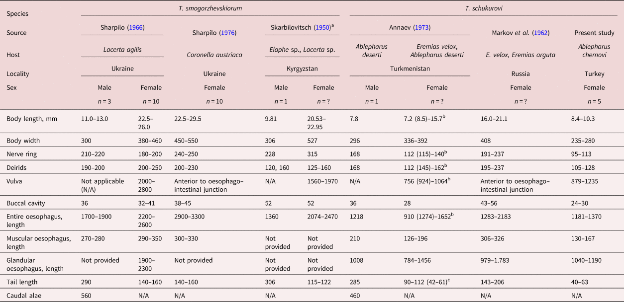
a Published as Foleyella schikhobalowi (not valid name) in Skarbilovitsch (Reference Skarbilovitsch1950).
b The smallest value of the range in the textual description differs from that provided in the table by Annaev (Reference Annaev1973); in our Table 1 the latter is given in parentheses.
c The length of the contracted tails is given in parentheses.
Thubunaea schukurovi seems to be a smaller nematode than T. smogorzhewskiorum (table 1). In addition to the size differences, scrutiny of the descriptions of the two species and the material studied here reveals several characters that can be useful for their differentiation. Tubercles composing the area rugosa of T. schukurovi are distributed between the level anteriorly twice as distant from the cloaca than the first pair of precloacal papillae, which posteriorly reach the level of phasmids. In contrast, the area rugosa of T. smogorzhewskiorum extends from a level that is slightly anterior to the first pair of caudal papillae to the tip of the tail. The vulva of T. schukurovi is situated more anteriorly than that of T. smogorzhewskiorum. Females of both species have a rounded tail with a conical posterior part; however, in T. schukurovi, the conical part is half as long as the base of the tail, whereas in T. smogorzhewskiorum the length of the conical distal part is about one-quarter of the length of the rounded part. Our record of T. schukurovi from A. chernovi in xerophytic and meso-xerophytic habitats also confirms the conclusion of Sharpilo (Reference Sharpilo1976) that this species is adapted as a parasite of lacertids and scincids in dry habitats.
This is the first record of T. schukurovi from Turkey. To the best of our knowledge, the only other record of the genus Thubunaea in Turkey is that of an unidentified species collected from Phoenicolacerta laevis (Gray, 1838) (Lacertidae) by Birlik et al. (Reference Bırlık, Yildirimhan, Sümer, Kumlutaş, Ilgaz, Durmuş, Güçlü and Candan2016). Jablonski et al. (Reference Jablonski, Bursey, Basit, Farooqi, Masroor and Goldberg2021) listed Thubunaea baylisi as a parasite known also from Laudakia caucasia (Eichwald, 1831) in Turkey referring to the study by Yildirimhan et al. (Reference Yildirimhan, Goldberg and Bursey2006); however, in the latter paper, the authors reported the oxyurid nematode Telandros baylisi Chatterji, 1935 (Pharyngodonidae) from the same host but not T. baylisi.
Below we discus some taxonomic issues concerning members of the genus Thubunaea from the Palaearctic and Indomalayan realms.
Thubunaea pudica Seurat, 1914, the type-species of the genus, was described from unidentified species of chameleon, Cerastes vipera (L.) (Viperidae) and Scincus scincus (L.) (=Scincus officinalis Laur.) (Scincidae) (type host was not designated) in Algeria by Seurat (Reference Seurat1914). Only the nominotypical subspecies of the common chameleon, Chamaeleo chamaeleon chamaeleon (L.), occurs in Algeria (see Sindaco, Reference Sindaco1998; Bauer et al., Reference Bauer, Deboer and Taylor2017) hence it should be regarded as one of the hosts of the species.
Thubunaea baylisi Akhtar, 1939 was described from a host referred to as ‘Agama sp.’ in Afghanistan (Akhtar, Reference Akhtar1939). All members of the genus Agama Daudin, as currently defined, are sub-Saharan species, whereas the type host of T. baylisi could belong to one of the few other genera of the subfamily Agaminae occurring in Afghanistan (see Wagner et al., Reference Wagner, Bauer, Leviton, Wilms and Böhme2016). Therefore, the type host of the species should be referred to as Agaminae gen. sp. Sharpilo (Reference Sharpilo1976) re-examined the material reported as Foleyella skrjabini Skarbilovitsch, 1948 in Skrjabin et al. (Reference Skrjabin, Shikhobalova and Sobolev1949), from unidentified terrestrial species of Colubridae in Kyrgyzstan and recognized it as conspecific with T. baylisi. In Skrjabin et al. (Reference Skrjabin, Shikhobalova and Sobolev1949), F. skrjabini was illustrated but textual description and differential diagnosis were not provided; a reference was made to an unpublished work of Skarbilovitsch from 1947. Thus, the F. skrjabini in Skrjabin et al. (Reference Skrjabin, Shikhobalova and Sobolev1949) does not satisfy the International Code of Zoological Nomenclature (1999, Article 13.1.1) and should be treated as nomen nudum.
Thubunaea smogorzhewskiorum was originally spelt as ‘smogorzhewskii’; however, the species was named after husband and wife Leonid A. Smogorzhevski and Lidiya A. Smogorzhevskaya (see Sharpilo, Reference Sharpilo1966). Therefore, we amended the spelling based on Art. 31.1.2 of the International Code of Zoological Nomenclature (1999).
Foleyella schikhobalowi Skarbilovitsch in Skrjabin et al. (Reference Skrjabin, Shikhobalova and Sobolev1949) from Elaphe sp. was published as a nomen nudum. Skarbilovitsch (1950) provided textual description of the material and listed as its hosts Elaphe sp. and Lacerta sp. collected from Kyrgyzstan, however a differential diagnosis was not provided and thus the requirements of the International Code of Zoological Nomenclature (1999, Article 13.1.1.) for a valid publication of a new species were not met. Sharpilo (Reference Sharpilo1976) considered this material conspecific to T. smogorzhewskiorum.
Bursey & Goldberg (Reference Bursey and Goldberg1991) and Ramallo et al. (Reference Ramallo, Goldberg, Bursey, Castillo and Acosta2017) listed Thubunaea mirzai Narayan, 1941 as a species known from the Indomalayan realm. The name T. mirzai seems to be published only in an abstract from a conference, which does not constitute a published work within the meaning of the International Code of Zoological Nomenclature (1999, Article 9.10) and should be treated as a nomen nudum.
Thubunaea dessetae Barus & Tenora, 1976, a parasite of Saara hardwickii (Gray, 1827) (=Uromastyx hardwickii Gray, 1827) (Agamidae) in Afghanistan, as described by Barus & Tenora (Reference Barus and Tenora1976), is characterized by a pair of well-developed and rounded pseudolabia each armed with a single large lateral tooth and a pair of smaller sublateral teeth, cuticular cervical inflation posterior to the pseudolabia, sclerotized buccal cavity, female reproductive system with four uteri and a peri-cloacal area rugosa in males. These characters correspond to the morphology of Pseudabbreviata Lichtenfels & Quigley, 1968 (see Lichtenfels & Quigley, Reference Lichtenfels and Quigley1968), members of which are also parasites of reptiles, mainly agamids (Sharpilo, Reference Sharpilo1976; Moravec & Baruš, Reference Moravec and Baruš1990). Therefore, we transfer T. dessetae to the genus Pseudabbreviata as Pseudabbreviata dessetae (Barus & Tenora, 1976) n. comb. Whereas the genus Thubunaea are distinct with not well-defined rounded pseudolabia, each armed with three similar in size teeth, absence of cervical inflation, none-sclerotized buccal cavity, didelphic female reproductive system and males with an area rugosa composed of irregularly distributed tubercles covering most of the ventral side of the caudal alae.
Thubunaea hemidactylae Oshmarin & Demshin, 1972 from Hemidactylus frenatus Duméril & Bibron, 1836 (Gekkonidae) in Vietnam was described with two lateral pseudolabia, one rounded and unarmed and the other bearing one large apical tooth and two smaller submedian teeth which do not project out from the pseudolabia; a weakly-developed cephalic collaret behind the pseudolabia; a short buccal cavity, a relatively short muscular oesophagus and a long and thick glandular oesophagus; a nerve situated slightly anterior to the junction between the muscular and glandular oesophagus; caudal allae with an area rugosa composed of numerous rounded tubercles; males with three precloacal pairs of papillae and six pairs of postcloacal papillae, and two spicules dissimilar in size; and females with a vulva situated anterior to the mid-length of the glandular oesophagus and with didelphic uteri (Oshmarin & Demshin, Reference Oshmarin and Demshin1972). This morphology corresponds to that of the genus Physalopteroides Wu & Liu, 1940 (see Chabaud & Brygoo, Reference Chabaud and Brygoo1960), hence we treat this species as Physalopteroides hemidactylae (Oshmarin & Demshin, 1972) n. comb. Indeed, Oshmarin & Demshin (Reference Oshmarin and Demshin1972) considered the species morphologically close to Thubunaea dactyluris [now accepted as Physalopteroides dactyluris (Karve, 1938) Chabaud & Brygoo, 1960] described from Hemidactylus flaviviridis Rüppell, 1835 and Calotes versicolor (Daudin, 1802) (Agamidae) in India (see Karve, Reference Karve1938).
Deshmukh (1969) described four species of Thubunaea from Gekkonidae in India, that is, Thubunaea singhi and Thubunaea brooki both from Hemidactylus brookii Gray, 1845 and Thubunaea aurangabadensis and Thubunaea syedi both from Hemidactylus giganteus Stoliczka, 1871. Deshmukh (1969) textually described the four species as having two symmetrical lips, each bearing three conical teeth and according to the author they should be regarded as members of the genus Thubunaea as defined by Chabaud & Brygoo (Reference Chabaud and Brygoo1960). The descriptions of T. aurangabadensis, T. syedi and T. brooki were accompanied with illustrations of the anterior extremities in dorsoventral view, which reveals that their pseudolabia are not symmetrical. Further illustrations of the cephalic end in apical view of T. aurangabadensis and T. syedi were provided showing that one large apical (lateral) tooth and two smaller sub-median teeth present only on one of the two pseudolabia. The anterior extremity of T. singhi was illustrated in lateral view and one large apical (lateral) tooth and two smaller sub-median teeth are present at least on one of the two pseudolabia. These likely correspond to the three interno-lateral teeth following the terminology in Chabaud (Reference Chabaud, RC, AG and S1975). In addition, all four species described by Deshmukh (Reference Deshmukh1969a) were illustrated with well-defined pseudolabia demarked with a groove or possibly a cephalic collarette at their base. The asymmetrical pseudolabia, armed with more pronounced and of different size teeth on one of the pseudolabia is a characteristic of the genus Physalopteroides, members of which are often described with well-defined pseudolabia and a cephalic collarette at their base (see Bursey & Goldberg, Reference Bursey and Goldberg2001; Bursey & Goldberg, Reference Bursey and Goldberg2016). All other species within the genus Thubunaea are characterized by two rounded, inconspicuous pseudolabia each bearing three similar in size teeth and there is no groove of cephalic collarette at their base (Seurat, Reference Seurat1914; Baylis, Reference Baylis1926; Ortlepp, Reference Ortlepp1931; Telford Jr, Reference Telford1965; Sharpilo, Reference Sharpilo1966; Babero & Matthias, Reference Babero and Matthias1967; Annaev, Reference Annaev1973; Bursey & Goldberg, Reference Bursey and Goldberg1991; Moravec et al., Reference Moravec, Salgado-Maldonado and Mayen-Peña1997; Pazoki & Rahimian, Reference Pazoki and Rahimian2014; Garduño-Montes de Oca et al., Reference Garduño-Montes de Oca, López-Caballero and Mata-López2017; Ramallo et al., Reference Ramallo, Goldberg, Bursey, Castillo and Acosta2017). Based on the above discussion, we consider the species described by Deshmukh (Reference Deshmukh1969a) as belonging to the genus Physalopteroides and propose the new combinations Physalopteroides singhi, Physalopteroides brooki, Physalopteroides aurangabadensis and Physalopteroides syedi. Their validity needs further taxonomic revision along with Physalopteroides versicoloris Deshmukh, 1968 described from Calotes versicolor (Daudin) in India in a separate study by Deshmukh (Reference Deshmukh1969b). It should be noted that P. versicoloris has pseudolabia very similar in appearance with those of P. brooki, P. aurangabadensis and P. syedi.
Acknowledgements
We are grateful to Dr Elena S. Ivanova (Centre of Parasitology of Severtsov Institute of Ecology and Evolution, Russian Academy of Sciences), Dr Riccardo P. Lia (Università degli Studi di Bari), Dr Bronwyn E. Campbell (Royal Melbourne Institute of Technology University) and Dr Ognyan Sivilov (Sofia University) for kindly providing some important literature. We are also grateful to Dr Heinz Grillitsch and Dr Silke Schweiger (Naturhistorisches Museum Wien) for the access to the material of Ablepharus chernovi.
Financial support
The scanning electron microscope study was supported by the National Endowment Fund ‘13 Centuries Bulgaria’.
Competing interests
None.
Ethical standards
Not applicable.


