


Carl Zeiss was born in Weimar on September 11, 1816. His mother’s name was Friederike (1786–1856), née Schmith (Figure 1). His father August Zeiss (1785–1849) was a respected artistic wood turner who turned mother-of-pearl, amber, ivory, and other raw materials into luxury goods and toys. Carl attended the local grammar school. He was interested in technical things from an early age and attended lessons at the Grand Duchy’s vocational college in Weimar during his school days before deciding to become a mechanic [Reference Paetrow and Wimmer1].

Figure 1 Carl Zeiss aged 50, 1866 (ZEISS archives).
To achieve this goal he went to Jena where, at Easter 1834, he began an apprenticeship under Dr. Friedrich Körner, court mechanic and private lecturer at the University of Jena (1778–1847). From the second year of his apprenticeship onward, he attended lectures on mathematics and the natural sciences at the university. After completing his apprenticeship, Carl Zeiss initially devoted his attention to mechanical engineering. From 1838 to 1845 he became a traveling tradesman, and his journeys took him to such destinations as Stuttgart, Darmstadt, and Berlin. He returned to Jena in 1845.
Biologist Matthias Schleiden (1804–1881) had been working in Jena since 1839 (Figure 2). He was one of the developers of the cell theory, which states that all life is made up of cells. This theory turned the microscope into an indispensable tool for biologists, physicians, and hygienists. The need for such instruments in research and teaching, but also for doctors’ surgeries and in the food industry, would henceforth grow steadily.

Figure 2 Matthias Jakob Schleiden from the book Studien: Populäre Vorträge, 1855.
The first chapter of Schleiden’s popular work Die Pflanze und ihr Leben, published in several editions after 1848, was titled Das Auge und das Mikroskop. In it, Schleiden delves into the significance of the microscope for biology: “The microscope is the necessary instrument without which the botanist can expect to make no advances in his scientific endeavors.” Further on, he states, “Our century was destined to make use of the possibilities afforded by the microscope in the study of nature, and it is very pleasing to see how the application of this instrument opens ever more doors and how, in ever larger circles, yields the most interesting results.” Also: “... how scientific processing in botany … is almost unfathomable without the consistent use of a microscope” [Reference Schleiden2]. In 1845 he jointly established the Physiological Institute with mineralogist Ernst Erhard Schmid (1815–1885, professor since 1843). They intended to teach students skills such as how to work with a microscope.
Schleiden contradicted the view that microscopic investigations required very expensive instruments. “In terms of the greatest advancements in optical technology, one is able to procure very workable instruments at relatively low prices from any reasonably adroit lensmaker and no one, not even the youngest of our contemporaries, will live to see the day when one such instrument can no longer be used to do anything to further the cause of science” [Reference Schleiden3]. Here he is referring to Friedrich Körner, the erstwhile employer of Carl Zeiss. Körner began producing telescopes and other optical instruments early on. He even conducted his own tests for producing optical glass. He therefore found himself following in the footsteps of Joseph von Fraunhofer (1787–1826), even if he was less successful. Schleiden then urged him to construct simple microscopes with magnifying lenses. These instruments featured a magnifying system that was composed of a single lens, rather than an objective lens and an eyepiece.
Schleiden wrote this in his 1845 textbook: “I would like to draw your attention in particular to the pocket microscopes now being produced by Dr. Körner in Jena. … This instrument is perfectly sufficient for all entomological, pharmacognostic and botanical investigations, and even for wholly adequate anatomical observations ... At my instigation, Dr. Körner always keeps a number of these finished instruments in stock, and I believe I can recommend them, and certainly with a clean conscience, to all of those who are enamored by proper natural science studies, as I myself inspect each individual instrument before it leaves the workshop, and can say that it certainly deserves its excellent reputation” [Reference Schleiden3].
Having returned to Jena in 1845, Carl Zeiss began attending events at the Physiological Institute. Before he established a company, he wanted to acquaint himself with the day-to-day work of natural scientists. A report written by Schleiden about Carl Zeiss states that he “has familiarized himself, especially in recent times, with the needs of the nature researchers at the Physiological Institute and has thus put himself in a much better position to meet their needs than anyone else” [4].
In 1846 Carl Zeiss founded a workshop for precision mechanics in Jena. He assumed the basic construction of simple microscopes from his employer Friedrich Körner, who passed away in 1847. At the time, these microscopes were employed primarily for preparatory work. When used, the instrument was secured on its storage case, thus offering the observer a suitable viewing height (Figure 3). It was, however, also possible to mount it on a wooden block, with its raised side sections serving as an armrest during preparatory work. These simply constructed microscopes were very well received by experts on account of their exceptional mechanical and optical features. In 1852, Zeiss began to incorporate new design features (fixed stage and movable lens holder) into his microscopes.
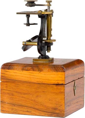
Figure 3 Dissection microscope with dovetail mount, produced in this form until 1848, from the collection of Timo Mappes. Photograph taken by Manfred Stich.
After Körner’s death, Carl Zeiss also continued the approach of collaborating with a scientist, Friedrich Wilhelm Barfuss (1809–1854). In 1839, Barfuss wrote a textbook on dipotrics in which he summarized all that was known about the construction of optical instruments at the time [Reference Barfuss5]. We can assume that this book, and the personal advice provided by the expert, had a considerable impact on Carl Zeiss’s early products. Because Barfuss only based his assumption on geometric optics, in other words did not take the wave character of light into consideration, his approaches were not very promising for the improvement of microscope optics—but he had no way of knowing this. It is occasionally reported that Carl Zeiss himself attempted to understand the problem of how to improve microscope optics through an understanding of science. As a trained mathematician, he had the skills to do so. That said, there is no evidence of any such studies.
In 1857 Carl Zeiss marketed his first compound microscopes, which quickly found favor among experts. Bonn-based botanist Hermann Schacht (1814–1864), who studied under Schleiden and in 1846 purchased the first-ever microscope from Carl Zeiss, wrote the following in his textbook in 1862: “Carl Zeiss in Jena, whose simple microscopes have long enjoyed a good reputation, recently also began producing excellent compound instruments… .” His instruments also were revered outside of Germany, for example by Utrecht-based biologist Pieter Harting, who noted that “the lenses [made by Carl Zeiss] are among the best that Germany has to offer” [Reference Paetrow and Wimmer1].
These early compound microscopes often varied in appearance until Zeiss developed his own particular style (Figure 4). The first large instruments were based on tried-and-true microscopes [6]. Inspiration was derived primarily from the stands of Georg Johann Oberhäuser (1798–1868) and his successor Edmund Hartnack (1826–1891). Carl Zeiss made no secret of his source of inspiration, instead honoring the first originator by name, as was the custom among craftsmen at the time. In the early years from 1857 until 1874, Zeiss produced eight different stands (O, I, Ib, II, IIIb, IIIc, IV, V). Different combinations of lens systems (A, B, C) and eyepieces (1, 2, 3, 4) could be selected. Carl Zeiss constantly sought to improve the microscope design, for instance in terms of the shape of the stand base and column. Over time, the aperture and the fine adjustment were also changed [Reference Paetrow and Wimmer1].
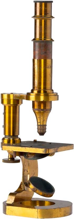
Figure 4 One of the earliest compound microscopes from 1862, from the collection of Timo Mappes. Photograph taken by Manfred Stich.
In 1842, Schleiden had written that key scientific advancements were only possible with such compound microscopes [Reference Schleiden7], in which early attempts at corrected objective lenses were made by combining lenses of crown and flint glass possessing convex and concave shapes, in order to minimize chromatic and spherical aberrations. So why did it take Carl Zeiss so long to bring a microscope like this to market? One explanation can be found in a letter he wrote to a friend of his youth, K.O. Beck, in 1855 [8]:
All the same, you will be surprised to hear that I have not yet had a flint lens ground for a compound microscope. Do not think that I would not be ready to make further endeavors in this area—indeed I have experimented with many other classes of microscope—yet I believe that the usual compositum cannot take us much further, and I have something of an aversion to this relentless rigmarole of trial and error that is so common among us lensmakers, with people such as Oberhäuser attempting to make one good objective lens from among hundreds of lenses.
Carl Zeiss had no qualms about working on corrected lenses. His successful market launch two years later proves that he was up to the task. Yet he was still suspicious of the traditional production methods involving trial and error. He wanted to get to grips with the problem by considering the appropriate theory. He continued his search for a scientific solution. It was therefore no coincidence that he approached physicist Ernst Abbe (1840–1905) in 1865 to help him (Figure 5). “He found what he wanted … because he sought it out” wrote Abbe 30 years later [Reference Abbe9].
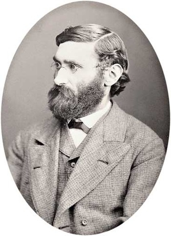
Figure 5 Ernst Abbe in 1876 (ZEISS archives).
Ernst Abbe grew up in very humble circumstances in Eisenach and studied physics and mathematics in Jena and Göttingen [10]. After spending a short time in Frankfurt, he returned to Jena in 1863 to earn his doctorate. In such a small town (estimated population of 7,200), it is very likely that the pair had already crossed paths. They got to know one another better when Abbe asked Zeiss to construct instruments for his lectures. We know nothing about what motivated Abbe to turn his attention to physical optics, an area with which he had previously not concerned himself in the slightest. It has been speculated that he may have underestimated the problem of optics in microscope construction and believed that the topic could be dealt with quickly.
Meanwhile, Edmund Hartnack had launched water immersion lenses that were superior to all others. Zeiss could have attempted to simply mimic the design of these lenses. But Abbe’s aim was not only to produce microscopes that were better than the competition—he wanted to tackle the problem by considering the appropriate theory. His approach was thus theory-driven and analytical, and his initial attempt to solve the problem geometrically led only to major setbacks in the beginning. He was only successful once he took the wave character of light into account. From 1872 onwards, all of Zeiss’s microscope optics were based on calculations made by Abbe [11]. In catalog 19, immersion systems were offered for the first time alongside new dry lens systems. Homogeneous oil immersion lenses followed in 1879 (Figure 6).
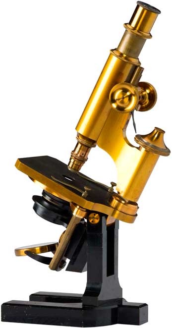
Figure 6 Stand with micrometer movement and tilt from 1882, from the collection of Timo Mappes. Photograph taken by Manfred Stich.
What is now known as Abbe’s theory, which stated that image resolution was related to diffraction effects, was a big disappointment as far as microscopists were concerned. It showed that the resolution capacity of light microscopes is limited by the wavelength of light. While Abbe did ruminate on using UV light with a shorter wavelength to push this limit [12], his musings were initially confined to the theoretical realm. It was not until 1904 that August Köhler (1866–1948), who headed the microphotography and projection department at Carl Zeiss, launched a UV microscope. The first microscope to clearly overcome the light microscope employed a medium with a still shorter wavelength; the electron microscope would not be brought to market by Siemens and AEG until more than 30 years later.
Abbe also revised microscope stand design. In 1877 the round fine-adjustment control knob was replaced by a prism control knob on the large- and medium-sized stands. The same year also saw the publication of the first bound catalog, which was reprinted in the following year and featured illustrations. This catalog reflected the considerable progress made in development at the workshop. It included a total of 11 microscope models. Large, powerful instruments were added. The horseshoe stand was then incorporated into the most important models. A new kind of micrometer movement was introduced in 1886. In terms of appearance, these new microscopes only differed slightly from the previous models.
Some competitors in the 1870s advertised by saying that their lenses were “not produced as they are in Jena,” which meant that the simple optics produced through trial and error no longer stood a chance. The year 1879 marked the beginning of the collaboration with chemist Otto Schott (1851–1935) that aimed to produce a better kind of optical glass. A systematic, scientific approach was taken here, too. The new glass types were used to launch the apochromats in the 1880s. In these lenses, secondary color deviation was eliminated, and spherical aberration was consistently remedied for light of different colors. From then on, all microscope manufacturers that wanted to remain competitive had to hire their own scientists and lens designers.
Zeiss employed academics in product development from 1879 onward. They were physicists, engineers, biologists, and physicians, all of whom contributed their practical experience. Some of them had attended lectures given by Abbe at the University of Jena. This practice was later described as the “Abbe school.” Initially, they all worked in microscopy: it is said of Karl Bratuschek (1865–1913; hired in 1892) that only his poor health prevented him from discovering phase contrast. The names Hermann Ambronn (1856–1927; hired in 1899), Henry Siedentopf (1872–1940; hired in 1899), and August Köhler (1866–1948; hired in 1900) are associated with major advancements in microscopy. For example, by the First World War the company produced the slit ultra-microscope for particle examination below the Abbe resolution limit and the UV microscope (Figure 7), which laid the foundation for fluorescence microscopy. All microscopists today are familiar with Köhler’s illumination principle. Starting in the 1890s, new fields of business were created for innovative products: for example, photographic lenses, binoculars, astronomical instruments, and optical instruments for chemical analysis. Advances were made in line with a systematic, scientific approach in microscopes, given the successful collaboration between Carl Zeiss and Ernst Abbe in the field of microscopy.
Figure 7 1904 Ultraviolet microscope by August Köhler and Moritz von Rohr (ZEISS archives).
At the beginning of his business, Carl Zeiss took his cue from the top European manufacturers, but later the company gained a leading position as a result of its well-developed scientific foundation. Innovative optical systems were brought to market, and Zeiss shaped microscopes in terms of their functions and look. The company became a leader in the development of new microscopic techniques.


