


Short bowel syndrome (SBS) is defined as the malabsorptive state in which the functional gut mass is reduced below the level necessary for adequate digestion and absorption of food and fluid and that eventually leads to clinical nutrient deficiencies and prolonged parenteral nutrition support( Reference Kaufman, Pehlivanova and Fennelly 1 , Reference Rossi, Kadamba and Hugosson 2 ). Previous studies have reported that up to 65 % of SBS infants develop intestinal failure-associated liver disease( Reference Kaufman, Pehlivanova and Fennelly 1 ). The only proven treatment for intestinal failure-associated liver disease is to increase enteral nutrition and reduce parenteral nutrition to achieve sufficient adaptive growth of the remnant bowel( Reference Rossi, Kadamba and Hugosson 2 ). Small-bowel bacterial overgrowth and translocation are common in SBS patients, which are not only related to liver complications but also might further impair the rehabilitation process( Reference Geier, Fickert and Trauner 3 , Reference El Kasmi, Anderson and Devereaux 4 ). In rodent models, massive small-bowel resection (mSBR) induces early small-bowel adaptive responses within several hours to a few days, and studies have found that several nutrients help to accelerate this adaptive process( Reference Tian, Hao and Chandra 5 , Reference Ben Lulu, Coran and Mogilner 6 ). Gut barrier dysfunction, with the translocation of luminal microbes to mesenteric lymph nodes (MLN) and subsequently to blood and organs such as the liver and spleen( Reference Tian, Hao and Chandra 5 , Reference Mogilner, Srugo and Lurie 7 ), has also been well documented in SBS animal models.
Lactoferrin (LF) is a milk glycoprotein present at highest concentrations in the colostrum and at low concentrations in mature milk. Many studies have indicated that bovine and human LF have anti-inflammatory effects( Reference Kuhara, Tanaka and Yamauchi 8 , Reference Kruzel, Harari and Mailman 9 ). Animal studies have found that bovine lactoferrin (bLF) down-regulates the lipopolysaccharide (LPS)-induced concentrations of TNF-α, IL-1β and IL-6 in mice via NF-κB modulation, thus exhibiting a potential to be used in the prophylaxis of inflammation( Reference Kruzel, Harari and Mailman 9 ). Research has also shown that bLF may be effective in the prevention of late-onset sepsis and necrotising enterocolitis in preterm neonates( Reference Walker 10 , Reference Venkatesh and Abrams 11 ). Moreover, ex vivo studies have found that LF could directly induce enterocyte growth and proliferation, indicating that LF might regulate the early postnatal intestinal development and might be a growth factor in selected intestinal diseases( Reference Buccigrossi, de Marco and Bruzzese 12 ).
The present study was designed to investigate the effect of enteral bLF supplementation on the indices of gut barrier function in a rat model of mSBR, an established translational model for human SBS, as well as to demonstrate whether bLF supplementation could help to promote intestinal adaptation.
Experimental design and methods
Experimental design and animal model
Male Sprague–Dawley rats aged 4 weeks and weighing about 150 g were obtained from the National Rodent Laboratory Animal Center, Shanghai Branch (Shanghai, China). The rats were housed in individual cages and allowed to acclimatise for 3 d under a 12 h light–12 h dark cycle at constant temperature (22 ± 2°C) with ad libitum access to water and chow (M01-F; Shanghai Slac Laboratory Animal Company Limited) before the start of the experiment.
Experimental protocols used in the present study were approved by the local Animal Care Committee (no. XHEC-F-2012-23) and conformed to the Guide for the Care and Use of Laboratory Animals published by the Science and Technology Commission of the People's Republic of China (STCC Publication no. 2, revised 1988).
A total of thirty rats were randomly divided into the following three experimental groups of ten rats each: Sham group (rats submitted to bowel transection with reanastomosis); SBS group (rats submitted to 80 % mSBR); SBS-bLF group (rats submitted to 80 % mSBR plus treatment with bLF by oral administration at a dosage of 0·5 g/kg per d)( Reference Sherman, Bennett and Hwang 13 ). DMV International provided bLF, with a purity of 98·0 % and a Fe content of 120 mg/kg. The experimental diets were not further balanced for Fe provided by bLF as the amount of Fe was negligible ( < 0·3 % of that consumed with the food).
The rats were fasted 12 h before the operation, with free access to water. All operative procedures were performed as described previously( Reference Chen, Wen and Cai 14 ). After recovery, the rats were transferred back to individual cages and given water ad libitum overnight and were then fed a regular diet (M01-F, Fe content 321 mg/kg).
Every morning from day 2 to day 20, each rat of the SBS-bLF group was orally administered with 0·8–1·2 ml of warm water containing bLF. Body weight and food intake were recorded twice a week, and bLF dosage was adjusted according to the change in body weight. The Sham and SBS groups were given 1 ml of warm water by oral administration every morning. The rats were monitored for 20 d and killed on day 21 by intraperitoneal injection of pentobarbital (75 mg/kg) before sample collection.
Histological analysis
Pieces of small intestine samples were first rinsed in cold normal saline and then immediately fixed in 10 % neutral buffered formalin (pH 7·2). After 72 h of fixation, the tissue was embedded in paraffin wax on the oriented edge and cut into 5 μm-thick sections for haematoxylin and eosin staining. Villus height and crypt depth were determined in the longitudinal sections using an analysis system (NIS-elements suite; Nikon). Villus height and crypt depth of ten vertically well-orientated villi and crypts were measured in each sample. Villus height was determined from the crypt opening to the top of the villus and crypt depth from the base of the crypt to its opening using ocular micrometers.
Assessment of intestinal epithelial cell proliferation and apoptosis
Intestinal epithelial cell proliferation was assessed by immunohistochemical staining for proliferating cell nuclear antigen( Reference Barnes, Hartmann and Holst 15 ). The index of proliferation is expressed as the number of proliferating cell nuclear antigen-positive cells per crypt in each slide( Reference Barnes, Hartmann and Holst 15 ).
Terminal deoxynucleotidyl transferase-mediated biotinylated 2′-deoxyuridine 5′-triphosphate nick end labelling staining was performed to measure the rate of apoptosis of intestinal epithelial cells using the In Situ Cell Death Detection Kit (Roche Applied Science). Apoptosis is expressed as the number of epithelial cells positive for the apoptotic marker per villus( Reference Barnes, Hartmann and Holst 15 ).
Determination of bacterial translocation
On day 21, after anaesthesia, blood samples (1 ml) obtained from the left ventricle were injected into blood culture vials and bacterial growth was detected using the BACTEC 9240 automated blood culture system (Becton Dickinson Diagnostic Instrument Systems). Homogenised samples of MLN, spleen and liver were inoculated into thioglycollate broth and blood, chocolate and MacConkey agar plates. The cultures were incubated at 37°C for 72 h, and a positive result was determined as three or more clones being detected( Reference Wu, Wang and Hong 16 ). Gram-negative bacteria were further identified using VITEK 2 bacteria identification cards in the VITEK 2 Compact system (BioMérieux, Inc.). The same microbiologist, who was blind to the experiment grouping, performed all the procedures and reported the culture results. Bacterial translocation (BT) was defined as the presence of bacteria of enteric origin, and the translocation rate was calculated as the ratio of the number of positive samples of each site:the number of animals. BT was stratified into three levels: (1) local, level 1 (MLN); (2) regional, level 2 (liver and spleen); (3) systemic, level 3 (peripheral blood)( Reference Mogilner, Srugo and Lurie 7 ).
Determination of intestinal permeability
On day 20, after an overnight fast, each rat was orally administered with permeability markers (2 ml) containing 100 mg of lactulose and 50 mg of mannitol (Wako Company)( Reference Chang, Chen and Ma 17 ). Urine samples were collected for 24 h and stored at − 80°C for analysis. For this analysis, urine samples were thawed at room temperature and then centrifuged at 151 g for 10 min. Later, 5 ml of the supernatant were taken into a new microcentrifuge tube. To this tube, 10 ml of acetic acid were added and boiled for 5 min and centrifuged at 15 139 g for 10 min, and the tube was cooled later. The samples were first filtered through a 0·45 mm filter and then through Microsep centrifugal devices (Pall Life Sciences) according to the manual instructions before analysis. The pre-treated samples were analysed using Dionex ICS 3000 anion exchange liquid chromatography (Dionex Corporation), with pulsed amperometric detection (Waters Associates Inc.)( Reference Fleming, Kapembwa and Laker 18 ). The results are expressed as the lactulose:mannitol (L:M) ratio.
Measurement of secretory IgA concentrations in ileal contents
Ileal contents were collected from the proximal ileum within 10 cm to the terminal ileum and dried for 30 min in a vacuum dryer at room temperature. Later, 0·1 g of a dried sample was weighed and vortexed in 1 ml of normal saline and centrifuged at 1358 g at 4°C for 10 min. Then, the supernatant was collected and centrifuged at 21 733 g for 15 min. The supernatant containing secretory IgA (sIgA) was stored at − 20°C. The concentrations of sIgA in ileal contents were determined by ELISA according to the protocol of Bethyl Laboratories (catalogue no. E111-102).
Quantification of tight junction proteins by Western blotting
The distal ileum of each rat was collected, flushed with ice-cold saline solution and opened along the mesenteric border. The tissue samples were homogenised in a pre-cold radioimmunoprecipitation assay buffer (25 mm-Tris–HCl (pH 7·6), 150 mm-NaCl, 1 % NP-40, 1 % sodium deoxycholate and 0·1 % SDS) containing a protease inhibitor cocktail (1 mm-4-(2-aminoethyl) benzenesulfonyl fluoride hydrochloride, 0·8 mm-aprotinin, 0·05 mm-bestatin, 0·015 mm-E-64, 0·02 mm-leupeptin hemisulphate and 0·01 mm-pepstatin), a phosphatase inhibitor cocktail (1 mm-sodium fluoride and 1 mm-sodium orthovanadate) and 1 mm-phenylmethylsulfonyl fluoride. Protein concentration was then determined using the Bradford method and a commercially available kit (Pierce). Tissue lysates (20 μg of total protein per lane) were separated by electrophoresis on a 10 % SDS–PAGE gel and transferred onto nitrocellulose membranes. The primary antibodies used were rabbit anti-claudin-1 (catalogue no. Ab15098, dilution 1:200; Abcam), rabbit anti-claudin-2 (catalogue no. Ab53032, dilution 1:200; Abcam), rabbit anti-claudin-4 (catalogue no. 364800, dilution 1:250; Invitrogen), rabbit anti-occludin (catalogue no. Ab31721, dilution 1:200; Abcam) and horseradish peroxidase-labelled mouse anti-glyceraldehyde-3-phosphate dehydrogenase (catalogue no. KC-5G5, dilution 1:10 000; Kangchen). The following horseradish peroxidase-conjugated secondary antibodies were used: anti-mouse IgG (catalogue no. KC-MM-035, dilution 1:5000; Kangchen) and anti-rabbit IgG (catalogue no. KC-RB-035, dilution 1:5000; Kangchen). Protein bands were detected using a chemiluminescence reagent (catalogue no. KC-420; KangChen) and visualised by exposure to X-ray films. Protein band intensity was quantified using ImageJ software (National Institutes of Health), and the relative expression was normalised to that of glyceraldehyde-3-phosphate dehydrogenase.
Statistical analysis
Data are presented either as means with their standard errors or as medians with inter-quartile ranges depending on the distribution. The differences among the three groups were evaluated by one-way ANOVA or Kruskal–Wallis test. Differences between the groups were further determined by Fisher's protected least significant difference test or Mann–Whitney U test. BT rates were compared using Fisher's exact test. Correlation was determined using Spearman's or Pearson's correlation analysis. The statistical software SPSS for Windows version 17.0 (SPSS, Inc.) was used for analysis, and P< 0·05 was defined as statistically significant.
Results
Body weight change and food intake
The mean daily food intake during the 21 d study period was similar among the three groups (Sham: 18·7 (sem 0·2) g; SBS: 18·2 (sem 0·2) g; SBS-bLF: 18·3 (sem 0·3) g; NS; ANOVA). All rats had similar initial body weight. The body weight of the SBS and SBS-bLF groups was lower than that of the Sham group during the 3-week post-operative period, which might be due to the removal of intestinal mass and diarrhoea induced by malabsorption after mSBR. No difference was observed between the SBS and SBS-bLF groups at each time point (Fig. 1(a)).

Fig. 1 (a) Changes in the body weight of rats. Sham (![]() ), rats submitted to small-bowel transection as the operative control; SBS (
), rats submitted to small-bowel transection as the operative control; SBS (![]() ), rats submitted to 80 % small-bowel resection; SBS-bLF (
), rats submitted to 80 % small-bowel resection; SBS-bLF (![]() ), rats submitted to 80 % small-bowel resection plus treatment with enteral supplementation of 0·5 g/kg per d bovine lactoferrin. (b) Changes in villus and crypt architecture.
), rats submitted to 80 % small-bowel resection plus treatment with enteral supplementation of 0·5 g/kg per d bovine lactoferrin. (b) Changes in villus and crypt architecture. ![]() , Sham;
, Sham; ![]() , SBS;
, SBS; ![]() , SBS-bLF. (c) Intestinal epithelial cell proliferation expressed as the number of proliferating cell nuclear antigen (PCNA)-positive cells per crypt. (d) Intestinal epithelial cell apoptosis expressed as the number of apoptotic cells per villus. No difference was observed in the number of apoptotic cells among the groups (determined by the Kruskal–Wallis test). (e) Intestinal permeability expressed as the urinary lactulose:mannitol (L:M) ratio. (f) Secretory IgA (sIgA) concentrations in ileal contents. Values are means, with their standard errors or medians with inter-quartile ranges represented by vertical bars for group depending on the distribution. * Mean values were significantly different from that of the Sham group (P< 0·05; least significant difference test or Mann–Whitney U test). † Mean values were significantly different from that of the SBS group (P< 0·05; least significant difference test or Mann–Whitney U test).
, SBS-bLF. (c) Intestinal epithelial cell proliferation expressed as the number of proliferating cell nuclear antigen (PCNA)-positive cells per crypt. (d) Intestinal epithelial cell apoptosis expressed as the number of apoptotic cells per villus. No difference was observed in the number of apoptotic cells among the groups (determined by the Kruskal–Wallis test). (e) Intestinal permeability expressed as the urinary lactulose:mannitol (L:M) ratio. (f) Secretory IgA (sIgA) concentrations in ileal contents. Values are means, with their standard errors or medians with inter-quartile ranges represented by vertical bars for group depending on the distribution. * Mean values were significantly different from that of the Sham group (P< 0·05; least significant difference test or Mann–Whitney U test). † Mean values were significantly different from that of the SBS group (P< 0·05; least significant difference test or Mann–Whitney U test).
Morphology of small-intestinal tissues
As revealed by histological staining, the intestinal villi were longer in rats after mSBR. Compared with the Sham group, both the SBS and SBS-bLF groups exhibited significant increases in villus height and crypt depth, but no significant difference was observed between the groups (Figs. 1(b) and 2).

Fig. 2 Structure of villi in the ileum as revealed by haematoxylin and eosin staining (magnification × 100). As revealed by histological staining, the intestinal villi were longer in the SBS and SBS-bLF groups. (a) Sham, rats submitted to small-bowel transection; (b) SBS, rats submitted to 80 % small-bowel resection; (c) SBS-bLF, rats submitted to 80 % small-bowel resection plus treatment with enteral supplementation of 0·5 g/kg per d bovine lactoferrin. (A colour version of this figure can be found online at http://www.journals.cambridge.org/bjn).
Intestinal epithelial cell proliferation and apoptosis
As shown in Fig. 1(c) and (d), both the SBS and SBS-bLF groups had higher proliferation indices (expressed as the number of proliferating cell nuclear antigen-positive cells per crypt) than the Sham group (P< 0·05; the least significant difference test/ANOVA). The proliferation index was positively correlated with villus height (r 0·452, P= 0·012; Pearson's correlation analysis), but not with crypt depth. The number of apoptotic cells in the villi varied within each group, and no differences were observed among the three groups by the Kruskal–Wallis test (5·4 (3·1–9·5), 6·9 (1·4–10·8) and 8·4 (4·9–11·4) apoptotic cells per villus in the Sham, SBS and SBS-bLF groups, respectively).
Bacterial translocation
No BT was detected in either the tissue or blood cultures of the Sham group. In the SBS group, 30 % BT to the MLN (level 1) and 20 % BT to the spleen (level 2) with Escherichia coli and Enterococcus faecalis were detected, with a significant difference being observed when compared with the Sham group (P< 0·05). In the bLF-supplemented rats, 20 % BT to the MLN with E. faecalis and no level 2 BT were detected (Table 1). The BT rate of both the SBS and SBS-bLF groups was higher than that of the Sham group. The BT rate of the SBS-bLF group was lower than that of SBS group, but no significant difference was observed. No level 3 BT was detected in all the three groups.
Table 1 Frequency of bacterial translocation
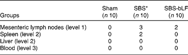
Sham, rats submitted to small-bowel transection as the operative control; SBS, rats submitted to 80 % small-bowel resection; SBS-bLF, rats submitted to 80 % small-bowel resection plus treatment with enteral supplementation of 0·5 g/kg per d bovine lactoferrin.
* The bacterial translocation frequency of SBS rats was significantly different from that of the Sham group (P< 0·05; Fisher's exact test).
Intestinal permeability
The urinary L:M ratio was significantly increased in the SBS group (3·0 (2·6–3·4)) compared with that in the Sham group (1·5 (1·2–2·3), P< 0·05). Supplementation with 0·5 g/kg per d bLF reduced intestinal permeability (L:M ratio: 1·7 (1·3–2·3)) when compared with that in the SBS group, which had values similar to those of the Sham group (Fig. 1(e)).
Secretory IgA concentrations in ileal contents
Enteral bLF supplementation significantly increased sIgA concentrations in ileal contents (30·0 (23·8–33·0) ng/ml saline extract), when compared with those in the SBS and Sham groups, with the latter two groups having similar sIgA concentrations (17·5 (12·6–29·1) and 19·3 (11·5–27·0) ng/ml saline extract, respectively). No difference was observed between the Sham and SBS groups (Fig. 1(f)).
Tight junction protein expression levels
The relative expression levels of both occludin and claudin-4 in the SBS-bLF group were significantly higher than those in the SBS group (P< 0·05), but did not exhibit any significant differences when compared with those in the Sham group. No difference was observed in the expression levels of claudin-1 and claudin-2 among the three groups (Fig. 3).

Fig. 3 Relative expression levels of tight junction proteins. (a) Western blot of occludin and claudin-1. (b) Relative expression levels of occludin normalised to those of glyceraldehyde-3-phosphate dehydrogenase (GAPDH). Sham, rats submitted to small-bowel transection; SBS, rats submitted to 80 % small-bowel resection; SBS-bLF, rats submitted to 80 % small-bowel resection plus treatment with enteral supplementation of 0·5 g/kg per d bovine lactoferrin. (c) Relative expression levels of claudin-1 normalised to those of GAPDH. No difference was observed among the groups (determined by ANOVA). (d) Western blot of claudin-2 and claudin-4. (e) Relative expression levels of claudin-2 normalised to those of GAPDH. No difference was observed among the groups (analysed by ANOVA). (f) Relative expression levels of claudin-4 normalised to those of GAPDH. Values are means, with their standard errors or medians with inter-quartile ranges represented by vertical bars for group depending on the distribution. * Mean values were significantly different from that of the Sham group (P< 0·05; least significant difference test or Mann–Whitney U test). † Mean values were significantly different from that of the SBS group (P< 0·05; least significant difference test or Mann–Whitney U test).
A significant negative correlation was observed between the intestinal L:M ratio and the relative concentration of occludin (r − 0·462, P= 0·030; Spearman's correlation analysis), but no such correlation was observed for the other three tight junction (TJ) proteins.
Discussion
LF is an Fe-binding protein that is abundantly present in human milk. It can be found in most exocrine secretions including milk, tears, saliva, intestinal mucus and genital secretions and in the specific granules of neutrophils. Studies have demonstrated that both bovine LF and human LF exert antibacterial, immune-modulating and anti-inflammatory effects( Reference Kuhara, Tanaka and Yamauchi 8 , Reference Kruzel, Harari and Mailman 9 , Reference Manzoni, Rinaldi and Cattani 19 ). In 2009, a prospective, double-blind, placebo-controlled, randomised trial revealed that bLF supplementation alone or in combination with Lactobacillus GG reduced the incidence of a first episode of late-onset sepsis in very-low-birth-weight neonates( Reference Manzoni, Rinaldi and Cattani 19 ). These previous studies have suggested that bLF might be beneficial in the setting of SBS. Thus, we investigated the effect of bLF supplementation on intestinal adaptation and intestinal barrier function using a previously established rat model of mSBR( Reference Chen, Wen and Cai 14 ).
Following massive intestinal resection, the remaining bowel undergoes a pattern of well-described morphological adaptation, which begins within 24–48 h of the operation. Similar to previous studies( Reference Ben Lulu, Coran and Mogilner 6 , Reference Weale, Edwards and Bailey 20 ), the present study demonstrated that massive intestinal resection results in increased villus height and crypt depth, coupled with an increase in epithelium proliferation, indicating an increased absorptive surface area as the structural adaptation. Buccigrossi et al. ( Reference Buccigrossi, de Marco and Bruzzese 12 ) found that LF stimulates the proliferation of immature Caco-2 cells in a dose-dependent manner. Such dose–response effects have seldom been demonstrated in animal models. A single dosage (0·5 g/kg per d) of bLF was used in the present study, and bLF supplementation did not have any effect on the enhancement of structural intestinal adaptation and no additional increase in body weight was detected. Whether a higher dosage of bLF could stimulate intestinal epithelial cell growth needs to be investigated further.
Intestinal BT in SBS has been demonstrated by a lot of studies( Reference El Kasmi, Anderson and Devereaux 4 , Reference Tian, Hao and Chandra 5 , Reference Dibaise, Young and Vanderhoof 21 ). In the present study, both the SBS and SBS-bLF groups exhibited higher BT rates than the Sham group. Both level 1 (MLN) and level 2 (spleen) BT were detected in the SBS group, while only level 1 (MLN) BT was detected in the SBS-bLF group, indicating LF to have potential protective effects. Multiple aspects may be involved in enteric bacterial invasion in SBS, including small-bowel bacterial overgrowth and impairment of gut-associated anatomical and immune barriers, e.g. the net loss of gut-associated lymphoid tissue. Ziegler et al. ( Reference Ziegler, Luo and Estívariz 22 ) found a high proportion of detectable LPS in the serum of SBS patients. The same group also demonstrated Gram-negative BT from the gut and a concomitant adaptive immune response to LPS in a rat model( Reference Tian, Hao and Chandra 5 ). A lot of studies have demonstrated the protective effect of LF against LPS( Reference Doursout, Horton and Hoang 23 – Reference Hirotani, Ikeda and Kato 26 ). Doursout et al. ( Reference Doursout, Horton and Hoang 23 ) found that pre-treatment with LF can protect intestinal tissues from histopathological damage after LPS administration. A recently published review has concluded that the anti-Gram negative bacteria activity of LF is due to its interaction with enterobacterial LPS on the bacterial surface, which might partly explain the relatively low rate of BT in the bLF-supplemented group( Reference Drago-Serrano, de la Garza-Amaya and Luna 24 ).
On day 21, the concentrations of sIgA in ileal contents were also increased by bLF supplementation, when compared with those in the SBS and Sham groups. As an important part of the intestinal barrier, sIgA plays a critical role in mucosal immunity. Several studies have shown that enteral supplementation of bLF helps to increase the sIgA release in the intestinal fluid( Reference Sfeir, Dubarry and Boyaka 27 ). The underlying mechanism for this remains unclear.
TJ are multifunctional proteins that form a seal between adjacent epithelial cells near the apical surface. They seal the paracellular space between epithelial cells, thus preventing the paracellular diffusion of micro-organisms and other antigens across the epithelium( Reference Ulluwishewa, Anderson and McNabb 28 ). In the present study, higher intestinal permeability and decreased expression of TJ proteins, e.g. occludin, manifested through an impaired gut barrier function in the SBS group. However, reduced L:M ratio and higher expression of occludin and claudin-4 were detected in the bLF-supplemented group, which indicates better intestinal barrier integrity compared with that in the SBS group. An inverse correlation was found between the intestinal L:M ratio and the concentration of occludin (r − 0·462, P= 0·030), which indicates that the improvements observed in the SBS-bLF group might be associated with changes in TJ structure via changes in TJ protein expression. Until now, there has been no report on the impact of LF on TJ protein expression. Studies have shown that LF up-regulates the production in cultures of MLN cells, while the latter strengthens the intestinal barrier by up-regulating the expression of claudin-4 gene( Reference Hering, Andres and Fromm 29 , Reference Zimecki, Artym and Chodaczek 30 ). However, further studies focusing on the underlying mechanisms involving cytokines and the signalling pathways should be conducted.
In summary, the present study showed that enteral supplementation of bLF up-regulates small-bowel luminal sIgA concentrations and TJ protein expression and reduces intestinal permeability and could thus support intestinal barrier integrity and provide protection against bacterial infections in SBS.
Acknowledgements
The authors thank Ms Cuihua Huang for her assistance with the statistical analysis.
The present study was supported by the National Nature Science Foundation of China (J. W., grant no. 81200652), Shanghai Key Laboratory (W. C., grant no. 11DZ2260500) and Mead Johnson Nutrition (W. C.).
The authors' contributions are as follows: J. W. and W. C. designed the study; J. C., J. W., W. W., J. S. and Q. T. conducted the study; J. W. wrote the article; Y. Z. and E. A. F. v. T. revised the article.
None of the authors has any conflicts of interest to declare.


