
Book contents
- Frontmatter
- Contents
- List of contributors
- Foreword by Alan Daneman
- Foreword by Phyllis A. Dennery
- Foreword by Avroy A. Fanaroff
- Preface
- 1 Introduction to principles of the radiological investigation of the neonate
- 2 Evidence-based use of diagnostic imaging: reliability and validity
- 3 The chest, page 11 to 40
- The chest, page 41 to 69
- 4 Neonatal congenital heart disease
- 5 Special considerations for neonatal ECMO
- 6 The central nervous system
- 7 The gastrointestinal tract
- 8 The kidney
- 9 Some principles of in utero and post-natal formation of the skeleton
- 10 Metabolic diseases
- 11 Catheters and tubes
- 12 Routine prenatal screening during pregnancy
- 13 Antenatal diagnosis of selected defects
- Index
- References
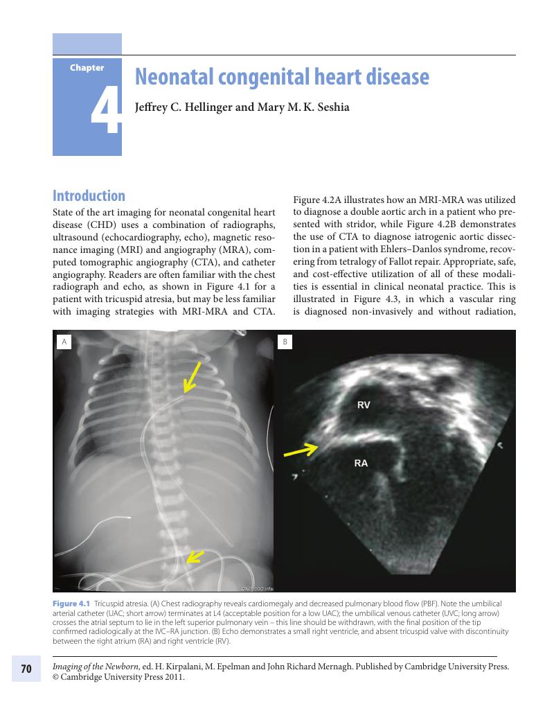
4 - Neonatal congenital heart disease
Published online by Cambridge University Press: 05 March 2012

Book contents
- Frontmatter
- Contents
- List of contributors
- Foreword by Alan Daneman
- Foreword by Phyllis A. Dennery
- Foreword by Avroy A. Fanaroff
- Preface
- 1 Introduction to principles of the radiological investigation of the neonate
- 2 Evidence-based use of diagnostic imaging: reliability and validity
- 3 The chest, page 11 to 40
- The chest, page 41 to 69
- 4 Neonatal congenital heart disease
- 5 Special considerations for neonatal ECMO
- 6 The central nervous system
- 7 The gastrointestinal tract
- 8 The kidney
- 9 Some principles of in utero and post-natal formation of the skeleton
- 10 Metabolic diseases
- 11 Catheters and tubes
- 12 Routine prenatal screening during pregnancy
- 13 Antenatal diagnosis of selected defects
- Index
- References
Summary
A summary is not available for this content so a preview has been provided. Please use the Get access link above for information on how to access this content.
- Type
- Chapter
- Information
- Imaging of the Newborn , pp. 70 - 97Publisher: Cambridge University PressPrint publication year: 2011
References
1. , , , et al. Three dimensional left atrial and esophagus reconstruction using cardiac C-arm computed tomography with image integration into fluoroscopic views for ablation of atrial fibrillation: accuracy of a novel modality in comparison with multislice computed tomography. Heart Rhythm 2008; 5(12):1651–7.Google Scholar
2. , , , , . Pediatric computed tomographic angiography: imaging the cardiovascular system gently. Radiol Clin North Am 2010; 48(2):439–67.Google Scholar
5. , . First thoracic vertebral body as reference for endotracheal tube placement. Arch Dis Child 1994; 71:F32–5.Google Scholar
6. , , , et al. Segmental approach to imaging of congenital heart disease. RadioGraphics 2010; 30:397–411.Google Scholar
7. , , , et al. Significant correlations between the flow volume of patent ductus venosus and early neonatal liver function: possible involvement of patent ductus venosus in postnatal liver function. Arch Dis Child Fetal Neonatal Ed 2006; 91:F175–9.Google Scholar
8. , , , et al. CT and MR imaging of pericardial disease. Radiographics 2003; 23:S167–80.Google Scholar
9. , , , et al. Fetal cardiomyopathies: pathogenic mechanisms, hemodynamic findings, and clinical outcome. Circulation 2002; 106(5):585–91.Google Scholar
10. , , , et al. The diagnosis, clinical course, and follow-up of children with cardiac tumours – a single center experience. Kardiol Pol 2010; 68(3):304–9.Google Scholar
11. , , , et al. Identification, imaging, functional assessment, and management of congenital arterial abnormalities in children. Cardiol Young 2007; 17(Suppl. 2):56–67.Google Scholar
12. . Diagnosis and management of cyanotic congenital heart disease: Part I. Indian J Pediatr 2009; 76(1):57–70.Google Scholar
13. . Diagnosis and management of cyanotic congenital heart disease: Part II. Indian J Pediatr 2009; 76(3):297–308.Google Scholar
14. , , , et al. Neonates with left-sided obstructive heart disease: clinical manifestation and management at primary care hospitals. Bratisl Lek Listy 2007; 108(7):316–19.Google Scholar
15. , , (2009) Congenital disease of the aortic arch: coarctation and arch anomalies. In , , (Eds.) Evidence-Based Imaging in Pediatrics: Optimizing Imaging in Pediatric Patient Care, New York, Springer, pp. 359–80.Google Scholar
16. , , , et al. Cardiovascular shunts: MR imaging evaluation. Radiographics 2003; 23(Spec No):S181–94.Google Scholar
17. . Conotruncal cardiac defects: a clinical imaging perspective. Pediatr Cardiol 2010; 31(3):430–7.Google Scholar