


Biological Applications
A Simple and Rapid Staining Technique for Sex Determination of Trichinella Larvae Parasites by Confocal Laser Scanning Microscopy by I Gavarane, E Kirilova, I Rubeniņa, L Mežaraupe, S Osipovs, G Deksne, A Pučkins, I Kokina, A Bulanovs, and M Kirjušina, Microsc Microanl | doi:10.1017/S1431927619015046
A novel fluorescent benzanthrone dye 3-N-[2-(4-morpholinyl)acetamido] has been developed for simple and rapid staining of parasitic roundworms such as Trichinella, a parasite of carnivorous and omnivorous animals. Trichinella can be transmitted to humans by consumption of uncooked infected meat products resulting in Trichinellosis, which can be fatal depending on the infective dose. Current staining protocols for Trichinella are time-consuming and labor-intensive. Using fixed T. spiralis and T. britovi larvae, the new preparation method takes approximately 5 minutes. Use of confocal microscopy and the new benzanthrone fluorescent dye makes examination of internal and external structures possible, and the sex of larval samples can be determined by measurement of the rectal length (Figure). Results confirmed that the larval male rectum is approximately two times larger than the female rectal length.

Confocal image of Trichinella spiralis female larva indicating the region of the rectum (<->).
Material Applications
Hollow Silver Nanostructures: The Role of Capping Agents in Tailoring the Shape, Structure, and Plasmonic Properties by BK Dadhich, B Bhushan, and A Priyam, Microsc Microanl | doi:10.1017/S1431927619000473
Due to single, intense, and highly tuneable plasmon peaks, hollow metal nanostructures have caught the imagination of scientists worldwide. Nanoshells of noble metals, particularly those of gold and silver, have a wide range of applications including catalysis and theranostics. In this work, the shape- and structure-directing ability of folic acid (FA) and acetic acid (AA) capping agents were investigated. FA transformed both the shape and structure of solid spherical Ag2O nanoparticles (NPs) resulting in hollow nanocubes (HAgNCs). In contrast, acetic acid (AA) acted only as a structure-directing agent resulting in the transformation of solid Ag2O nanospheres into hollow nanospheres (HAgNSs) (Figure). FA-capping leads to enhanced plasmon tunability ranging from 535–640 nm in the hollow Ag2O NPs. High-resolution transmission electron microscopy revealed that the outer diameter of AA-capped HAgNSs was 50 ± 10 nm, while the edge-length for FA-capped HAgNCs was 100 ± 15 nm. The diameter of the inner void space was 30 ± 5 nm and 43 ± 5 nm for the HAgNSs and HAgNCs, respectively.

Spherical Ag2O nanoparticles were transformed into hollow spheres (left) or cubes (right) by AA or FA. The transformation from spherical to cubical is attributed to the shape- and structure-directing ability of FA.
Techniques
HAADF-STEM Image Resolution Enhancement Using High-Quality Image Reconstruction Techniques: Case of Fe3O4(111) Surface by G Bárcena-González, MP Guerrero-Lebrero, E Guerrero, A Yañez, B Nuñez-Moraleda, D Kepaptsoglou, VK Lazarov, and PL Galindo, Microsc Microanal | doi: 10.1017/S1431927619014788
Microscopes can now provide multiple images of the same sample region in a very short period of time, and methods that rely on the use of a series of images are being developed. Rather than using a single image to extract information, the underlying idea is to take advantage of the entire series of images to allow details to be localized more precisely using super-resolution (SR) techniques. SR was applied to a series of HAADF-STEM images of Fe3O4 (111) to improve the signal-to-noise ratio (SNR) by a factor of 5 and roundness metrics (variability in the shape of the atomic columns) resulting in images with less noise and more detail than the original images. Zooming the SR image of Fe3O4 (111) in the surface region (Figure) reveals that the magnetite (111) surface consists of a full monolayer of Fe with the topmost Fe atoms in tetrahedral position (FeA2).
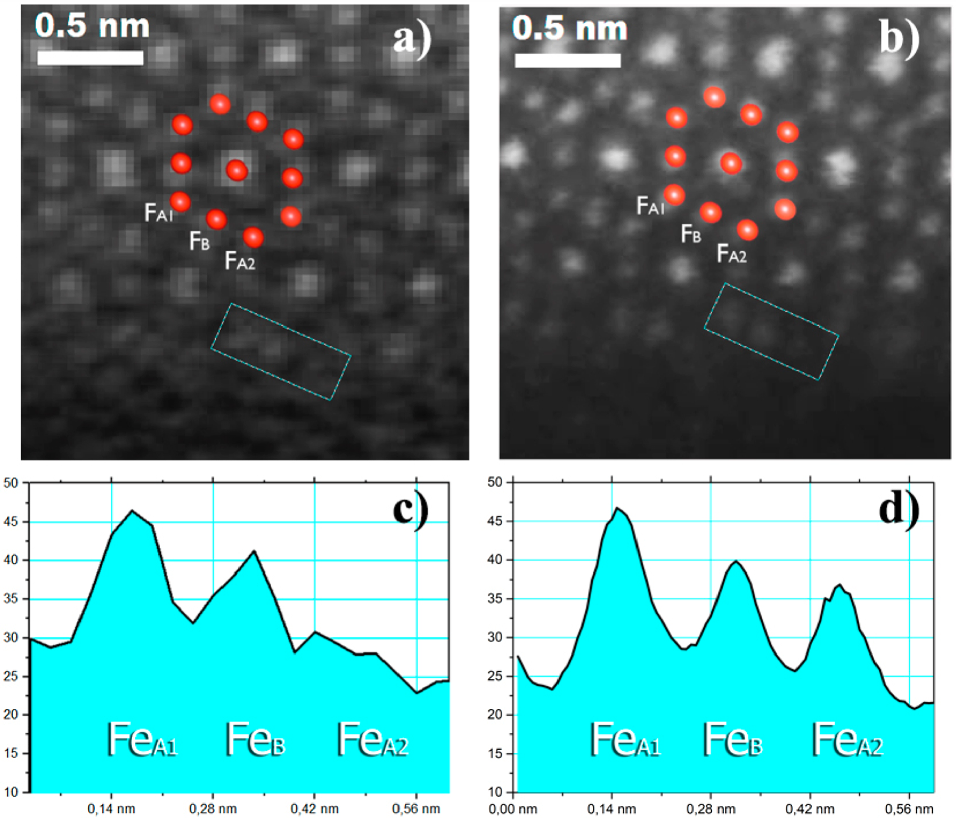
Images before (a) and after (b) SR processing. FeA1 and FeB denote Fe atomic columns in tetrahedral and octahedral positions. (c) and (d) show intensity profiles of the boxed areas in (a) and (b). In (c), two intensity peaks are observed, while in the SR image (d) three atomic columns are identified.
