


Introduction
Andreas Vesalius's De Humani Corporis Fabrica Libri Septum (On the Fabric of the Human Body in Seven Books) was a revolutionary publication that not only paved the way for modern anatomy, but changed the way we think about knowledge itself.
Andreas Vesalius (Figure 1) was born in Brussels in 1514 and was part of a medical family.Reference Nutton1,Reference Cunningham2 He enrolled at the University of Padua, where he found he had a flair for anatomy. At the time, Italy was the epicentre of the Renaissance; Ancient Roman and Greek texts had been revived and retranslated, and the classical civilisations were seen as the pinnacle of human achievement, influencing everything from art and literature to how the natural world was perceived. The main authority for medicine, and anatomy in particular, was the Roman Galen. Anatomy teaching was based on Galen's works, and these closely guided tutors’ dissection demonstrations and interpretations.

Fig. 1. Portrait of Vesalius, unnumbered page of De Humani Corporis Fabrica.Reference Vesalius3
At Padua, Vesalius quickly gained a reputation as a very accomplished dissector. In particular, he drew pictures of what his dissections revealed to him; his colleagues found these useful and so he decided to bring these illustrations together in a book. However, although an admirer of Galen, Vesalius’ meticulous observations were often at odds with Galen's texts, with Vesalius concluding that Galen's descriptions were often presented as human but garnered from animal vivisection (human cadavers being in short supply in the Roman Empire). This had brought Vesalius into conflict with more senior anatomists, who accused him of having contempt for Galen, consequently making him a controversial figure. However, De Humani Corporis Fabrica was an opportunity to show that Galen's work, and by extrapolation that of the other Ancients, could be analysed, amended and built upon, rather than rote learned and accepted at face value.
This article examines a second edition De Humani Corporis Fabrica held in John Ryland's Library,Reference Vesalius3 Manchester, with a particular focus on ENT, in order to trace the influence this book has had on our present-day practices, from nomenclature to shaping the surgical perspective.
Materials and methods
John Rylands Library, part of the University of Manchester, was opened in 1902 by Enriquetta Rylands in memory of her husband, a textiles entrepreneur and philanthropist.4 This imposing Victorian Gothic building houses an extensive collection of books and other manuscripts, including 2 second editions of De Humani Corporis Fabrica out of a known 113 copies.Reference Joffe5 Being a very large and delicate book, it is generally viewed on a purpose-made cushion and takes two people to manoeuvre it safely. However, the Library has recently digitised one of their copies, producing high-definition images. Therefore, various images of De Humani Corporis Fabrica will be used here, with permission of the Library, supplemented by secondary literature to better understand what these images depict.
Firstly, the title page will be examined as a general introduction to the themes of the book, followed by anatomy of the skull where pertinent to otolaryngology, the ear and nose, and finally the throat.
Results and discussion
Title page of De Fabrica
The title page of De Humani Corporis Fabrica itself is a work of art, demonstrating Vesalius as the ultimate Renaissance man, strewn with symbolism alluding to Aristotle's philosophy, royal destiny, Christian saints and Roman Emperors (Figure 2). In the centre, we have the great teacher, showman and visionary Vesalius: with a fresh perspective, he is championing meticulous observation and independent analysis over Galen's edicts, which are relegated to guidance. Vesalius's left hand pointing heavenward reminds students they are examining God's greatest creation.Reference Cunningham2 It is evident how he has his right hand on the cadaver – he is advocating a much more hands-on approach to anatomy.

Fig. 2. Title page of De Humani Corporis Fabrica.Reference Vesalius3
Rather than looking at Galen and ‘seeing’ his texts in the body, he is inviting us to learn through our own observations. Vesalius had realised Galen did not actually have access to many bodies and performed a lot of vivisection; therefore, much of his work had been presenting animal anatomy as human. Vesalius builds on this to show human anatomy only. Two characters are visible at the front in togas and sandals; these are Aristotle and Galen. Vesalius is putting himself on a par with these greats; he is the next chapter in the advancement of medical knowledge – not just following the classics, but building on them.Reference Siraisi6 He is not disrespecting Galen; the skeleton in the middle of the page represents the fact that Galen always used the bones as the starting point in anatomy, a practice replicated by Vesalius.
Further evidence of Vesalius’ ambition is in his dedication of the book to the Holy Roman Emperor. This is evident from the inscription at the top of the page, but also the cadaver. This is a female cadaver, with an incision in her abdomen, bringing to mind a Caesarean section – the method of Julius Caesar's birth.Reference Park7 As mentioned earlier, the classics were seen as the absolute pinnacle of civilisation at this time, and the royal family in particular liked to align themselves with the Roman Emperors. Hence, Vesalius is paying tribute to them and presenting himself as the obvious choice for court physician. In the Renaissance, the majority of funding for the arts and sciences came from rich, influential dynasties, and if Vesalius wanted to be thought of as one of the greats of medicine, he had to ingratiate himself accordingly.
Thus, this title page sets out Vesalius’ intention to revolutionise the field of anatomy, which is borne out in the rest of the text, as we will examine in the case of otolaryngology.
The skull pertinent to ENT in De Fabrica
Figures 3 and 4 show two pictures of the skull base, from above and below. Here we see the first depiction on the eponymous foramen of Vesalius indicated in the image – marked with an arrow in Figure 4 – transporting the emissary veins posteriorly from the face, through which infection can cause cavernous sinus thrombosis.Reference Saunders and O'Malley8

Fig. 3. Skull base, inferior view, page 28 of De Humani Corporis Fabrica.Reference Vesalius3

Fig. 4. Skull base, superior view, page 29 of De Humani Corporis Fabrica,Reference Vesalius3 with arrow indicating foramen of Vesalius.
Figure 5 depicts Vesalius’ description of the maxilla, which is very different to Galen's description, hence he has included a picture of a canine skull as well to remind us that Galen was not likely to have been using human maxilla for his description.Reference Saunders and O'Malley8

Fig. 5. Comparing the human and canine maxilla, page 46 of De Humani Corporis Fabrica.Reference Vesalius3
Otology in De Fabrica
Vesalius describes the ear as having two membranes: the first is the tympanic membrane and the second is the lining of the labyrinth. The auditory nerve then fans out over this membrane, as seen in Figure 6. The disconnect between this illustration and contemporary anatomy shows that Vesalius was unsure of otological structures. Although the first two ossicles had previously been recognised, we have the first naming of them as incus and malleus, to the right of the middle ear; however, the stapes is not featured yet at this point.Reference Saunders and O'Malley8,Reference Simpson9

Fig. 6. Middle and inner ear, with ossicles to the right, page 43 of De Humani Corporis Fabrica.Reference Vesalius3
This work paved the way for Vesalius’ follower Gabriele Fallopio/Fallopius, who studied under him in Padua, and went on to greatly further our understanding of the ear in Observations Anatomicae, including distinguishing between the facial nerve and the auditory nerve.Reference Mortazavi, Adeeb, Latif, Watanabe, Deep and Griessenauer10 Together with Vesalius, he mentored Giovanni Filippo Ingrassia, who has been credited with the first description of the lesser wings of the sphenoid and, by some, the stapes.Reference Cappello, Gerbino and Zummo11 However, others attribute the stapes to Vesalius’ Rome-based rival Bartholomew Eustachi/Eustachius, along with the tensor tympani and the cervical sympathetic chain.Reference Simpson9
Rhinology in De Fabrica
Firstly, we see the sphenoid in Figure 7. Without access to fixative, it was very difficult to dissect out the various neural structures at the skull base, but Vesalius attempted to give as complete a picture of this as possible.

Fig. 7. Sphenoid, page 31 of De Humani Corporis Fabrica.Reference Vesalius3
Figure 8 is an illustration of the pituitary. Vesalius thought that the pituitary collected the phlegm from the brain and then siphoned it into the nose.Reference Saunders and O'Malley8 He writes here about the ‘organ of smell’, which is very similar to Galen's treatise of the same name.

Fig. 8. Pituitary gland, showing drainage channels of phlegm, page 64 of De Humani Corporis Fabrica.Reference Vesalius3
Although Vesalius did not carry out much more dissection after the second edition of De Humani Corporis Fabrica, because of his duties at the royal court, his own copy of the book has been found with his annotations in the margins, where he was planning a third edition, which has been examined by Vivian Nutton.Reference Nutton12 In it, he changes the term ‘organ of smell’ to ‘the instrument which distinguishes smells’, a very subtle shift that nonetheless implies that he is moving away from Galen to a more complex pathway. He also starts to expand more on the foramina of the skull and their contents.
His notes here also describe people occasionally being able to taste in their mouth what is put into their ears, showing that he is expanding his understanding of the Eustachian tubes (although Eustachius is mentioned above as a contemporary of Vesalius, the Eustachian tubes were actually described much earlier than this time period, possibly around 500 BC, by the GreeksReference Simpson9).
Laryngology and the neck in De Fabrica
We now turn our attention to the trachea and laryngeal cartilages pictured in Figures 9 and 10. The epiglottis is depicted as very small – possibly because of the way the specimens were prepared by boiling them in water, which may have distorted the cartilages.
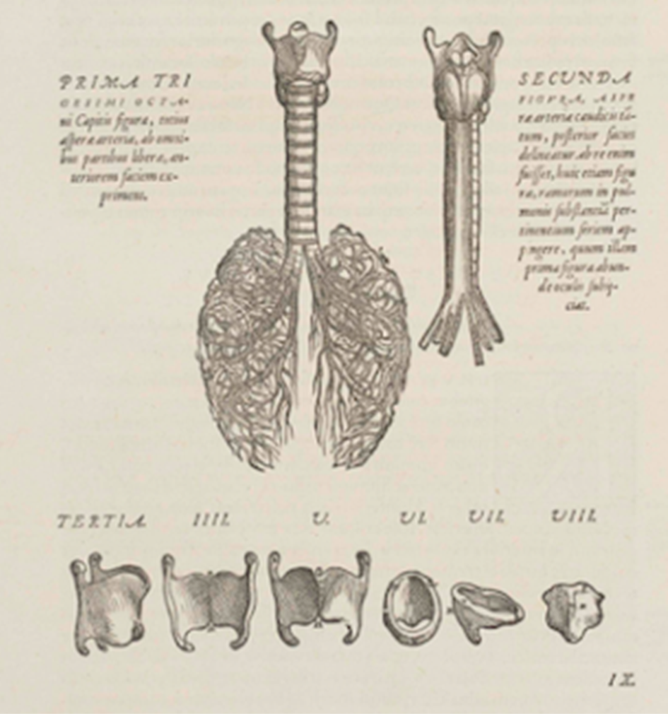
Fig. 9. Trachea and laryngeal cartilages, page 184 of De Humani Corporis Fabrica.Reference Vesalius3

Fig. 10. The larynx and its musculature, page 304 of De Humani Corporis Fabrica.Reference Vesalius3
Figure 11 shows the cricoid – it is not yet known as the cricoid (coming from the Greek for ‘ring’: ‘kricoid’). However, although Vesalius names it with the widely used, contemporary term ‘innominate’, he is the first to point out that it looks like a ring, likening it in the accompanying text to the rings the Turks use for archery.Reference Lydiatt and Bucher13 Later on, William Harvey (another University of Padua alumnus) adopted the term ‘ring’ also.

Fig. 11. The cricoid, with an inscription describing it as being shaped like the Turks’ archery rings, page 187 of De Humani Corporis Fabrica.Reference Vesalius3
Vesalius’ other work on the upper airway is similarly precise. He determines that ‘larynx’ cannot be simply interchanged with ‘pharynx’ or ‘throat’. Likewise, the glottis makes up but a small part of the airway, rather than its whole. The thyroid cartilage is shield-like (scutiform), rather than the commonly used (at the time) shield-shaped (scutalis). In addition, the ‘arytenoids' (derived from the Greek ‘arytaina’, or ladle or vessel) refer specifically to the spout shape, rather than the whole vessel as implied by other anatomists.Reference Lydiatt and Bucher13,Reference Garrison and Hast14
There is also arguably the first mention of the thyroid gland in modern anatomy – it can be seen in Vesalius' drawing, just inferior to the lateral thyroid cartilage in Figure 12. It is visible within Figure 9 too, if one looks closely at the same location. In the text, Vesalius describes the two fleshy glandulae laryngis at the second laryngeal cartilage.Reference Lamberg and Solin15 However, Galen had previously alluded to a set of glands in the pharynx and near the larynx in animal studies. In addition, Mondino de’ Luizzi (also known as Mundius Liucius) at the University of Bologna, a follower of Galen's teachings, published Anothomia Mundini in 1316, in which the thyroid does convincingly appear to be mentioned as almond-shaped glands beneath what we now call the strap muscles. Furthermore, Leonardo da Vinci's human anatomy drawings, known to be influenced by Mondino, do feature thyroid glands. Confusingly, though, Mondino calls them amigdalae, which was the contemporary term for tonsils, and Galen only writes about these glands in connection to those in the pharynx also, which is perhaps why his successors seemed to prefer to gloss over this portion of his work, making Vesalius’ description appear novel.
• Vesalius’ De Humani Corporis Fabrica is a celebrated Renaissance book that changed our approach to teaching and learning in anatomy, and, by extrapolation, medicine and surgery
• Vesalius showed that ancient knowledge was not set in stone, but could be analysed and built upon
• This article shows how this plays out in ENT, with detailed illustrations of the skull base, and the emergence of the thyroid as a significant structure
• The incus, malleus and cricoid are also named
• Vesalius may have minimised others’ contributions to anatomy, but his reputation has prevailed through the centuries

Fig. 12. Trachea and vessels, demonstrating the position of the thyroid, page 523 of De Humani Corporis Fabrica.Reference Vesalius3
Vesalius would have been familiar with the works of Mondino and his amygdalae. Furthermore, he had often been accused of minimising the influence of recent and contemporary anatomists, something highlighted by Nutton in his examination of Vesalius’ editing of the first and second versions of De Humani Corporis Fabrica, where he appears to be phasing out others’ contributions in this area of the neck.Reference Nutton12 He postulates that this may in part be because of his increasing rivalries with Eustachius, mentioned earlier, and Realdo Colombo, whom Vesalius had considered an assistant at Padua but who had probably been on a more equal standing,Reference Lydiatt and Bucher13 referred to in the first edition of De Humani Corporis Fabrica but subsequently erased after a conflict over the composition of the laryngeal structures (ultimately, Vesalius’ conviction that they were made of cartilage and not bone prevailed). He was known to later acknowledge Fallopius’ and Ingrassius’ ‘discoveries’ of levator palpebrae superioris and the stapes respectively, and corroborate these with his own dissections, but this was perhaps after the editing of the text had occurred.Reference Nutton12
In summary, Vesalius likely knew that these glands in the neck he was highlighting were not his ‘discovery’, and a different author might possibly have credited Mondino at this point. Nevertheless, Vesalius certainly brought more attention to the thyroid, and a more accurate depiction, than those before him.
Conclusion
Vesalius was talented and ambitious, and very confident in his own abilities, which is reflected well in De Humani Corporis Fabrica. His emphasis on observation, a hands-on approach, and his quest for the greatest accuracy in naming and defining structures, gave his work longevity, with its influence persisting to the twenty-first century. His detailed illustrations of the skull base are striking in their resemblance to modern diagrams. Although he found it difficult to access the minutiae of the middle-ear structures, he named two ossicles and paved the way for his protegees’ greater understanding. He hinted at more complex pathways for smell in his later work. He consolidated work on the upper airway, including the nature of its cartilages and delineation of its boundaries. Whilst the thyroid gland might not have been a ‘discovery’ of his, he brought it definition and made it a focus of study compared to its previous vague and intermittent appearances in the anatomical literature.
Vesalius perhaps minimised the work of his immediate predecessors and contemporaries, and we can only guess at the reasons for this; maybe it was a result of fierce professional rivalries, as recorded in his correspondence, or a burning desire to set himself apart as the most eminent anatomist of his time, as evidenced by his grandiose title page. Therefore, De Humani Corporis Fabrica may not have been as revolutionary as Vesalius would like us to have thought it was. Nevertheless, we can still appreciate how his precise annotations, and emphasis on using Galen as a guide rather than a hard and fast rule of anatomy, changed perspectives of anatomy and medicine as a whole, and even influenced how knowledge itself is gained.
Acknowledgements
I would like to thank the staff at John Rylands Library for their help in accessing the images and granting permission for their use. I would also like to thank Dr James Sumner and Dr Carsten Timmermann at the Centre for the History of Science, Technology and Medicine, University of Manchester, for introducing me to the work of Vesalius and suggesting a number of the secondary sources; also Dr Sumner for providing feedback on an earlier draft of work on the title page.
Competing interests
None declared


