Hostname: page-component-76fb5796d-22dnz Total loading time: 0 Render date: 2024-04-25T10:36:17.679Z Has data issue: false hasContentIssue false

Using Correlative Light and Electron Microscopy (CLEM) to Understand Ultrastructural Changes Induced by Salmonella typhimurium Infection in a Calf Ileum Loop Model

Published online by Cambridge University Press:  05 August 2019

Sophia Antonioli-Schmit
Affiliation:
Montana State University, Microbiology, Bozeman, Montana, USA.
Audrey Chong
Affiliation:
NIH/NIAID/ RML/Laboratory of Bacteriology, Hamilton, Montana, USA.
Bryan T Hansen
Affiliation:
NIH/NIAID/ RML/RTB/EM Unit, Hamilton, Montana, USA.
Mikayla Pulliam
Affiliation:
NIH/NIAID/ RML/RTB/EM Unit, Hamilton, Montana, USA.
Vinod Nair*
Affiliation:
NIH/NIAID/ RML/RTB/EM Unit, Hamilton, Montana, USA.
*
*Corresponding author: nairv@niaid.nih.gov

Abstract

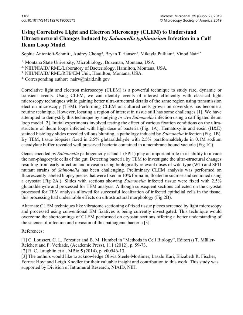
Image of the first page of this content. For PDF version, please use the ‘Save PDF’ preceeding this image.'
Type
Utilizing Microscopy for Research and Diagnosis of Diseases in Humans, Plants and Animals
Copyright
Copyright © Microscopy Society of America 2019 

References

[1]Loussert, C., Forestier, C. L. and Humbel, B. M. in “Methods in Cell Biology”, Editor(s) Müller-Reichert, T. and Verkade, P., (Academic Press), 111 (2012), p. 59-73.Google Scholar
[2]Laughlin, R. C. et al. MBio 5 (2014), p. e00946-13.Google Scholar
[3]The authors would like to acknowledge Olivia Steele-Mortimer, Laszlo Kari, Elizabeth R. Fischer, Forrest Hoyt and Leigh Knodler for their valuable insight and contribution to this work. This study was supported by Division of Intramural Research, NIAID, NIH.Google Scholar