


No CrossRef data available.
Article contents
Ultrastructural and Molecular Development of the Myotendinous Junction Triggered by Stretching Prior to Resistance Exercise
Published online by Cambridge University Press: 08 March 2022
Abstract
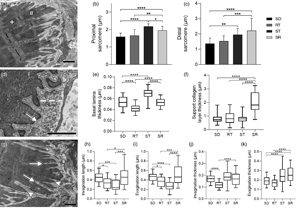
The myotendinous junction (MTJ) is a highly specialized region of the locomotor apparatus. Here, we investigated the ultrastructural and molecular effects in the MTJ region after static stretching prior to the ladder-based resistance training. Thirty-two male, 60-day old Wistar rats were divided into four groups: Sedentary, Resistance Training, Stretching, and Stretching-Resistance Training. The gastrocnemius muscle was processed for transmission electron microscopy techniques and Western blot assay. We observed that the static stretching prior to the ladder-based resistance training increased the MTJ components, the fibroblast growth factor (FGF)-2 and FGF-6 protein expression. Also, we demonstrated the lower transforming growth factor expression and no difference in the lysyl oxidase expression after combined training. The MTJ alterations in response to combined training demonstrate adaptive mechanisms which can be used for the prescription or development of methods to reduce or prevent injuries in humans and promote the myotendinous interface benefit.
- Type
- Biological Applications
- Information
- Copyright
- Copyright © The Author(s), 2022. Published by Cambridge University Press on behalf of the Microscopy Society of America
Footnotes
†
These authors contributed equally to this study.
References
Barbosa, GK, Jacob, CS, Rodrigues, MP, Rocha, LC, Pimentel Neto, J & Ciena, AP (2021). Morphological changes in the motor endplate and in the belly muscle induced by previous static stretching to the climbing protocol. Microsc Microanal 23, 1–9.Google Scholar
Bengtsson, H, Ekstrand, J, Waldén, M & Hägglund, M (2019). Few training sessions between return to play and first match appearance are associated with an increased propensity for injury: A prospective cohort study of male professional football players during 16 consecutive seasons. Br J Sports Med 54, 427–432.10.1136/bjsports-2019-100655CrossRefGoogle ScholarPubMed
Bouvier, T, Opplert, J, Cometti, C & Babault, N (2017). Acute effects of static stretching on muscle–tendon mechanics of quadriceps and plantar flexor muscles. Eur J Appl Physiol 117, 1309–1315.10.1007/s00421-017-3618-9CrossRefGoogle ScholarPubMed
Bradford, MM (1976). A rapid and sensitive method for the quantitation of microgram quantities of protein utilizing the principle of protein-dye binding. Anal Biochem 72, 258–254.10.1016/0003-2697(76)90527-3CrossRefGoogle ScholarPubMed
Cai, Q, Wu, G, Zhua, M, Gea, H, Xuea, C, Zhanga, Q, Chenga, B, Zuc, S & Wua, P (2020). FGF6 enhances muscle regeneration after nerve injury by relying on ERK1/2 mechanism. Life Sci 248, 117465.10.1016/j.lfs.2020.117465CrossRefGoogle Scholar
Chlebova, K, Bryja, V, Dvorak, P, Kozubik, A, Wilcox, WR & Krejci, P (2009). High molecular weight FGF2: The biology of a nuclear growth factor. Cell Mol Life Sci 66, 225–235.10.1007/s00018-008-8440-4CrossRefGoogle ScholarPubMed
Ciena, AP, Almeida, SRY, Alves, PHM, Bolina-Matos, RS, Dias, FJ, Issa, JPM, Iyomasa, MM & Watanabe, I (2011). Histochemical and ultrastructural changes of sternomastoid muscle in aged wistar rats. Micron 42, 871–876.10.1016/j.micron.2011.06.003CrossRefGoogle ScholarPubMed
Ciena, AP, Luques, IU, Dias, FJ, Almeida, SRY, Iyomasa, MM & Watanabe, I (2010). Ultrastructure of the myotendinous junction of the medial pterygoid muscle of adult and aged wistar rats. Micron 41, 1011–1014.10.1016/j.micron.2010.04.006CrossRefGoogle ScholarPubMed
Curzi, D (2016). Ultrastructural study of myotendinous junction plasticity: From disuse to exercise. Sport Sci Health 12, 279–286.10.1007/s11332-016-0301-1CrossRefGoogle Scholar
Curzi, D, Salucci, S, Marini, M, Esposito, F, Agnello, L, Veicsteinas, A, Buratini, S & Falcieri, E (2012). How physical exercise changes rat myotendinous junction: An ultrastructural study. Eur J Histochem 56, 117–122.CrossRefGoogle Scholar
De Palma, L, Marinelli, M, Pavan, M & Bertoni-Freddari, C (2011). Involvement of the muscle–tendon junction in skeletal muscle atrophy: An ultrastructural study. Rom J Morphol Embryol 52, 105–109.Google Scholar
Fleming, JW, Capel, AJ, Rimington, RP, Wheeler, P, Leonard, AN, Bishop, NC, Davies, OG & Lewis, MP (2020). Bioengineered human skeletal muscle capable of functional regeneration. BMC Biol 18, 145.10.1186/s12915-020-00884-3CrossRefGoogle ScholarPubMed
Gianelo, MCS, Polizzelo, JC, Chesca, D & Mattiello-Sverzut, AC (2016). Three days of intermittent stretching after muscle disuse alters the proteins involved in force transmission in muscle fibers in weanling rats. Braz J Med Biol Res 49, e4118.10.1590/1414-431x20154118CrossRefGoogle ScholarPubMed
Grillo, BAC, Rocha, LC, Martinez, GZ, Pimentel Neto, J, Jacob, CS, Watanabe, I & Ciena, AP (2021). Myotendinous junction components of different skeletal muscles present morphological changes in obese rats. Microsc Microanal 27, 598–560.10.1017/S1431927621000313CrossRefGoogle Scholar
Guo, D, Li, H, Liu, Y, Yu, X, Zhang, X, Chu, W, She, Y, Wang, D & Chen, G (2020). Fibroblast growth factor-2 promotes the function of tendon-derived stem cells in achilles tendon restoration in an achilles tendon injury rat model. Biochem Biophys Res Commun 521, 91–97.10.1016/j.bbrc.2019.10.082CrossRefGoogle Scholar
Heinemeier, KM, Olesen, JL, Haddad, F, Langberg, H, Kjaer, M, Baldwin, KM & Schjerling, P (2007). Expression of collagen and related growth factors in rat tendon and skeletal muscle in response to specific contraction types. J Physiol 582.3, 1303–1316.10.1113/jphysiol.2007.127639CrossRefGoogle Scholar
Hornberger, TA & Farrar, RP (2004). Physiological hypertrophy of the FHL muscle following 8 weeks of progressive resistance exercise in the rat. Can J Appl Physiol 29, 16–31.10.1139/h04-002CrossRefGoogle ScholarPubMed
Ismaeel, A, Kim, J, Kirk, JS, Smith, RS, Bohannon, WT & Koutakis, P (2019). Role of transforming growth factor-β in skeletal muscle fibrosis: A review. Int J Mol Sci 20, 2446.10.3390/ijms20102446CrossRefGoogle ScholarPubMed
Jacob, CS, Rocha, LC, Pimentel Neto, J, Watanabe, I & Ciena, AP (2019). Effects of physical training on sarcomere lengths and muscle-tendon interface of the cervical region in an experimental model of menopause. Eur J Histochem 63, 131–135.10.4081/ejh.2019.3038CrossRefGoogle Scholar
Jakobsen, JR & Krogsgaard, MR (2021). The myotendinous junction—A vulnerable companion in sports. A narrative review. Front Physiol 2021, 635561.10.3389/fphys.2021.635561CrossRefGoogle Scholar
Jiang, C, Shao, L, Wang, Q & Dong, Y (2012). Repetitive mechanical stretching modulates transforming growth factor-β induced collagen synthesis and apoptosis in human patellar tendon fibroblasts. Biochem Cell Biol 90, 667–674.10.1139/o2012-024CrossRefGoogle ScholarPubMed
Kamonseki, DH, Gonçalves, GA, Yi, LC & Lombardi Júnior, I (2014). Effect of stretching with and without muscle strengthening exercises for the foot and hip in patients with plantar fasciitis: A randomized controlled single-blind clinical trial. Man Ther 23, 76–82.10.1016/j.math.2015.10.006CrossRefGoogle Scholar
Kraemer, WJ & Ratamess, NA (2004). Fundamentals of resistance training: Progression and exercise prescription. Med Sci Sports Exerc 36, 674–688.10.1249/01.MSS.0000121945.36635.61CrossRefGoogle ScholarPubMed
Laczko, R & Csiszar, K (2020). Lysyl oxidase (LOX): Functional contributions to signaling pathways. Biomolecules 10, 1093.10.3390/biom10081093CrossRefGoogle ScholarPubMed
Leite, FS & Rassier, DE (2020). Sarcomere length nonuniformity and force regulation. Biophys J 199, 2372–2377.10.1016/j.bpj.2020.11.005CrossRefGoogle Scholar
Martinez, GZ, Grillo, BAC, Rocha, LC, Jacob, CS, Pimentel Neto, J, Tomiate, NA, Barbosa, GK, Watanabe, I & Ciena, AP (2021). Morphological changes in the myotendinous junction of mdx mice. Microsc Microanal 27, 1290–1294.10.1017/S1431927621012496CrossRefGoogle Scholar
Martins, HRF, Zotz, TGG, Messa, SP, Capriglione, LGA, Zotz, R, Noronha, L, Azevedo, MLV & Gomes, ARS (2020). Morphometric and molecular muscle remodeling after passive stretching in elderly female rats. Clinics 75, e1769.10.6061/clinics/2020/e1769CrossRefGoogle ScholarPubMed
Miller, AJ, Brandon, R & Norstrom, EM (2016). Protein electrophoretic migration data from custom and commercial gradient gels. Data Brief 9, 1–3.10.1016/j.dib.2016.08.018CrossRefGoogle ScholarPubMed
Moo, EK & Herzog, W (2018). Single sarcomere contraction dynamics in a whole muscle. Sci Rep 8, 15235.10.1038/s41598-018-33658-7CrossRefGoogle Scholar
Moo, EK, Leonard, TR & Herzog, W (2017). In vivo sarcomere lengths become more non-uniform upon activation in intact whole muscle. Front Physiol 8, 1015.10.3389/fphys.2017.01015CrossRefGoogle ScholarPubMed
Moo, EK, Leonard, TR & Herzog, W (2020). The sarcomere force–length relationship in an intact muscle–tendon unit. J Exp Biol 25, jeb215020.10.1242/jeb.215020CrossRefGoogle Scholar
Morton, SK, Whitehead, JR, Brinkert, RH & Caine, DJ (2011). Resistance training vs. static stretching: Effects on flexibility and strength. J Strength Cond Res 25, 3391–3398.10.1519/JSC.0b013e31821624aaCrossRefGoogle ScholarPubMed
Nunes, JP, Schoenfeld, BJ, Nakamura, M, Ribeiro, AS, Cunha, PM & Cyrino, ES (2020). Does stretch training induce muscle hypertrophy in humans? A review of the literature. Clin Physiol Funct Imaging 40, 148–156.10.1111/cpf.12622CrossRefGoogle ScholarPubMed
Pawlikowski, B, Vogler, TO, Gadek, K & Olwin, BB (2017). Regulation of skeletal muscle stem cells by fibroblast growth factors. Dev Dyn 246, 359–367.CrossRefGoogle ScholarPubMed
Peviani, SM, Guzzoni, V, Pinheiro-Dardis, CM, Silva, YPD, Fioravante, ACR, Sagawa, AH, Delfino, GB, Durigan, JLQ & Salvini, TF (2018). Regulation of extracellular matrix elements and sarcomerogenesis in response to different periods of passive stretching in the soleus muscle of rats. Sci Rep 8, 9010.10.1038/s41598-018-27239-xCrossRefGoogle ScholarPubMed
Pimentel Neto, J, Rocha, LC, Barbosa, GK, Jacob, CS, Krause Neto, W, Watanabe, I & Ciena, AP (2020). Myotendinous junction adaptations to ladder-based resistance training: Identification of a new telocyte niche. Sci Rep 10, 14124.10.1038/s41598-020-70971-6CrossRefGoogle ScholarPubMed
Rassier, DE (2017). Sarcomere mechanics in striated muscles: From molecules to sarcomeres to cells. Am J Physiol Cell Physiol 313, 134–145.CrossRefGoogle ScholarPubMed
Rocha, LC, Barbosa, GK, Pimentel Neto, J, Jacob, CS, Knudsen, AB, Watanabe, I & Ciena, AP (2021). Aquatic training after joint immobilization in rats promotes adaptations in myotendinous junctions. Int J Mol Sci 22, 6983.10.3390/ijms22136983CrossRefGoogle ScholarPubMed
Rocha, LC, Jacob, CS, Barbosa, GK, Watanabe, I & Ciena, AP (2020). Repercussions on sarcomeres of the myotendinous junction and the myofibrillar type adaptations in response to different trainings on vertical ladder. Microsc Res Tech 83, 1190–1197.Google ScholarPubMed
Schmelzer, CEH, Hedtke, T & Heinz, A (2019). Unique molecular networks: Formation and role of elastin cross-links. IUBMB Life 72, 842–854.10.1002/iub.2213CrossRefGoogle ScholarPubMed
Sierra, LR, Fávaro, G, Cerri, BR, Rocha, LC, Almeida, SRY, Watanabe, I & Ciena, AP (2018). Myotendinous junction plasticity in aged ovariectomized rats submitted to aquatic training. Microsc Res Tech 81, 816–822.CrossRefGoogle Scholar
Wood, LK & Brooks, SV (2015). Ten weeks of treadmill running decreases stiffness and increases collagen turnover in tendons of old mice. J Orthop Res 34, 346–353.CrossRefGoogle ScholarPubMed