Hostname: page-component-76fb5796d-r6qrq Total loading time: 0 Render date: 2024-04-25T09:29:03.868Z Has data issue: false hasContentIssue false

Structural Investigation of Epididymal Microvasculature and Its Relation to Telocytes and Immune Cells in Camel

Published online by Cambridge University Press:  15 July 2020

Manal T. Hussein*
Affiliation:
Department of Anatomy and Histology, Faculty of Veterinary Medicine, Assiut University, Assiut71526, Egypt
Fatma M. Abdel-Maksoud
Affiliation:
Department of Anatomy and Histology, Faculty of Veterinary Medicine, Assiut University, Assiut71526, Egypt
*
*Author for correspondence: Manal T. Hussein, E-mail: manaltawfik22@gmail.com
Get access

Abstract

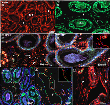
The vascular and perivascular cells, including telocytes (TCs) and immune cells, play an important role in male fertility. The current study intended to describe in detail the microvascular structures harboring special regulatory devices in addition to the interstitial cellular components of the one-humped camel epididymis. The samples were collected from 10 clinically healthy mature camels (Camelus dromedarius). The distribution and characteristics of TCs, peripheral blood vessels of the epididymis, and immune cells were investigated using the light, immunohistochemistry, immunofluorescence, and transmission electron microscopy analyses. Frequent occlusive or throttle arterioles were demonstrated in the epididymal interstitium and their tunica media consisted of glomus cells. In addition, some vein walls consisted of one or two layers of glomus cells. TCs, fibroblasts, muscle cells, and tunica media of the blood vessels, that present in the loose connective tissue surrounding the intertubular interstitium of camel epididymis, showed a positive reaction with vimentin. The endothelium of blood vessels and veins showed positive immunoreactivity for CD34 and vascular endothelial growth factor (VEGF). Furthermore, VEGF, CD34, and S100 proteins were expressed in dendritic cells (DCs) as well as TCs. The current data suggest the involvement of DCs and TCs in angiogenesis and a possible role for the interstitial components in creating an appropriate milieu for the full maturation of sperms.

Type
Micrographia
Copyright
Copyright © Microscopy Society of America 2020

Access options

Get access to the full version of this content by using one of the access options below. (Log in options will check for institutional or personal access. Content may require purchase if you do not have access.)

References

Abd-Elhafeez, HH, Mokhtar, DM & Hassan, AH (2017). Effect of melatonin on telocytes in the seminal vesicle of the Soay Ram: An immunohistochemical, ultrastructural and morphometrical study. Cells Tissues Organs 203, 2954.CrossRefGoogle ScholarPubMed
Abdel-Maksoud, FM, Abd-Elhafeez, HH & Soliman, SA (2019 a). Morphological changes of telocytes in camel efferent ductules in response to seasonal variations during the reproductive cycle. Sci Rep 9, 4507.CrossRefGoogle ScholarPubMed
Abdel-Maksoud, FM, Hussein, MT & Attaai, A (2019 b). Seasonal variation of the intraepithelial gland in camel epididymis with special reference to autophagosome. Microsc Microanal 25, 10521060.CrossRefGoogle ScholarPubMed
Abd El-Maksoud, FM (2010). Morphological studies on the seasonal changes in the epididymis of the one-humped camel (Camelus dromedarius). M.V.Sc. Assiut University, Faculty of Veterinary Medicine.Google Scholar
Alkafafy, M, Ebada, S, Rashed, R & Attia, H (2012). Comparative morphometric and glycohistochemical studies on the epididymal duct in the donkey (Equus asinus) and dromedary camel (Camelus dromedarius). Acta Histochem 114, 434447.CrossRefGoogle Scholar
Ambartsumian, N, Grigorian, M & Lukanidin, E (2005). Genetically modified mouse models to study the role of metastasis-promoting S100A4(mts1) protein in metastatic mammary cancer. J Dairy Res 72 Spec No:27-33.CrossRefGoogle Scholar
Atanasova, DY, Iliev, ME & Lazarov, NE (2011). Morphology of the rat carotid body. Biomed Rev 22, 4155.CrossRefGoogle Scholar
Bancroft, JD, Layton, C & Suvarna, SK (2013). Bancroft's Theory and Practice of Histological Techniques, 7th ed.. London: Elsevier/Churchill Livingstone.Google Scholar
Ceafalan, L, Gherghiceanu, M, Popescu, LM & Simionescu, O (2012). Telocytes in human skin – Are they involved in skin regeneration? J Cell Mol Med 16, 14051420.CrossRefGoogle ScholarPubMed
Cismaşiu, VB & Popescu, LM (2015). Telocytes transfer extracellular vesicles loaded with microRNAs to stem cells. J Cell Mol Med 19, 351358.CrossRefGoogle ScholarPubMed
Cretoiu, D, Radu, BM, Banciu, A, Banciu, DD & Cretoiu, SM (2017). Telocytes heterogeneity: From cellular morphology to functional evidence. Semin Cell Dev Biol 64, 2639.CrossRefGoogle ScholarPubMed
Cretoiu, SM, Cretoiu, D & Popescu, LM (2012). Human myometrium – The ultrastructural 3D network of telocytes. J Cell Mol Med 16, 28442849.CrossRefGoogle ScholarPubMed
Cretoiu, SM & Popescu, LM (2014). Telocytes revisited. Biomol Concepts 5, 353369.CrossRefGoogle ScholarPubMed
Cretoiu, SM, Simionescu, AA, Caravia, L, Curici, A, Cretoiu, D & Popescu, LM (2011). Complex effects of imatinib on spontaneous and oxytocin-induced contractions in human non-pregnant myometrium. Acta Physiol Hung 98, 329338.CrossRefGoogle ScholarPubMed
Díaz-Flores, L, Gutiérrez, R, García, MP, González, M, Sáez, FJ, Aparicio, F, Díaz-Flores, L Jr., & Madrid, FJ (2015). Human resident CD34+ stromal cells/telocytes have progenitor capacity and are a source of αSMA+ cells during repair. Histol Histopathol 30, 615627.Google ScholarPubMed
Donato, R (2003). Intracellular and extracellular roles of S100 proteins. Microsc Res Tech 60, 540551.CrossRefGoogle ScholarPubMed
Eidenschink, L, Dizerega, G, Rodgers, K, Bartlett, M, Wells, DA & Loken, MR (2012). Basal levels of CD34 positive cells in peripheral blood differ between individuals and are stable for 18 months. Cytometry B Clin Cytom 82, 1825.CrossRefGoogle ScholarPubMed
En El Camello Bactriano, C (2016). Ultrastructure features of coronary artery endothelium in Bactrian camel (Camelus bactrianus). Int J Morphol 34, 280284.Google Scholar
Fath-Elbab, MR & Abou-Elhamd, AS (2016). Special cutaneous vascular elements in one-humped camel (Camelus dromedarius). J Adv Vet Anim Res 3, 106111.CrossRefGoogle Scholar
Fina, L, Molgaard, HV, Robertson, D, Bradley, NJ, Monaghan, P, Delia, D, Sutherland, DR, Baker, MA & Greaves, MF (1990). Expression of the CD34 gene in vascular endothelial cells. Blood 75, 24172426.CrossRefGoogle ScholarPubMed
Gherghiceanu, M & Popescu, LM (2012). Cardiac telocytes - their junctions and functional implications. Cell Tissue Res 348, 265279.CrossRefGoogle ScholarPubMed
Gorgas, K, Bock, P, Tischendorf, F & Curri, SB (1977). The fine structure of human digital arterio-venous anastomoses (Hoyer-Grosser's organs). Anat Embryol (Berl) 150, 269289.CrossRefGoogle Scholar
Hicklin, DJ & Ellis, LM (2005). Role of the vascular endothelial growth factor pathway in tumor growth and angiogenesis. J Clin Oncol 23, 10111027.CrossRefGoogle ScholarPubMed
Hirai, S, Naito, M, Terayama, H, Ning, Q, Miura, M, Shirakami, G & Itoh, M (2010). Difference in abundance of blood and lymphatic capillaries in the murine epididymis. Med Mol Morphol 43, 3742.CrossRefGoogle ScholarPubMed
Hume, DA (2006). The mononuclear phagocyte system. Curr Opin Immunol 18, 4953.CrossRefGoogle ScholarPubMed
Hussein, MM (2019). Structural and functional characteristics of the special regulatory devices in the peripheral pulmonary circulation in rabbits. Protoplasma 257, 755766.CrossRefGoogle ScholarPubMed
Hussein, MM & Hassan, AHS (2018). Special histological features of the angioarchitecture of the ovaries in the Egyptian Buffaloes (Bubalus bubalis). J Adv Microsc Res 13, 298305.CrossRefGoogle Scholar
Hussein, MM & Mokhtar, DM (2018). The roles of telocytes in lung development and angiogenesis: An immunohistochemical, ultrastructural, scanning electron microscopy and morphometrical study. Dev Biol 443, 137152.CrossRefGoogle ScholarPubMed
Hussein, MT, Mokhtar, DM & Hassan, AHS (2020). Melatonin activates the vascular elements, telocytes, and neuroimmune communication in the adrenal gland of Soay rams during the non-breeding season. Protoplasma 257, 353369.CrossRefGoogle ScholarPubMed
Kameda, Y (2005). Mash1 is required for glomus cell formation in the mouse carotid body. Dev Biol 283, 128139.CrossRefGoogle ScholarPubMed
Karnovsky, MJ (1965). A formaldehyde-glutaraldehyde fixative of high osmolarity for use in electron microscopy. J Cell Biol 27, 137A138A.Google Scholar
Lin, CS, Ning, H, Lin, G & Lue, TF (2012). Is CD34 truly a negative marker for mesenchymal stromal cells? Cytotherapy 14, 11591163.CrossRefGoogle ScholarPubMed
Marini, M, Manetti, M, Rosa, I, Ibba-ManneschI, L & Sgambati, E (2018). Telocytes in human fetal skeletal muscle interstitium during early myogenesis. Acta Histochem 120, 397404.CrossRefGoogle ScholarPubMed
Mital, P, Hinton, BT & Dufour, JM (2011). The blood-testis and blood-epididymis barriers are more than just their tight junctions. Biol Reprod 84, 851858.CrossRefGoogle ScholarPubMed
Mokhtar, DM & Abd-Elhafez, EA (2016). Morphological studies on the peripheral circulation of the ovary in one-humped camel (Camelus dromedarius). Anat Histol Embryol 45, 319328.CrossRefGoogle Scholar
Mokhtar, DM, Hussein, MT & Hassan, AHS (2017). Melatonin elicits stimulatory action on the adrenal gland of Soay Ram: Morphometrical, immunohistochemical, and ultrastructural study. Microsc Microanal 23, 11731188.CrossRefGoogle ScholarPubMed
Nielsen, JS & Mcnagny, KM (2008). Novel functions of the CD34 family. J Cell Sci 121, 36833692.CrossRefGoogle ScholarPubMed
Popescu, BO, Gherghiceanu, M, Kostin, S, Ceafalan, L & Popescu, LM (2012). Telocytes in meninges and choroid plexus. Neurosci Lett 516, 265269.CrossRefGoogle ScholarPubMed
Popescu, LM, Ciontea, SM & Cretoiu, D (2007). Interstitial Cajal-like cells in human uterus and fallopian tube. Ann N Y Acad Sci 1101, 139165.CrossRefGoogle ScholarPubMed
Popescu, LM, Ciontea, SM, Cretoiu, D, Hinescu, ME, Radu, E, Ionescu, N, Ceausu, M, Gherghiceanu, M, Braga, RI, Vasilescu, F, Zagrean, L & Ardeleanu, C (2005). Novel type of interstitial cell (Cajal-like) in human fallopian tube. J Cell Mol Med 9, 479523.CrossRefGoogle Scholar
Potter, SJ & Defalco, T (2017). Role of the testis interstitial compartment in spermatogonial stem cell function. Reproduction 153, R151R162.CrossRefGoogle ScholarPubMed
Rafii, S, Butler, JM & Ding, BS (2016). Angiocrine functions of organ-specific endothelial cells. Nature 529, 316325.CrossRefGoogle ScholarPubMed
Reynolds, ES (1963). The use of lead citrate at high pH as an electron-opaque stain in electron microscopy. J Cell Biol 17, 208212.CrossRefGoogle ScholarPubMed
Richardson, KC, Jarett, L & Finke, EH (1960). Embedding in epoxy resins for ultrathin sectioning in electron microscopy. Stain Technol 35, 313323.CrossRefGoogle ScholarPubMed
Romeis, B (1989). Mikroskopische Technik.Urban und Schwarzenberg. München, Baltimore: Wien.Google Scholar
Rusu, MC, Jianu, AM, Mirancea, N, Didilescu, AC, Manoiu, VS & Paduraru, D (2012). Tracheal telocytes. J Cell Mol Med 16, 401405.CrossRefGoogle ScholarPubMed
Sidney, LE, Branch, MJ, Dunphy, SE, Dua, HS & Hopkinson, A (2014). Concise review: Evidence for CD34 as a common marker for diverse progenitors. Stem Cells 32, 13801389.CrossRefGoogle ScholarPubMed
Skidmore, JA (2018). Reproduction in dromedary camels: An update. Anim. Reprod 2, 161171.Google Scholar
Sullivan, R & Mieusset, R (2016). The human epididymis: Its function in sperm maturation. Hum Reprod Update 22, 574587.CrossRefGoogle ScholarPubMed
Turner, TT (2008). De Graaf's thread: The human epididymis. J Androl 29, 237250.CrossRefGoogle ScholarPubMed
Voisin, A, Saez, F, Drevet, JR & Guiton, R (2019). The epididymal immune balance: A key to preserving male fertility. Asian J Androl 21 (6), 531539.Google ScholarPubMed
Zayed, AE, Aly, K, Ibrahim, IA & Abd El-Maksoud, FM (2012). Morphological studies on the epididymal duct of the one-humped camel (Camelus dromedaries). J Vet Med 2, 245254.Google Scholar
Zheng, Y, Bai, C & Wang, X (2012 a). Telocyte morphologies and potential roles in diseases. J Cell Physiol 227, 23112317.CrossRefGoogle ScholarPubMed
Zheng, Y, Cretoiu, D, Yan, G, Cretoiu, SM, Popescu, LM, Fang, H & Wang, X (2014). Protein profiling of human lung telocytes and microvascular endothelial cells using iTRAQ quantitative proteomics. J Cell Mol Med 18, 10351059.CrossRefGoogle ScholarPubMed
Zheng, Y, Zhu, T, Lin, M, Wu, D & Wang, X (2012 b). Telocytes in the urinary system. J Transl Med 10, 188.CrossRefGoogle ScholarPubMed
Zhou, J, Zhang, Y, Wen, X, Cao, J, Li, D, Lin, Q, WanG, H, Liu, Z, Duan, C, Wu, K & Wang, C (2010). Telocytes accompanying cardiomyocyte in primary culture: Two- and three-dimensional culture environment. J Cell Mol Med 14, 26412645.CrossRefGoogle ScholarPubMed